The community site for and by
developmental and stem cell biologists

Post-doc position: Canada

Posted by , on 28 July 2011

Closing Date: 15 March 2021

I currently have an opening in my research group for a post-doc to investigate the development of the vertebrate skeleton.  Our lab studies the development of the neural crest derived skeleton in a comparative manner in chicken and fish embryos (zebrafish and Mexican tetra).   This position will focus on the signals involved in the patterning of skeletal elements in one or more of these animals and the interactions between neural crest and mesodermal tissues.  Applicants who have recently completed a PhD, have experience in molecular biology, developmental and cell biology are strongly encouraged to apply.  

MSVU is an undergraduate university on the East Coast of Canada in beautiful Nova Scotia.  Although a small university, I have a large research group.  This position offers opportunities to interact with a growing research group of undergraduates and graduate students in a well equipped CFI funded lab.  In addition, opportunities to teach, train undergraduates/graduate students and to help manage my lab will be available.  

If you want to hone your teaching and management skills, as well as engage in exciting research then this position might be for you!  Please email a CV, a one page statement of your research experience and interests, and the contact information for three referees to:

Dr Tamara Franz-Odendaal    Tamara.franz-odendaal@msvu.caBiology Dept, Mount Saint Vincent University,

166 Bedford Highway, Halifax, Nova Scotia, B3M 3E4,CANADA

Thumbs up (No Ratings Yet)
Loading...

Tags: , , , , , , , ,
Categories: Careers, Jobs

Navigate the archive

Use our Advanced Search tool to search and filter posts by date, category, tags and authors.

Live imaging of stem cell maintenance, loss, and renewal in the Drosophila testis

Posted by , on 28 July 2011

Stem cells have often been imaged live in culture, but very few stem cell systems are conducive to live imaging within their native tissues.  An essential property of adult stem cells that they are maintained at specific anatomical locations called niches.  The interactions between stem cells and their niche are crucial, but are often disrupted when stem cells are studied in vitro.  In our recent Development paper, we use live imaging to directly watch male Drosophila germline stem cells (GSCs) in action within their native niche by ex-vivo imaging of intact testes.  We find that while wild type GSC divisions usually result in the production of a stem cell and daughter cell, they occasionally produce two stem cells or two daughter cells.  Our findings highlight a flexibility in stem cell output that is modulated during regeneration similar to mouse spermatogonial stem cells (Nakagawa et al., 2007, Nakagawa et al., 2010).

Niches are usually composed of stromal cells that activate developmental signaling pathways in nearby stem cells, thus maintaining them in an undifferentiated state.  The cluster of non-dividing stromal cells to which germline and somatic stem cells in the Drosophila testis apex are attached is appropriately called the “hub” and secretes cytokines required for the maintenance of both stem cell populations.  Cells adjacent to the hub have activated Jak-STAT signaling, and remain undifferentiated.  Cells further away from the hub initiate differentiation and eventually form sperm.

After running into similar issues as what Lucy Morris described in her previous post, we developed a live imaging protocol using either spinning-disk or 2-photon microscopy that enabled us to image stem cells within intact testes for up to 12 hours – about half of the GSC cell cycle.  By manually tracking individual stem cells and their daughter cells, we saw the most GSCs divide with a stereotypical spindle orientation perpendicular to the hub, thus displacing daughter cells out of the niche.  This phenomenon had been seen many times previously in immunostained, fixed testes and in testes studied by short-term live imaging, and had been the only observed mode of GSC divisions (Yamashita et al., 2003).  Surprisingly, in wild type young testes, we saw a few cases where GSCs, after first displacing their daughter cells away from hub, then swiveled such that the daughter cell gained and maintained contact with the hub until the end of imaging.  This suggested that GSCs can switch from their normal mode of division where one GSC and one daughter cell re produced, to one where two GSCs are produced.  We also saw the converse, where GSCs-daughter pairs lost contact with the hub and appeared to directly differentiate into a pair of spermatogonia.  Thus, our live imaging captured three different modes of division – asymmetric division, symmetric renewal, and symmetric differentiation – that occur simultaneously within a stem cell niche to balance its output during steady-state (click here to see a movie of this!).

By using the same imaging technique, we were able to look at cells undergoing the process of dedifferentiation, where a more differentiated cell reverts into a less differentiated cell or stem cell.  Our lab previously demonstrated that spermatogonia (transit-amplifying stem cell daughters) are able to revert into stem cells when they encounter a niche depleted of stem cells (Brawley and Matunis, 2004).  While examining dedifferentiation, we stumbled upon a scenario where we thought that the normally immobile spermatogonia were able to move towards the hub in order to gain physical contact with the hub before reverting into stem cells.  Whether the cells were gaining a migratory morphology during the process was unknown.  While we never attained a satisfactory answer to whether Drosophila spermatogonia gain intrinsic migratory properties, we saw many examples of spermatogonia initially not in contact with the hub gain (click here to see a movie of this).  The caveat is that we were not able to image the somatic cells within the niche, and these somatic cells could easily be shuttling the dedifferentiating spermatogonia in a manner similar to that of escort stem cells in the Drosophila ovary (Morris and Spradling, 2011).  We also noticed that the modes of GSC divisions changed greatly from that seen during steady-state (movie of this too!).  The balance of the different modes of stem cell division was now tipped to that of producing more GSCs in an effort to replenish the niche.

Our findings that stem cells are frequently replaced and regenerated in the Drosophila testis are in line with recent studies in mammalian tissues using lineage tracing and mathematical modeling of clone behavior.  These studies suggest that the classical stem cell definition of a long-lived, slow cycling cell may no longer be accurate.  While this view has yet to be challenged in the mammalian hematopoietic system, it has been demonstrated recently that mammalian gut, interfollicular epidermis, and testes all contain stem cells that not only undergo turnover on the scale of weeks, but also divide frequently (reviewed in Klein and Simmons, 2011).  I believe that more and more stem cell systems will be demonstrated to have these properties, and that diseases such as cancer may eventually be similarly modeled.

ResearchBlogging.orgSheng, X., & Matunis, E. (2011). Live imaging of the Drosophila spermatogonial stem cell niche reveals novel mechanisms regulating germline stem cell output Development, 138 (16), 3367-3376 DOI: 10.1242/dev.065797

Thumbs up (7 votes)
Loading...

Tags: , , , , , , ,
Categories: Research

From the Embryology 2011 Class: 4th of July Parade

Posted by , on 26 July 2011

Because Woods Hole, MA is home to both an Oceanographic Institute and the Marine Biological Laboratory, most of the people in this small town have some connection to the scientific community. As a result, the fourth of July festival in Woods Hole, MA is a celebration of uninhibited science-geekery.

Some of the highlights included the neurobiology students:

Microbial diversity, with their giant squid:

And my personal favorite, Mendel with his peas:

Bringing up the rear was our own Embryology course.  We have a time-honored tradition of performing gastrulation through interpretive dance, which is quite possibly the best way to show gastrulation:

If that wasn’t self explanatory, let me help.

The three different colors of our shirts represent the three germ layers: blue for ectoderm, red for mesoderm, yellow for endoderm.  In this particular display, we are performing gastrulation as it occurs in the sea urchin (that was obvious, right?) We started out as a blastula (a hollow ball of cells), and then invaginated to create the three layers of tissue. Finally, some of the mesoderm cells start to form spicules, which create the skeleton of the urchin.

Sea urchins were only the beginning. We also performed gastrulation as it occurs in frogs, nematode worms, fruit flies, as well as chaotic cleavage (like you find in some sea anemones and jellyfish) and chicken neurulation for good measure.  The parade lasted less than an hour, and only traveled a few blocks, but we got as many gastrulations as we could in there.

If you’re ever in the area during the fourth of July, I highly recommend you check out the Woods Hole parade.  Their were a lot of people, and a lot of energy (including an epic water gun fight).  You can get a more detailed explanation of gastrulation and some more pictures over at the blog BioBlueprints.

Thumbs up (4 votes)
Loading...

Categories: Events

In Development this week (Vol. 138, Issue 16)

Posted by , on 26 July 2011

Here are the research highlights from the current issue of Development:

Pushing the nuclear envelope

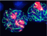
Not all nuclei are regular spheres as is often shown in textbooks. For example, in Drosophila embryos, nuclei are initially spherical but they elongate and acquire an irregular lobulated morphology during cellularisation. These morphological changes coincide with transcriptional activation of the zygotic genome and reflect poorly understood changes in nuclear envelope (NE) mechanics. Here (see p. 3377), Thomas Lecuit and co-workers provide new insights into NE morphogenesis in early Drosophila embryos. Microtubule (MT) polymerisation events produce the forces necessary for NE dynamics, they report, and the large-scale NE deformations associated with lobulation require both a concentration of MT polymerisation in bundles that are organised by dynein and the presence of the farnesylated inner nuclear membrane protein Kugelkern. The researchers also show that MT-induced NE deformations control the dynamics of chromatin and its organisation at steady state. They suggest, therefore, that the mechanical regulation of chromatin dynamics by MT-induced NE fluctuations might be important for gene regulation in Drosophila embryos.

Many roads lead to stem cell renewal

Tissue maintenance relies on adult stem cells that both self-renew and produce differentiating progeny in specialised niches. But stem cells are not immortal, so how are lost stem cells replaced? On p. 3367, Rebecca Sheng and Erika Matunis use extended live imaging of the Drosophila testis niche to investigate this question. Germline stem cells (GSCs) in the Drosophila testis are attached to somatic hub cells and divide asymmetrically to produce a stem cell that remains attached to the hub and a daughter cell that is displaced away from the hub. Unexpectedly, Sheng and Matunis show that ‘symmetric renewal’, a process in which GSC daughter cell pairs swivel so that both cells contact the hub, generates new GSCs in the testis niche. Moreover, after severe genetically induced GSC loss, the rate of symmetric renewal increases and, in addition, spermatogonia de-differentiate. Thus, asymmetric stem cell divisions do not always lead to an asymmetric cell fate, and lost stem cells can be regenerated by multiple mechanisms.

CaMK-II: the missing link in kidney development

Ca2+ signalling influences many processes during early development, including organogenesis, but the pathways through which intracellular Ca2+ acts remain elusive. On p. 3387, Rob Tombes and colleagues show that, during pronephric kidney development in zebrafish, the conserved calmodulin-dependent protein kinase CaMK-II is an effector of the Ca2+ channel PKD2 (a polycystin that is mutated in the ciliopathy autosomal dominant polycystic kidney disease, ADPKD). The researchers show that activated CaMK-II is present during early zebrafish development in the pronephric kidney and in other ciliated tissues. Pronephric duct formation fails in both PKD2-deficient and CaMK-II-deficient embryos, they report, and both types of embryo develop kidney cysts and have destabilised cloacal cilia. Importantly, PKD2 suppression inactivates CaMK-II in pronephric cells and cilia, whereas constitutively active CaMK-II restores pronephric duct formation in PKD2-deficient embryos. The researchers conclude that CaMK-II is a crucial PKD2 target that promotes pronephric kidney development and stabilises primary cloacal cilia, and suggest that CaMK-II could provide a therapeutic target for ADPKD and other ciliopathies.

Hear, hear: Kif3a and auditory hair cell polarisation

In the mammalian cochlea, V-shaped hair bundles (rows of actin-based stereocilia) on sensory hair cells convert sound energy into electrical signals. The hair cells display uniform planar polarity, which is necessary for correct sound perception and is controlled by non-canonical Wnt/planar cell polarity (PCP) signalling at the tissue level. But how is the V-shape of hair bundles established? On p. 3441, Conor Sipe and Xiaowei Lu report that the microtubule motor subunit Kif3a regulates hair cell planar polarisation in mice through both ciliary and non-ciliary mechanisms. They show that Kif3a disruption in the inner ear leads to the absence of the kinocilium (a specialised primary cilium), flattened hair bundle morphology and uncoupling of hair bundle orientation from basal body positioning. Moreover, they report, Kif3a coordinates the planar polarity of hair bundles and hair cell centrioles through localised p21-activated kinase (PAK) activation on the hair cell cortex. These results suggest that Kif3-mediated hair cell intrinsic polarity pathways and PCP signalling converge on PAK to regulate hair cell polarity.

Fishing for ways to mend broken hearts

In heart failure, which is characterised by exercise intolerance, shortness of breath and oedema, the heart muscle is unable to pump a sufficient blood supply around the body. Cardiac muscle regeneration might thus restore function to a failing heart but how can cardiomyocyte regeneration be achieved? A zebrafish model of cardiac injury developed by Kenneth Poss and colleagues (see p. 3421) could provide valuable clues. It is known that adult zebrafish can regenerate cardiac muscle after surgical removal of about 20% of the ventricle. To study heart regeneration after larger injuries, the researchers created transgenic zebrafish in which destruction of more than 60% of the ventricular myocardium can be genetically induced. This massive myocardial loss triggers exercise intolerance in the fish, they report, but is completely reversed within 30 days through de-differentiation and proliferation of surviving cardiomyocytes. This new model of heart injury can now be used to understand why heart regeneration occurs in zebrafish – information that might help efforts to reverse human heart failure.

INCENP goes to seed

In plants, gametes and the accessory cells that support them are formed from haploid gametophytes during a tightly regulated developmental program that involves cell division, cell specification and cell differentiation. Now, on p. 3409, Ueli Grossniklaus and colleagues report that WYRD (WYR), which encodes a putative plant ortholog of the inner centromere protein (INCENP, a protein that controls chromosome segregation and cytokinesis in yeasts and animals), is required for cell specification in the female gametophyte and for seed development in Arabidopsis. The wyr mutant, which was identified in a screen for mutations affecting egg cell differentiation, produces additional egg cells at the expense of accessory cells. Disruption of WYR, the researchers report, also affects mitotic divisions in the male gametophyte (pollen) and the endosperm, and has a parental effect on embryo cytokinesis, which suggests that WYR is involved in cell-cycle regulation. Finally, WYR expression is upregulated in gametic cells. Together, these results reveal a new developmental function for the conserved cell-cycle-associated INCENP protein in plant reproduction.

Plus…

The mammalian target of rapamycin (mTOR) responds to an array of signals to regulate cell metabolism and growth. Recent studies, reviewed by Guan and colleagues, highlight a role for mTOR signaling in metabolically sensitive tissues and in stem cells.
See the Review article on p. 3343.

Thumbs up (No Ratings Yet)
Loading...

Tags: , , , , , , , ,
Categories: Research

Top Developmental Biologists meet in Chile for a laboratory and lecture course: open to Latin American and International applicants

Posted by , on 25 July 2011

Eric Wieschaus , John Gurdon, Claudio Stern, Alejandro Sanchez-Alvarado, among others, will teach, hands-on, the paradigms, problems and technologies of modern Developmental Biology.  The course will take place in the beautiful fishing village of Quintay, at the Centre for Marine Biological Research (CIMARQ).   

The course is intended primarily for Latin American student but it is also open to no-Latin American applicants. We believe that the interaction between the students will establish links and promote a culture of international collaboration that will further contribute to the field.

More information about the course in:

http://biodesarrollo.unab.cl/

Thumbs up (4 votes)
Loading...

Tags: ,
Categories: Education, Events, News

Biology Open – Be one of the first to submit

Posted by , on 22 July 2011

Biology Open is a new journal, launching in September 2011, from The Company of Biologists, publishers of Development, Disease Models & Mechanisms, Journal of Cell Science and The Journal of Experimental Biology.

The journal is online-only and Open Access and will publish peer-reviewed research across all aspects of the biological sciences. Biology Open will aim to ensure the timely publication of valid research, avoid the pain of repetitive submission and the excessive demand for additional experiments and decrease the burden of peer review.

The journal is now open for submissions and as well as accepting direct submissions, it will also offer a transfer option from papers originally submitted to the other Company of Biologists’ journals, such as Development.

By focusing on the timely publication of good-quality, sound research rather than its perceived impact, Biology Open is designed to facilitate dialogue and build a valuable body of work supporting the efforts of the research community. The journal will also consider useful reports of negative results.

For more information about Biology Open, please see the journal homepage.

Thumbs up (2 votes)
Loading...

Categories: News

Visualize it!

Posted by , on 19 July 2011

Here’s a short “behind the scenes” on our paper about visualizing gene expression data, published in Nucleic Acids Research.

When I joined James Briscoe‘s lab as a postdoc, my project’s ultimate goal was (and still is) to decipher and model the gene regulatory network underlying neural tube patterning. For this, I need to know where, when and which genes are expressed in the neural tube. Having spent the first few months optimising a protocol to isolate manipulated neural tube cells, I started generating transcriptome data using Affymetrix microarrays. I quickly realised that the amount of the data these assays produce accumulates very rapidly – we were analysing dozens of samples at the same time and measuring tens of thousands of genes. The complexity of these data made their analysis very difficult. Fortunately around about this time I met Chris Watkins, a computer scientist at Royal Holloway, University of London.

Chris happens to be an expert in the field of “machine learning”, the branch of computer science that aims to develop algorithms that identify patterns in complex data and make decisions based on these patterns. Over the years, he has worked in various fields including epidemiology and artificial intelligence and for a while he was employed by hedge funds analysing financial data. The connection between these fields is the desire to find patterns in large sets of data. So although Chris had never seen or worked with transcriptome data before he was instantly familiar with the problem I faced.

One of the goals with transcriptome data is to define sets of co-regulated genes and identify patterns of gene expression. For this clustering algorithms (e.g. hierarchical clustering, k-means clustering) are often used. These are powerful techniques, however, while using them I realised they have limitations. I was struck by two major drawbacks. First, the algorithms tend to be “black boxes”. They are designed to produce sharp delineations between the clusters of genes and it was noticeable that different methods often produced quite different classifications. It was never clear which method produced the most valid clusters and why one algorithm partitioned the data in one way while another algorithm put the same genes in different clusters. The second drawback of these techniques is that they do not reveal global patterns in the data. You are left with a series of seemingly unconnected lists of genes – clusters – and you are not sure how these clusters relate to one another.

I explained my angst to Chris and he suggested developing a visualization of the data that would allow us to explore it in a more intuitive manner. After some false starts he came up with a method that proved useful. He was aware of a recently developed “dimensionality reduction” algorithm called t-distributed Stochastic Neighbor Embedding (t-SNE) and although it had never been used on transcriptome data before, Chris thought it might be suitable for our needs. What is “dimensionality reduction”? Perhaps the most familiar example is maps of the world – these are 2D representations of the 3D globe. The aim of these maps is to accurately represent the distances between locations on the surface of the earth. Of course it’s impossible to draw the perfect 2D map of the 3D world and depending on the compromises that are made by the map maker you end up with different types of maps – the most familiar are the Mercator and the Gall-Peters projections. Dimensionality reduction algorithms aim to generate 2D projections from data that has more than three dimensions. A set of transcriptome data typically has several different conditions and each of these conditions can be thought of as a dimension (e.g. 6 conditions = 6 dimensions). The expression level of a gene is measured in each condition and these measurements can be thought of as the position of that gene in each condition/dimension. The image you can think about to help understand the idea is a cloud of points in high dimensional space with each point representing a gene. The goal of dimensionality reduction algorithms is to project this cloud of points into two dimensions in a way that preserves the spatial relationships between the points as much as possible.

The initial tests of the t-SNE method seemed to indicate it worked nicely for transcriptome data. We then managed to persuade a computer science undergraduate, James Smith, to spend the summer in our lab turning the t-SNE computer code into a little package that made it much easier to use. This allowed me to try it out on many different data sets, some of my own and some published by other labs. As you can see in the paper (and in the image below of Fang et al.‘s data from developing human embryos) the method nicely projects complex gene expression datasets into a two-dimensional map in a way that makes the relationships between genes easy to visualize and understand.

Each gene is represented by a point on the map and is surrounded by genes that have similar expression patterns in the data set. The software tools that James wrote over the summer allowed easy interactions with the maps and provided ways to visually cluster genes. One of these support tools – Chris called it “neighbour plots” – produced some really beautiful images.

My experience with the t-SNE algorithm, so far, is that it makes identification of co-regulated genes much easier and more intuitive than other methods. One thing that I’ve found is that it is particularly useful when used in conjunction with established clustering methods. Overlaying onto a t-SNE map the cluster assignments, from a regular clustering algorithm, using different colours provides a way to assess and explore the partitioning decisions in the clustering. And it’s finally allowing me to start to make sense of some of the data I’ve been generating.

All the code for using the t-SNE algorithm for gene expression data is available from our website – here. It’s in the form of a MATLAB-implemented graphical user interface. If you try it out and have ideas of how to improve or change it, do let me know!

ResearchBlogging.org

Bushati N, Smith J, Briscoe J, & Watkins C (2011). An intuitive graphical visualization technique for the interrogation of transcriptome data. Nucleic acids research PMID: 21690098

Fang H, Yang Y, Li C, Fu S, Yang Z, Jin G, Wang K, Zhang J, & Jin Y (2010). Transcriptome analysis of early organogenesis in human embryos. Developmental cell, 19 (1), 174-84 PMID: 20643359

Thumbs up (7 votes)
Loading...

Tags: , , ,
Categories: Research

SCI Coordinator – vacancy at University of Cambridge, Wellcome Trust CSCR

Posted by , on 18 July 2011

Closing Date: 15 March 2021

The Cambridge Stem Cell Initiative encompasses 200 researchers spanning fundamental science through to clinical applications. Our goal is to advance disease modelling, drug discovery and regenerative medicine through understanding the genetic and biochemical mechanisms that control stem cell fate.

The Coordinator will enhance the performance and profile of Cambridge as a world-leading centre of excellence in stem cell biology and regenerative medicine. You will be responsible for developing resources to facilitate the coordination, networking and information exchange. You will have outstanding organisational and administrative experience and be comfortable working to tight deadlines with minimal supervision.

The post will be based in the Wellcome Trust Centre for Stem Cell Research and report directly to the Centre Director. You will have a degree (or equivalent) and have worked within the HE or research sector in a senior administrative role. You should have demonstrable experience in data management, report writing, in WEB-based communication and event organisation. You will have well-developed interpersonal skills with excellent verbal and written communications ability.

You will be IT literate with experience. You will be self motivated and able to work both on own your initiative and as part of a team.

Offers of employment will be conditional upon occupational health clearance and satisfactory proof of the right to work in the UK.

To apply, please visit our vacancies webpage: http://www.cscr.cam.ac.uk/vacancies.html
Informal enquiries are also welcome via email: cscrjobs@cscr.cam.ac.uk

Applications must be submitted by 17:00 on the closing date of 19th August 2011.

Interviews will be held on the morning of 26th August. If you have not been invited for interview by this date, you have not been successful on this occasion.

Thumbs up (No Ratings Yet)
Loading...

Categories: Jobs

In touch with your feminine side? Not like a butterfly (or a chicken)

Posted by , on 18 July 2011

Last week, the butterfly keepers at the Natural History Museum in London came across an incredibly rare phenomenon: a lateral gynandromorph. This butterfly is both male and female, and the division is bilateral, with the left half being male and the right female. This occurrence is almost certainly the result of chance improper separation of the sex chromosomes during the first division following fertilisation, hence its rarity. As well as being a great find for the lepidopterists at the NHM (and I would heartily encourage anyone even remotely interested in the diversity of animal form to visit the 9 million or so butterflies and moths at the museum), it illustrates wonderfully well that there is more than one developmental route to sexual identity.

The question of what makes males and females different from one another has an extremely long history (but I know better than to speculate on such differences and so will here refer only to their developmental basis), and has naturally been the subject of intense investigation by biologists. There are essentially two models of how this is achieved during somatic development: either through the concerted action of hormones emanating from a sex-specific gonad, or else through an inherently genetic mechanism where each cell knows what sex it is; as with so much else in development, the crucial distinction is between a non-cell autonomous and a cell autonomous process.

Mammals fall into the former category, with male phenotype specified by a genetic cascade beginning with the expression in the gonad primordium of the Sry gene, which sits on the Y chromosome. Remarkably, experimental introduction of the Sry gene can cause a female mouse embryo to develop as a male. This mode (if not the exact same genetic components) had been widely assumed to operate in most vertebrates. However, work published last year using elegant grafting experiments with transgenic GFP chickens, showed that in contrast, bird somatic cells know their sex. The motivation for this work was the presence of three lateral gynandromorphs amongst the flocks of the Roslin Institute in Edinburgh. Like the butterfly at the NHM, these curious creatures are male on one side and female on the other, and studying them showed that, contrary to expectations, this was not the result of aneuploidy, but rather was the result of them being genuine male/female chimaeras: one side composed largely of genetically normal male cells, and the other of genetically normal female cells. Their different responses to the hormones circulating during development were therefore cell-autonomous – like butterflies, the chick soma knows what sex it is. Thus, despite its rarity in nature (of the 9 million butterflies and moths at the NHM, only 200 are gynandromorphs, though only the new one is split down the middle), gynandromorphy can tell us fundamental things about the way animals determine their sexual identity. It also makes amateur lepidoptery an even more rewarding passtime.

References:

http://www.nhm.ac.uk/about-us/news/2011/july/dual-sex-butterfly-hatches-at-natural-history-museum99300.html

Zhao et al. (2010) Somatic sex identity is cell autonomous in the chicken. Nature 464: 237-242.

Kashimada & Koopman (2010) Sry: the master switch in mammalian sex determination. Development 137: 3921-3930.

Thumbs up (7 votes)
Loading...

Categories: News, Research

ISSCR 9th Annual Meeting (Toronto, June 2011) – Day 4: Saturday 18th

Posted by , on 15 July 2011

Welcome to my report from the 9th Annual ISSCR Meeting in Toronto, 2011. This is  the final part, covering the events of day four of the conference (Saturday); Follow these links to read my account of day one, day two, and day three.

Day 4 – Saturday 18th June

There is a great amount of interest in therapies involving the use of stem cells, but unfortunately it would seem that the case has been overstated, as was suggested by Irving Weissman on the first day of the conference. Nevertheless, it was very great to see some fantastic talks during the ‘Therapeutic Approaches to Stem Cells’ plenary, where the research behind already promising therapies was revealed.

To start off, Leonard Zon (Children’s Hospital and Dana-Farber Cancer Institute at Harvard) presented work studying haematopoiesis in zebrafish, which are used for screening drugs to increase HSC numbers (for exmaple pGE2). Ken Chien (Massachusetts General Hospital) showed his work directing in vivo heart cell fate using modified RNAs, a treatment shown in mice to repair infarcted heart muscle and increase coronary vasculature.

Chris Breuer from Yale School of Medicine described his work with engineering vascular grafts for use in reconstructive surgery. The main problem with using non-living vasculature for repairing congenital heart defects is that once implanted shortly after birth, they don’t grow, and so need to be replaced. Chris and his colleagues have managed to engineer large blood vessel grafts that contain living endothelium (generated from bone marrow derived mononuclear cells) that integrate seamlessly into the recipient’s existing blood vessels. The most interesting part of this reconstruction is that the cells that make up the new vessel, once the biodegradable polymer scaffold has disappeared, are not those bone-marrow derived cells that were originally implanted, but cells that have migrated from the neighbouring blood vessels, akin to how skin wounds heal.  One problem that they have encounted when using this in human patients has been the narrowing of the vessel. Whilst macrophage infiltration is essential for the forming of new vessel tissue (in addition to removing the scaffold), it appears that excessive infiltration is the cause of this narrowing. As such, current work which aims to design a ‘cell-free’ graft will have to take this balance into account.

Next, Michele De Luca (University of Modena and Reggio Emelia, Italy) presented amazing work repairing corneal injuries by using stem cells derived from the limbus, a region of the between the sclera and the cornea – video demonstrations of the delicate sugery already being performed on patients was not for the squeamish! Sheng Ding (Gladstone Institute of Cardiovascular Disease) gave a talk outling his work identifying small molecules that replace reprogramming factors and increase reprogramming efficiency, with a view to generating functional cardiomyocytes. Finally, renowned journalist Charles Sabine, addressing the ISSCR as a patient’s advocate of Huntington’s Disease, talked frankly about his life dealing with the consequences of his family’s history and his subsequent diagnosis of this condition – an incredibly moving speech which aimed to inspire stem cell researchers to strive for future cures.

For the final concurrent sessions, I attended the ‘Epigenetic Programming of Stem Cells’ track, which Yi Zhang (University of North Carolina) opened by talking about Tet proteins and the control of DNA methylation in ESCs. Next, Andrew Xiao (Yale) discussed the role of the histone variant protein H2A.X in ESCs, and Kevin Huang (UCLA) discussed his bioinformatics-based research into the regulation of the mESC transcriptome by DNA methylation.

Alessandra Giorgetti from Centre of Regenerative Medicine in Barcelona described her work involving the transdifferentiation of cord-blood derived CD133+ cells towards the neural lineage. This transdifferentiation process, which did not go through a pluripotent stem cell state, required only one transcription factor, Sox2, could be enhanced by using cMyc. The resultant neural progenitor cells gave rise to functional mature motor neurones in vitro and in vivo. Finally, Suneet Agarwal (Boston’s Children’s Hospital) returned to Tet proteins, this time focussing on the role of Tet2 in mESCs and myeloid tumourigenesis.

The final session of the meeting began with the presenation of the first McEwan Centre Award for Innovation to Shinya Yamanaka and Kazutoshi Takahashi, for their pioneering work to generate iPS cells from somatic cells using various combinations of pluripotency related transcription factors. Upon acceptance of the award (which names both senior and junior researchers of discoveries), Shinya described himself as a less than enthuastic physician before resorting to work as a scientist, and also recounted the early days of Kazutoshi’s PhD in his lab, where he consistently made mistakes that I’m sure most members of the audience are very familiar with! Kazutoshi, for his part, self-depricatingly played down his own contribution towards the innovation –  “all I did was transfection”. Shinya closed their acceptance speech by expressing gratitude for international support during the aftermath of the earthquake that hit Japan earlier in the year, and extending a warm invitation to next year’s ISSCR meeting in Yokohama.

In the closing plenary, ‘Regulatory Networks of Stem Cells’, Rick Young (Whitehead Institute, MIT) presented his lab’s work on the role of Oct4, Sox2, and Nanog in the transcriptional control circuitry of ESCs. Stuart Orkin (Harvard) presented work identifying the components of the Polycomb Repressive Complex-2 (PRC2)  and its role in controlling pluripotency in ESCs and cancer. Judi Lieberman (CBR Institute for Biomedical Research) described a genome-wide siRNA screen to identify selective inhibitors of triple negative breast cancer, which unexpected came up with proteosome components. Finally, in the Anne McLaren Memorial Lecture, Nicole Le Douarin (Academie Des Science, France) described her extensive research in the field of neural crest development, identifying the neural crest as a pluripotent organ which contributes cells to every tissue in the body.

And so ends four days of brilliant science. This year’s ISSCR meeting was my first, and it was a great experience, not least for the sense of community that the ISSCR has strived to create. I’d highly recommend anyone involved in stem cell research to head to Yokohama next year (or Boston the year after) for it.

~Richard Berks

PS. As a small cheeky plug, I’d like to point you towards a blog I write (on a regrettably infrequent basis), Coffee & Cake, Pizza & Beer, which has so far concerned itself with life as a PhD student in biological sciences. I hope to have an article about my experience from the ISSCR meeting up soon.

Thumbs up (3 votes)
Loading...

Tags:
Categories: Events, Research