The community site for and by
developmental and stem cell biologists

Marion Silies wins GfE thesis award

Posted by , on 31 March 2011

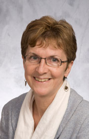
Every two years, the German society for developmental biology (Gesellschaft für Entwicklungsbiologie – GfE) hands out an award for the best PhD thesis of the previous two years. At their society meeting last week, this award went to Marion Silies, for her PhD thesis on glial cell migration.

I met up with Marion after her talk and asked a few questions about her PhD work in Christian Klämbt’s lab, and whether she had any tips for graduate students.

Congratulations on your award. What was your thesis about?
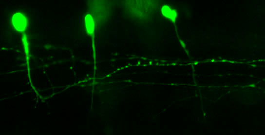
I worked on glial cell migration in the fly peripheral nervous system. I looked at how neurons and glial cells co-regulate their development. In a screen we found a cell cycle regulator with strong phenotypes in the migration of glial cells, but I showed that it has a post-mitotic function, so a function outside of its function in cell cycle: it controls glial cell migration from the neuron, by regulating distribution of a cell adhesion molecule.

What are you doing now? Are you still working on the nervous system?
For my PhD I studied developmental processes, but for my postdoc I moved on to understand how the nervous system functions. I’m in the lab of Tom Clandinin now, at Stanford University.

Did you have to come back to Germany just to pick up your award?
I would have loved to come just for this meeting, but I was actually in Germany anyway for another meeting, so this just fit very well.

Do you have any tips for current or new PhD students?
My tip for students about to start their PhD would be to pick something that they’re really excited about. I think this is the most important thing: A PhD takes a long time, and you put in a lot of work, so try to find something that you really like. A lot of people think that they have to be at a very prestigious university, or at a very well-known institute. I would say: go wherever you want – just find something that you really like to do, and find a nice boss.
(more…)

Thumbs up (6 votes)
Loading...

Tags: ,
Categories: Careers, Education, Interview

Navigate the archive

Use our Advanced Search tool to search and filter posts by date, category, tags and authors.

Seeing Further

Posted by , on 29 March 2011

The Royal Society has collected a series of images that illustrate the moment important scientific discoveries were made. This “Moments of Seeing Further” collection includes a notebook sketch from 1980, contributed by Sir John E. Sulston and depicting cell division in C. elegans – work that contributed to the discovery of the fate map of the worm.

Another, much older, image is a 17th century sketch of the process of bean sprouting, by Marcello Malpighi. He didn’t look at plants alone: His microscopy studies in many different organisms has contributed to the early study of development, and his name lives on in several microscopic structures, including the Malpighian corpuscles in the kidneys.

Have a look at the rest of the gallery as well. It even includes a photo of a letter to the Royal Society from Isaac Newton.

(Image © the Royal Society; used with permission)

Thumbs up (No Ratings Yet)
Loading...

Tags: , ,
Categories: Images, Research

GfE/JSDB meeting in Dresden

Posted by , on 24 March 2011

The joint GfE/JSDB meeting is currently underway in Dresden. The organizers have managed to keep the meeting going with a full schedule, despite some delegates from Japan being unable to attend after the earthquake and tsunami earlier this month. Three speakers had to cancel, but their speaking slots were taken over at the last moment. David Greenstein pulled up one slide at the start of his own talk to point the audience to a recent paper by Asako Sugimoto from Tohoku University, who was scheduled to speak at the conference herself, but obviously had to stay in Sendai now. She, and all other JSDB members, are doing all right, though.

Everyone at the meeting is in charge of nominating the best posters, which makes for busy and engaging poster sessions. It’s hard to get a look at all the posters, let alone choose!


Just a few of the posters – they are spread over three floors!

One of the things I’m doing at the meeting this week is interviewing Elisabeth Knust, the president of the GfE, so you’ll be able to read that in Development and on the Node in several weeks. For more up-to-date news from the meeting, have a look at our Twitter account. We’re not mentioning unpublished data, of course, so you’re missing out on some of the best things from the meeting, but maybe we’ll hear more from some of the attending researchers on the Node later.

Thumbs up (3 votes)
Loading...

Tags: , ,
Categories: Events

In Development this week (Vol. 138, Issue 8):

Posted by , on 22 March 2011

Here are the research highlights from the current issue of Development:

Fishing out adult neural stem cells

Adult neural stem cells (NSCs) hold great potential for the treatment of neurodegenerative diseases and nervous system injuries. To date, adult neurogenesis has been mainly studied in rodents but, on p. 1459, Laure Bally-Cuif and co-workers use GFP-encoding viruses and clonal analyses to characterise NSCs in the adult zebrafish brain. Zebrafish grow throughout life and maintain germinal centres in the brain that continually add new cells to the nervous system. The telencephalic germinal zone contains quiescent radial glial progenitors and actively dividing neuroblasts and the researchers now show that these progenitors have different division modes and fates. Thus, neuroblasts primarily undergo a limited amplification phase followed by symmetric neurogenic divisions, whereas radial glia self-renew and generate different cell types, a result that identifies them as bona fide NSCs. Importantly, the researchers also show that most radial glia divide symmetrically, which amplifies and maintains the NSC pool. These and other results establish zebrafish as an important model system for studying adult neurogenesis.

All change: stepwise in vivo transdifferentiation

Many differentiated cells can be reprogrammed to adopt new identities; reprogramming can occur through an embryonic stem cell-like state or by direct conversion to another cell type (transdifferentiation). The latter route is poorly understood but, here, Sophie Jarriault and colleagues provide detailed analyses of a natural direct reprogramming event – the in vivo transdifferentiation of a C. elegans rectal cell into a motoneuron (see p. 1483). The researchers show that when the rectal cell undergoes transdifferentiation, it adopts a temporary state that lacks the characteristics of both the initial and final cellular identities before undergoing stepwise redifferentiation into a motoneuron. Dedifferentiation, they report, can occur without cell division, and redifferentiation requires the conserved transcription factor UNC-3. Importantly, the intermediate dedifferentiated stage has restricted plasticity. Together, these results suggest that direct in vivo reprogramming in C. elegans (and possibly other species) involves transition through discrete stages and that tight control mechanisms restrict cell potential at each stage, a conclusion with important implications for regenerative medicine.

Apoptosis sets the speed of morphogenesis

During development, dynamic cell behaviours are carefully orchestrated to ensure that morphogenesis is completed within the correct developmental time frame, but how is this achieved? Erina Kuranaga and colleagues (p. 1493) have been examining genital morphogenesis in Drosophila and report that apoptosis controls the speed of looping morphogenesis in the fly’s male terminalia. The terminalia is an asymmetric looping organ in which the internal genitalia (spermiduct) loops around the hindgut. During maturation of the internal genitalia, the male terminalia rotates 360° clockwise. Previous work has shown that the adult male terminalia is incorrectly orientated in mutants for apoptotic signalling. Now, using time-lapse imaging, the researchers show that, in normal flies, genitalia rotation accelerates as development proceeds but that this acceleration is impaired when the activity of apoptotic signalling components is reduced. The researchers propose that apoptosis drives the movement of cell sheets during the morphogenesis of male terminalia, thereby ensuring that morphogenesis is completed within a limited developmental time frame.

Xist marks the spot

In XX female mammals, the inactivation of one X chromosome during development equalises the levels of X-linked gene products in females with levels in males. The Xist locus regulates X inactivation by producing Xist, a non-coding RNA that coats and silences the chromosome from which it is transcribed. Now, on p. 1541, Neil Brockdorff and co-workers analyse X inactivation in XistINV mice, which carry a mutation in which a conserved region of Xist exon 1 is inverted. Inheritance of XistINV on the maternal X chromosome in female embryos results in secondary non-random X inactivation, they report, which indicates that the inversion affects Xist-mediated silencing but not Xist gene regulation. Moreover, XistINV inheritance on the paternal X chromosome leads to embryonic lethality because of failed imprinted X inactivation in extra-embryonic tissues. Other analyses show that XistINV RNA localises in cis to the X chromosome but with reduced efficiency. Thus, the researchers conclude, conserved Xist exon 1 sequences are important for Xist RNA localisation and, consequently, X-linked gene silencing.

Muscle building: actin polymerisation drives myoblast fusion

Myoblast fusion, which is essential for skeletal muscle development, involves cell recognition and adhesion, followed by cell membrane breakdown and multinucleate syncitia formation. Here, Susan Abmayr and colleagues clarify the molecular mechanism of myoblast fusion in Drosophila embryos (see p. 1551). In Drosophila, the initial myoblast fusion event occurs asymmetrically between a founder cell (which patterns the musculature) and a fusion-competent myoblast (FCM). The researchers report that the non-conventional guanine nucleotide exchange factor Myoblast city (Mbc) is required in the FCMs but not in the founder cells for myoblast fusion, and that Mbc activates the small GTPase Rac1 in the FCMs. Notably, Mbc, active Rac1 and F-actin foci are concentrated in the FCMs at their site of contact with founder cells, and Mbc is essential for the formation and organisation of F-actin foci and the cytoskeleton in the FCMs. The researchers suggest, therefore, that Mbc-dependent actin polymerisation in FCMs may be one of the driving forces behind Drosophila myoblast fusion.

Arteriovenous malformations go with the flow

Arteriovenous malformations (AVMs) are direct connections between arteries and veins that arise during active angiogenesis. Most AVMs are sporadic but some are associated with mutations in genes involved in TGFβ signalling. For example, mutations in activin receptor-like kinase 1 (ALK1, a TGFβ receptor) are implicated in the vascular disorder hereditary haemorrhagic telangiectasia 2 (HHT2). But what are the molecular and cellular errors that lead to AVM formation? On p. 1573 Beth Roman and colleagues address this question by analysing AVM development in alk1 mutant zebrafish embryos. They report that blood flow triggers alk1 expression in nascent arteries exposed to high haemodynamic forces and that Alk1 normally limits vessel calibre. In alk1 mutants, however, Alk1-dependent arteries are enlarged, and the downstream vessels adapt to the consequent increases in blood flow by retaining normally transient arteriovenous drainage connections, which subsequently enlarge to form AVMs. This two-step model for AVM formation suggests that HHT2 treatments might focus on preventing arterial enlargement and/or abrogating flow-induced AVM development.

Plus…

As part of the Evolutionary crossroads in developmental biology series, Technau and Steele introduce Cnidaria and discuss how studies of this diverse phylum, which includes corals, sea anemones, jellyfish and hydroids, have informed our understanding of bilaterian evolution and development.

See the Primer article on p. 1447

Also have a look at the other articles in the Featured Topic on Evolutionary crossroads in developmental biology.

Thumbs up (No Ratings Yet)
Loading...

Tags: , , , , , , , , , ,
Categories: Research

PhD studentship: the role of chromatin in embryonic development

Posted by , on 21 March 2011

Closing Date: 15 March 2021

Nicoletta Bobola’s research group

Faculty of Medical and Human Sciences, University of Manchester

The objective of this fully-funded 4-year PhD project is to investigate how the composition of chromatin regulates embryonic development, with a focus on Hox transcription factors activity.

The local composition of chromatin is a major determinant of the transcriptional activity of a gene. Changes in the spatio-temporal expression of genes generate the different cell types, even though their genomes are identical. Understanding how this happens is a major challenge in biology, and may lead to important progress in our ability to generate specific cell types for regenerative purposes.

Hox transcription factors are highly conserved and have crucial roles in embryonic development. In this project we will generate genome wide location maps of transcription factors, chromatin proteins and selected histone modifications. These data will be integrated in a systematic map of chromatin in areas of the embryo whose development is instructed by Hox proteins, to understand how different chromatin environments help to target Hox proteins to specific genomic regions and regulate their target genes.

The lab is located at the University of Manchester, in the AV Hill building, a cutting-edge training environment incorporating both the life and biomedical sciences.

Any enquiries relating to the project and/or suitability should be directed to Nicoletta Bobola (nicoletta.bobola@manchester.ac.uk).

The project is due to commence October 2011 and is open to UK/EU nationals only due to the nature of the funding. If interested, please apply by following the details at http://www.mhs.manchester.ac.uk/postgraduate/studentships/

Closing date 4th April 2011

Thumbs up (No Ratings Yet)
Loading...

Categories: Jobs

Image competition finalists

Posted by , on 21 March 2011

In the Node’s recent image competition, we asked for images related to developmental biology and some sort of intersection – playing on the double meaning of the word “node” in the site’s name. We received as many different interpretations of this theme as submissions, which gave the judges a difficult task. But by looking both at the image itself and the idea behind it, they were able to narrow it down to the following three images.

Now it’s up to you to select your favourite image of the three nominees and determine who will win a TipArt commission. You have until April 4 (noon UK time) to cast your vote.

Click any of the images to see them full size. The descriptions are below each image, and the poll is at the bottom of the post.

—-


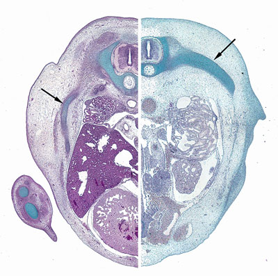
“This is a co-localization of slow myosin heavy chain (in magenta) with Sox6 (in blue) on a gut section of a E17.5 mouse embryo. The embryo itself spells “intersection”, at least it begins to do so…”

—-


“Scientific background of the image: Head process stage of chick/mammalian embryo.
Artistic background of the image: The asymmetric node is the point of interaction (between various personalities: gender/seniority/fame disparities, etc); The two sides of the streak are the bodies of interacting personas; The head process (the flame) is the idea/publication coming out of the interaction (made blurry for a purpose); The black background is the ignorance we all try to overcome (connected to the cross figure between the interaction partners, one can read religion into it); The unfinished circle is the quest for fullness/the big answer (one may be standing on it without realizing it).”

—-


“Metamorphosis is a drastic (and common) event in animal life histories intersecting larval and adult stages. The photo shows a sea biscuit during metamorphosis. The apparently amorphous mass of cells exposes the duality of this transformation moment. The larval body retracted and lost its form, but larval skeleton spicules are still attached (at the top). At the bottom developing podia and spines already move and interact with the substrate as the sea biscuit learns how to walk its first steps.”

Thumbs up (4 votes)
Loading...

Tags: , , , , , ,
Categories: Images

YEN Seminar Next Week – March 24

Posted by , on 17 March 2011

We are delighted to announce the first double-seminar session hosted by the Young Embryologist Network (http://www.ucl.ac.uk/cdb/yen) on March 24th from 4pm – 6pm (& refreshments afterwards) in the A.V. Hill Lecture Theatre, Medical Sciences Building, UCL. The theme of this session is visualising biological processes and there will be 2 speakers:

– Dr Florencia Cavodeassi (Steve Wilson Group, Dept of Cell and Developmental Biology, UCL).
Title TBC.

– Professor Scott Fraser (Anna L. Rosen Professor of Biology & Engineering and Applied Science, Beckman Institute, Caltech, USA).

Title: “New tools for imaging the motions and fates of embryonic cells.”

This session is run by PhD and PostDocs in the Network (CDB, UCL, NIMR, ICH, KCL) but is open to everyone. Through these seminars and our annual meeting we hope to open lines of communication and create a diverse, interactive research community for PhD students, Post-Doc and PI embryologists. So please come along and become part of the Network.

Directions:
AV. Hill Lecture Theatre can be reached via the Anatomy Building on Gower Street or through the Medical Sciences building ( http://crf.casa.ucl.ac.uk/screenRoute.aspx?s=1178&d=115&w=False ).
The Anatomy building route is the following: Enter through the main entrance on Gower Street and walk up the central staircase to the first floor, enter through the door marked “Physiology” and along the corridor. AV. Hill is to your right at the end of the corridor through 2 sets of double doors.

————

We look forwards to seeing you there!
Sorrel Bickley
YEM2011 Organising Committee
youngembryologistnetwork@gmail.com

Young Embryologist Network (Do you know about our annual meeting YEM2011 on May 6th? Check out our website)
http://www.ucl.ac.uk/cdb/yen/

Thumbs up (1 votes)
Loading...

Categories: Discussion, Events, Research

Travel funding

Posted by , on 17 March 2011

If you’re attending a conference later this year and need help funding your travel, you can apply for one of the Company of Biologists’ Direct Travel grants. The maximum amount of these grants has just been increased to £600 for international travel and £300 for travel in the same country as resident. (The website still lists the old amounts.)

-Deadline for the next round is March 31st.
-Applicants must be a PhD student or junior (in first three years post PhD and in first postdoctoral position) postdoctoral researcher.
-Applications will not be considered from those eligible to apply for travel grants from the British Society for Developmental Biology (BSDB) or any other Company of Biologists grant-aided source.

In addition, Development also offers fellowship awards of up to £2500 for PhD students and postdocs to travel to another lab for a collaborative exchange. You can find out more about those Travelling Fellowships on the Development website, and the application deadline for the next round of these awards is April 30.
We’ve featured some reports from previous recipients of the Travelling Fellowships on the Node, so you can read their experiences.

Both the Direct Travel Grants and the Travelling Fellowships are open to applicants from any country.

(image by Duncan Harris on Flickr.)

Thumbs up (2 votes)
Loading...

Categories: Funding

Bird’s-eye view: CoB workshop on Cancer as a Microevolutionary Process

Posted by , on 15 March 2011

During the past week, a group of scientists followed in the footsteps of Winston Churchill and convened at the lovely Wilton Park in Sussex for a Company of Biologists Workshop to challenge central dogmas and provoke new ideas – this time in a quest to understand cancer as a microevolutionary process. The conference organisers – Dr. Gerard Evan, Dr. Doug Green and Dr. Karen Vousden – invited an excellent range of speakers from across the fields of cancer and evolution to join them for intense discussions on key outstanding questions, including: the search for effective cancer therapeutics, the role of stem cells in metastasis, the importance of the tumour microenvironment and the value of research in more diverse species across the breadth of eukaryotes (did you know that only 6 model organisms are commonly used to probe biological questions, yet there are 50 million species?!).

This Workshop was the fourth in a series organised by The Company of Biologists. The format of these meetings aims to divert from the traditional presentation of primary research findings and instead provide a unique discussion-based forum for the exchange of new ideas between experts from diverse disciplines. As the Editorial Intern on Journal of Cell Science, I attended to represent the journal, and was joined by members of the editorial team from another Company of Biologists journal, Disease Models & Mechanisms. Personally, I feel very privileged to have had the opportunity to hear the new and thought-provoking ideas being exchanged across the conference room, during the delicious meals, and on into the evening in the library and the bar. I am sure that future collaborative projects will be spawned from this meeting, so keep your eye on the pages of JCS, DMM and the other journals published by The Company of Biologists to see how this interdisciplinary field develops.

If you have an idea for a ground-breaking biology-related workshop then please visit the Workshops website at http://workshops.biologists.com/ for more details.

Thumbs up (2 votes)
Loading...

Categories: Events

Japan Earthquake and Tsunami

Posted by , on 14 March 2011

We were deeply shocked to hear about the devastating earthquake and tsunami that hit Japan on March 11, and would like to express our sympathy with the people of Japan.

Throughout the weekend, we have been following news from colleagues in Japan, but we still don’t have a complete picture of where researchers in the affected area are and how they are doing. If you do have information or would like to enquire about biologists in Northern Japan, please feel free to use the comments area of this post to communicate and share information with other developmental biologists.

We do have an update at this moment from the CDB Epigenetics Symposium.
This symposium is currently underway in Kobe – a city no stranger to earthquakes itself. Despite many cancellations from participants, and a few requests to call the meeting off, the organizers made a decision to hold the symposium anyway. In an email to meeting participants, they explain why:

“[S]ome advised us not to hold the CDB symposium. However, we also learnt from the Kobe earthquake that maintaining our daily activity and maintaining a sense of normalcy, is the best what we can do in the unaffected areas.”

The symposium has, however, been reduced to two days only. Many attendees had to cancel, and flights in and out of any part of Japan are experiencing delays.

We are closely following news from the Japanese developmental biology community through contacts at RIKEN CDB and the JSDB. We’ll update this post with news, and encourage you to communicate in the comments, so you can bookmark this post and come back to it later to find out what’s going on with your colleagues in Japan.

Updates:
– From various sources (but first through Raj’s comment below) we heard that Tohoku University’s Dr. Koji Tamura, Dr. Harukazu Nakamura, Dr. Funahashi, Dr. Toshihiko Ogura, Dr. Noriko Osumi, and Dr. Asako Sugimoto and their lab members are all doing fine. The labs have suffered material damage, and they have lost freezer samples, but we hope to have more detail on how the research community can help them once they’re settling back into their labs.
– The joint GfE/JSDB meeting in Dresden will be held next week with a shortened programme, as not all Japanese speakers will be able to attend.
– German researchers have set up the Nippon Science Support Network, to offer space and resources to their Japanese colleagues. (via The Great Beyond, which has more updates).
– (31/3 update) The NIH has sent Japanese societies an offer to help: “Plans are underway to provide temporary scientific homes at NIH for Japanese colleagues and fellows who may have lost research facilities in Japan. Also, the Foundation for Advanced Education in the Sciences (FAES) has established a fund to help earthquake victims in Japan. Voluntary contributions may be sent to FAES, NIH Building 60, Suite 230 (One Cloister Court, Bethesda, MD 20814) and designated for the “Japanese Earthquake Fund.”” (as sent to the JSDB by Michael M. Gottesman of the NIH)
-(6/4 update) The NSF has grants available for researchers affected by either the New Zealand or Japan earthquakes. See their website for details.

Relevant links:

CDB epigenetic symposium (with updates)

Tohoku University emergency page for students and staff (Japanese).

Google Person Finder (English/Japanese)

Thumbs up (1 votes)
Loading...

Tags: ,
Categories: News