The community site for and by
developmental and stem cell biologists

Citing Data

Posted by , on 17 January 2011

I just got back from attending two meetings about academia and the internet – one in person and the second, in true internet style, virtually. Both meetings at one point or another discussed the growing trend toward archiving and citing data itself (on top of citing the papers written based on analysis of the data).

The first meeting was the HighWire publishers meeting. HighWire takes care of the online version of many journals, including Development, and their meeting was mostly about practical things for journals and not directly relevant to most of you yet. (You can still find some of the discussion on Twitter, although Twitter’s search function expires after about 10 days.)

The second meeting, which I followed over the web, was Science Online in North Carolina. That meeting is now in its fifth year, and started out as a meeting solely about science blogging, but has expanded to cover other aspects of science and the internet.

It was rather interesting to follow both meetings back to back, since the first was very practical and aimed at things that publishers can do and are doing right now, while the second was full of thoughts and ideas and the audience was very varied. There is still a lot of science blogging being discussed at the Science Online meeting, and I myself Skyped into a panel with 15 community managers of different science blog networks. (“Different” is the keyword here, because the Node has very little in common with, say, the Guardian or Discover blog networks, but it was interesting to hear some comparisons.)

A few topics, however, came up at both the HighWire meeting and at Science Online, and one of these was the new problem of citing data. As Benoit mentioned here back in August, the Journal of Neuroscience no longer publishes supplementary data. This journal was at the HighWire meeting to share how that is going so far, and one of the surprising reactions they had was a response from librarians: If journals don’t publish supplementary data, it has to go somewhere, and libraries are stepping up to claim the niche of data archiving and curating. The J. Neurosci. talk was followed by a librarian from Stanford who mentioned that they just hired a “data librarian” to look into things like this.

And they’re not the only ones: last fall at the Science Online London meeting, the British Library showed that they were very involved in data archiving as well. And as I mentioned, this weekend’s Science Online North Carolina meeting also included a few talks about data archiving and whether having your data cited will one day be valuable for your career (just like having your papers cited is now).

What do you think of this movement toward archiving and citing data? Are you happy that you’ll be able to find other people’s data? Annoyed that it’s yet another thing to keep on top of? And where are all your data at the moment? Can someone easily find them if they want to build upon your work? Have you ever cited a database in a paper?
(more…)

Thumbs up (No Ratings Yet)
Loading...

Tags: , , ,
Categories: Discussion, Events

Navigate the archive

Use our Advanced Search tool to search and filter posts by date, category, tags and authors.

Postdoc positions in Jim Smith’s lab

Posted by , on 16 January 2011

Closing Date: 15 March 2021

Two postdoctoral positions are available in Jim Smith’s lab at the National Institute for Medical Research in north London. One is is supported by the Leducq foundation, under a multidisciplinary programme designed to elucidate the role of bone morphogenetic protein (BMP) signalling in the pathogenesis of pulmonary and systemic vascular diseases. The work will use zebrafish embryos to study the activation and roles of BMP target genes identified by high-throughput sequencing. Further details, including salary and how to apply, are available at http://www.nimr.mrc.ac.uk/jobs/IRC11806/.

The second position will continue the laboratory’s work on evolutionary aspects of the genetic regulatory network that underlies mesoderm formation. The work will use Xenopus and zebrafish embryos and human and mouse ES cells as appropriate. Further details, including salary and how to apply, are available at http://www.nimr.mrc.ac.uk/jobs/IRC11805/.

The closing date for both positions is February 14, 2011. Informal enquiries can be made to Jim Smith (jim.smith@nimr.mrc.ac.uk).

Thumbs up (No Ratings Yet)
Loading...

Categories: Jobs

POSTDOCTORAL POSITION IN CELL and DEVELOPMENTAL BIOLOGY

Posted by , on 13 January 2011

Closing Date: 15 March 2021

Post doctoral position available to study the neural crest gene regulatory network (NC-GRN) in Xenopus and zebrafish. Neural crest cells are stem cell-like progenitors that migrate extensively and are essential to the establishment of the vertebrate body plan. Misregulation of components of the NC-GRN underlies numerous human diseases and congenital disorders. Studies involve post-translational regulation of known network components, and use of proteomics and next generation sequencing to identify novel components.

Highly motivated applicants with a PhD and strong background in cell and molecular biology and/or developmental biology are encourage to apply. Please send a CV, brief description of research interests, and the names of three references to:

Carole LaBonne, PhD (clabonne@northwestern.edu)
Department of Molecular Biosciences
Northwestern University, Evanston, IL 602028

Northwestern University’s main (Evanston) campus is on the shores of Lake Michigan, close to the heart of Chicago, one of the most beautiful and culturally rich cities in the US.

Thumbs up (No Ratings Yet)
Loading...

Categories: Jobs

PhD studentship: Analysis of progenitor cell differentiation programmes

Posted by , on 12 January 2011

Closing Date: 15 March 2021

Applications are invited for a PhD studentship investigating cell differentiation during development (closing date 14th January, 2011).

Cell differentiation programmes are central to the production of specialised tissues during development. Moreover, in-depth understanding of cell differentiation is essential for many applications, including stem cell technology and tissue repair. We study the programme that governs muscle formation. This is important not only because muscle is a major cell type and an established paradigm for cell differentiation, but also because of its significance for human health. You will analyse the control of when and where muscle differentiation occurs and how this differentiation programme is orchestrated. You will use the classic, genetically tractable, model organism Drosophila melanogaster, which has shaped much of our understanding of animal development and has an impressive history of informing human biology and medicine. You will analyse the differentiation of both embryo and adult progenitor cells, the latter in remodeling and regeneration during metamorphosis.

Progenitor cell differentiation is controlled by a balance of factors. A key promoting factor for muscle is the conserved Mef2 transcription factor. We found that expression of different muscle genes requires different levels of Mef2 activity (PNAS 105:918-923 (2008)). This highlights the importance of understanding how Mef2 activity is regulated, which is the focus of this project. We also recently identified a novel regulator, Him, that down-regulates Mef2 activity and inhibits muscle differentiation (Current Biology 17:1409-13 (2007)). You will analyse both Him and other Mef2 regulators, including those identified in an ongoing screen, and also assess Mef2 activity during muscle differentiation using in vivo Mef2 sensors. Together, this will indicate how Mef2 can co-ordinate the expression of diverse muscle genes and unravel mechanisms that maintain progenitor cells in an undifferentiated state.

The host lab in the School of Biosciences, Cardiff University offers valuable training possibilities through interactions with labs across Europe and the opportunity to use a broad range of techniques from molecular cell biology and genetics.

Interested candidates should send a CV and statement of research interests to Dr Mike Taylor (TaylorMV@cardiff.ac.uk) as soon as possible. You must also apply formally on-line by following the details at http://www.cardiff.ac.uk/biosi/degreeprogrammes/postgraduateresearch/index.html.

Informal enquiries are welcome. Please email Dr Mike Taylor at TaylorMV@cardiff.ac.uk or telephone on +44 (0)29 208 75881.

Thumbs up (No Ratings Yet)
Loading...

Categories: Jobs

Arrested Development in Plant miRNA Mutants

Posted by , on 11 January 2011

Animals and Plants have hundreds of miRNAs with diverse roles in gene regulation. In humans, each miRNA family can control up to several hundred genes (or up to 500 to be exact, in humans). A loss of function in one, can lead to an array of developmental defects & diseases. The same goes for plants. However, many plant miRNAs only have one target, which is frequently a transcription factor that in turn, controls many genes itself. It’s really like a house of cards. An mutant with a loss of function in one miRNA can have a full range of phenotypes.

Focusing on one miRNA pathway in plants:

Arabidopsis miR159, which only has 2 validated targets which are functionally redundant transcription factors (MYB33, MYB65).

mir159ab mutant v.s. wild type: (personal images)

The mir159ab mutant plant is smaller and the leaves curl upwards. Flowers are also affected, and fertility is reduced in the mutant. All because miR159 is no longer active, and it’s targets MYB33 and MYB65 are at free rein to meddle in regular development. miRNA mutants are quite revealing on how miRNAs ‘switch off’ target genes that would otherwise inhibit development.

miR159 targets actually have an important role in pollen development. Loss of function mutants in MYB33/MYB65 lead to male sterility in plants. Pollen is also the only tissues/cells where miR159 isn’t expressed, so the plant needs active MYB33 and 65 for fertility. Seems a bit wasteful to express MYB33/65 in the entire plant, if they appear to only have a role in the pollen. Also, what’s the point of expressing them and having a miR159 ‘switch’ em off?

It’s speculated that they have additional roles in programmed cell death (PCD) that’s associated with plant defense. It’s seemingly unrelated to pollen development. Bizarre, but MYB33/65 are actually transcription factors that up-regulate the genes involved in PCD. To connect the dots: PCD has a role in pollen development: it causes the degeneration of tissue that impede growth past a certain stage. Additionally, when a plant is challenged with a virus, infected cells & tissues will undergo PCD in an attempt to stop its spread. Many viruses are known to suppress siRNA and miRNA production. In addition to roles in its own development, gene silencing is used by plants to quell viral replication. If the virus attack the plant’s RNAi machinery, to halt defensive siRNA production, it also influences endogenous miRNAs. Thus, it could be possible that the miR159-MYB33/65 system is involved in this as a sort of viral sensor. If miR159 is no longer active, MYB33/65 will begin to trigger PCD.

All this, for one miRNA-target gene connection.

It’s a bit like playing Jenga. One piece may only touch 3-4 others, but if you remove the essential one, all 20 pieces fall down because they were all connected. (Image: Flikr CC, Jenga, by Paul_Carvill)

It gets more complex if key proteins in miRNA biogenesis & action are rendered function-less. Initially, before RNAi was more established, researchers believed they were dealing with a multitude of proteins with various functions..when really it was just a few participating in one show. Loss of one RNAi-related protein can translate into the loss of function in hundreds of miRNAs. Moreover, miRNAs aren’t actually the effectors of regulation, they are simply the guides. The proteins do the dirty work, from making miRNAs to making use of them..

Arabidopsis Dicer like 1 (DCL1) a one significant protein in miRNA biogenesis. It cuts out the mature miRNA strand from it’s precursory hairpin structure (the pri-miRNA). Furthermore, in plants at least, miRNA precursors come in many different hairpin shapes and lengths, so DCL1 is often forced to cut them up differently. And so a mutation in any of DCL1’s sequences can lead to diverse interruptions in plant development. For DCL1 alone, there are up to 10 different mutant alleles with variations in severity and phenotype. At first, research groups thought they were dealing with 3 different proteins and their mutant alleles. They all had different phenotypes and names, suspensor-1 (sus-1) was arrested in embryo development and was embryonic lethal. Carpel factory-1 (caf-1) overproduces carpels (female parts in the flowers) and has sterile anthers (male parts). Eventually, by virtue of gene mapping they began to connect the dots. Cloning of the DCL1 gene years later also verified this. The history of DCL1 and it’s mutants is summed up an article artfully called, “DICER-LIKE1: blind men and elephants in Arabidopsis development”.

Carpel factory-1, aka dcl1-9, flower versus a wild type flower (Images: Laufs et al. 2004, published in Development).

ResearchBlogging.org

Garzon, R., Marcucci, G., & Croce, C. (2010). Targeting microRNAs in cancer: rationale, strategies and challenges Nature Reviews Drug Discovery, 9 (10), 775-789 DOI: 10.1038/nrd3179

Allen, R., Li, J., Stahle, M., Dubroue, A., Gubler, F., & Millar, A. (2007). From the Cover: Genetic analysis reveals functional redundancy and the major target genes of the Arabidopsis miR159 family Proceedings of the National Academy of Sciences, 104 (41), 16371-16376 DOI: 10.1073/pnas.0707653104

Alonso-Peral, M., Li, J., Li, Y., Allen, R., Schnippenkoetter, W., Ohms, S., White, R., & Millar, A. (2010). The MicroRNA159-Regulated GAMYB-like Genes Inhibit Growth and Promote Programmed Cell Death in Arabidopsis PLANT PHYSIOLOGY, 154 (2), 757-771 DOI: 10.1104/pp.110.160630

Schwab, R., & Voinnet, O. (2009). miRNA processing turned upside down The EMBO Journal, 28 (23), 3633-3634 DOI: 10.1038/emboj.2009.334

SCHAUER, S., JACOBSEN, S., MEINKE, D., & RAY, A. (2002). : blind men and elephants in development Trends in Plant Science, 7 (11), 487-491 DOI: 10.1016/S1360-1385(02)02355-5

Laufs, P. (2004). MicroRNA regulation of the CUC genes is required for boundary size control in Arabidopsis meristems Development, 131 (17), 4311-4322 DOI: 10.1242/dev.01320

Thumbs up (1 votes)
Loading...

Tags: , , , ,
Categories: Research

Royal Society Stem Cell Meeting talks online

Posted by , on 11 January 2011

The Royal Society has uploaded audio files of almost all the talks of their Discussion Meeting last October: “2010: What Next for Stem Cell Biology?”.

Unfortunately there are no slides to look at (as far as I can tell) so some of the more technical talks may be hard to follow, but if you’re already familiar with the work of some of the speakers, hearing them speak may help put things in context, even without seeing the slides. The abstracts of the talks are all in the programme booklet, which you can download from their site as well.

I attended this meeting in person, and was impressed with both the variety of the talks as well as the overlap between them. If you’re trying to answer the RSc’s question posed in the meeting title, then the common thread of many of the talks suggests that what’s next for stem cell biology is mainly to get a complete picture of reprogramming. But the variety of the talks indicates that there are many ways to approach this: finding out how to make one particular cell type for therapeutic means (addressed by several speakers), finding out the mathematical concepts behind reprogramming to define a formal theory (Sui Huang’s talk), or even calculating the cost of reprogramming when setting up a company that relies on iPS cells for research (Cathy Prescott’s talk). There’s still a lot of work to be done, but hopefully the collective approaches will reveal a clear picture of reprogramming in the next few years.

Thumbs up (2 votes)
Loading...

Tags: , , , ,
Categories: Events

Open access video protocol: electroporating zebrafish ears

Posted by , on 10 January 2011

I’ll save the thousand words.  Here’s the link:

J Vis Exp. 2011;47 http://www.jove.com/details.stp?id=2466

Holmes KE, Wyatt MJ, Shen Y, Thompson DA, Barald KF. Direct Delivery of MIF Morpholinos Into the Zebrafish Otocyst by Injection and Electroporation Affects Inner Ear Development.  J Vis Exp. 2011;47 http://www.jove.com/details.stp?id=2466 doi: 10.3791/2466.

Thumbs up (4 votes)
Loading...

Categories: Video

modENCODE

Posted by , on 6 January 2011

The modENCODE project (model organism encyclopedia of DNA elements) is a collaborative effort to identify all sequence-based functional elements of Drosophila and C. elegans. The project has now produced almost a thousand data sets with information about transcription, epigenetics, replication and gene regulation across different developmental stages and multiple cell lines.

Just before the holidays, the modENCODE teams published several analyses of these data sets in a number of papers across four journals. Because the data were collected across multiple developmental stages, they give information that cannot be gathered from looking at just one point in development.

In a CSHL podcast, modENCODE team member Thomas Gingeras describes work from his group that was published in Science. They used the modENCODE data to confirm that 90% of all 17 thousand predicted protein coding regions in Drosophila indeed correspond to RNA expression. At any given point along the developmental timeline, this percentage would of course be a lot smaller, as genes switch on and off. In addition, they found almost two thousand previously unannotated genes.

Another result relevant to developmental biologists is covered in one of the Nature papers: An analysis of different chromatin states in Drosophila revealed a more complex pattern of Polycomb target regulation than was previously suspected.

Worm researchers also have a lot of new information to work with. For example, a Science paper describes the mapping of transcription factor binding sites in C. elegans and reports that some regions of the worm genome – which they called HOT regions – were bound by more than 15 transcription factors!

So what’s next for the modENCODE project? There is still a year of funding left, and much of that time will be spent adding more annotations for newly found functional regions, as well as integrating different types of data for a more complete picture.

Of course, the worm and fly are not the only organisms to be mapped. Let’s not forget ourselves! The (human) ENCODE project, which in fact precedes modENCODE, hopes to publish a full genome analysis next year.
(more…)

Thumbs up (3 votes)
Loading...

Tags: , ,
Categories: News, Research

Intestinal stem cell regeneration

Posted by , on 5 January 2011

Cancer and stem cells are two very loaded biology concepts, and more frequently can be found in the same discussion.  Stem cells within tumors are able to divide and provide the various differentiated cell types that a tumor requires to thrive.  And, identifying how a normal stem cell divides, or stops dividing, can help further the understanding of tumorigenesis.  Along these lines, a paper from the December 15 issue of Development describes a pathway involved in intestinal stem cell proliferation.

Intestinal stem cells (ISCs) normally divide to replace differentiated intestinal cells at a rate that supports tissue homeostasis.  This rate of ISC division quickly increases when intestinal cells suffer injury due to damage, disease, or exposure to pathogens or chemical agents.  Recently, Karpowicz and colleagues investigated this switch from normal to “acute regeneration” of intestinal cells in Drosophila midgut epithelium, a great model for ISC self-renewal.  In this paper, the authors find that ISC proliferation is constitutively controlled by Hippo, a member of a pathway involved in organ growth and cancer.  In addition, injury disrupts this regulation of Hippo, which in turn activates Yorkie, a Hippo pathway target.  The authors find that this cell-autonomous role for the Hippo pathway is crucial for regulation of ISC proliferation.

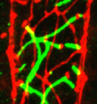
Images above show regions of control (top) and Yorkie-depleted (bottom) Drosophila midgut tissue.  Yorkie depletion causes fewer ISC divisions, as seen as fewer cells positive for Escargot (Esg; green), a known transcription factor expressed in ISCs.

For a more general description of this image, see my post on EuroStemCell, the European stem cell portal.

ResearchBlogging.orgKarpowicz, P., Perez, J., & Perrimon, N. (2010). The Hippo tumor suppressor pathway regulates intestinal stem cell regeneration Development, 137 (24), 4135-4145 DOI: 10.1242/dev.060483

Thumbs up (5 votes)
Loading...

Tags: ,
Categories: Images, Research

Funding Opportunity for BSDB members travelling to ES cell course

Posted by , on 5 January 2011

Message from the BSDB, reposted with permission. Keep in mind that the funding opportunities are for BSDB members only, and the final deadline is very, very soon. The poster for the course was posted on the Node earlier.

We have the unique opportunity to provide UK BSDB members with funded opportunities to attend an intensive practical and theoretical course on embryonic stem cells and mammalian development in Mexico this March. This was to be explained in the upcoming newsletter, but as it is late, we would like to make sure that BSDB members don’t miss this opportunity and therefore we are sending this unusual email to the membership. There are up to 8 funded places open to students, post docs, or young principle investigators. The website is www.escellslatinamerica.org and you can either apply on line by the 5th of January or submit applications as a single PDF file to either Josh Brickman (josh.brickman@ed.ac.uk) or Jenny Nichols (jn270@cscr.cam.ac.uk), by Saturday the 8th of January at 5 PM. If you apply via an email to either Josh or Jenny, then you will need to include your education and research experience (one page, include all degrees and classifications), a list of publications (for PhD students with little experience this is not important), a letter explaining why you want to attend the course (no more than 500 words) and a letter from your supervisor or department head explaining why you should be considered for the course. If you are unable to get in touch with your supervisor this week, because of the holiday, then please contact us for alternative directions.

The course, “ES Cells as a Model System for Embryonic Development,” is organized every two years at different sites in Latin America. It is a practical and theoretical course on mouse development and embryonic stem cell technologies. The course strives to simultaneously teach and build international collaborative relationships. This year it will be held in March 2011 in Mexico (Feb 27th to March 17th, 2011). The course has funding to send from 5-8 PhD students, post docs or young faculty members from the UK to Mexico to participate in both the course and its associated scientific symposium, at which all participants will be expected to give a short talk.

Course includes lectures and workshops with:
Alejandro Schinder (Buenos Aires, AR)
Alfonso Martinez-Arias (Cambridge, UK)
Andrew Smith (Edinburgh, UK)
Austin Smith (Cambridge, UK)
Heiko Lickert (Neuherberg, GER)
Ivan Velasco (Mexico City, Mexico)
James Briscoe (London, UK)
Janet Rossant (Toronto, CA)
Jennifer Nichols (Cambridge, UK)
José Xavier Neto (Campinas, BR)
Joshua Brickman (Edinburgh, UK)
Luis Covarrubias (Cuernavaca, Mexico)
Meng Li (London, UK)
Peter Andrews (Sheffield, UK)
Philippe Soriano (New York, USA)
Robin Lovell-Badge (London, UK)
Sally Lowell (Edinburgh, UK)
Simon Tomlinson (Edinburgh, UK)
Tariq Enver (London, UK)
Tetsuya Taga (Tokyo, Japan)
Tilo Kunath (Edinburgh, UK)
Wendy Bickmore (Edinburgh, UK)
Yann Barrandon (Lausanne, Switzerland)
Diana Escalante-Alcalde (Mexico City, Mexico)
Chris Wood (Cuernavaca, Mexico)
Guillermo Lanuza (Buenos Aires, AR)

Thumbs up (No Ratings Yet)
Loading...

Tags:
Categories: Events, Funding