“And that is one of the sort of worrying things about climate change; as we get more unpredictable weather patterns, can we actually design resilient wheat? So switching from a focus of just increasing wheat yields at any cost to having wheat that’s really robust to fluctuating weather conditions such as drought, but also flooding and unpredictable patterns basically.”
Dr Hannah Rees, Earlham Institute
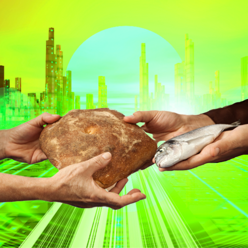
In the latest episode of the Genetics Unzipped podcast, we’re looking at the future of food. With climate change making crop harvests more unpredictable and fresh water becoming a more scarce resource, what are geneticists doing to make sure we will still have food on our plates? Dr Kat Arney chats with Dr Hannah Rees about giving wheat jet lag to create a more reliable crop, and Dr Sally Le Page talks to Dr Tarang Mehta about breeding genetically improved tilapia for fish farming.
The next Society for Developmental Biology Ethel Browne Harvey Postdoctoral Seminar will be held Friday, October 14, at 3 pm ET (9 pm CEST). This seminar featuring Hae Ryong Kwon from Oklahoma Medical Research Foundation and Leslie Slota-Burtt from Duke University is generously sponsored by The Node.
Hae Ryong Kwon did his undergraduate studies in Microbiology at Chungbuk National University in South Korea. He completed a Master’s in Microbiology and Biotechnology at Chugbuk National University and a Master’s in Genome Science and Technology at the University of Tennessee. Kwon went on to earn his doctorate in Molecular, Cellular, Developmental, and Neural Biology at the University of Albany, State University of New York where he studied the function of endothelial cells in early salivary gland development in Melinda Larson’s lab. In 2016, Kwon joined Lorin E. Olson’s lab at the Oklahoma Medical Research Foundation where he studies the roles of platelet-derived growth factor signaling in human pathogenesis driving genetic diseases such as Kosaki overgrowth syndrome, Penttinen syndrome, and infantile myofibromatosis. Kwon was the recipient of an NIH National Research Service Award (F32) from the National Heart, Blood and Lung Institute.
Leslie Slota-Burtt did her undergraduate studies in Chemistry at the University of Florida. She earned her doctorate in the Developmental and Stem Cell Biology Program at Duke University where she studied cell type specification and evolution of the developing sea urchin nervous and digestive systems in Dave McClay’s lab. In 2019, Slota-Burtt joined Kenneth Poss’ lab at Duke University where she studies adult brain regeneration, specifically how genes and signaling pathways are activated after brain injury in the zebrafish. Slota-Burtt is the recipient of the NIH National Research Service Award (F32) from the Eunice Kennedy Shriver National Institute of Child Health and Human Development.
What’s in a name? From defining ‘epigenetics’, to naming nervous system organoids and assembloids, #SciTwitter has been alive with debate over the last two weeks. We bring you some of our favourite Twitter threads on these topics.
What’s in a name, part one
The lively discussion from #EMBOepigenome on what is real and hearsay in epigenetics spilled over onto Twitter. What does epigenetics mean to you and where do you sit on Zack Chiang’s epigenetics alignment chart? As always, click on the Tweets to read the full thread!
Things are getting intense! @ericmiska is leading a roundtable about what is real and what is heresy in epigenetics. As you can imagine, a lot of nuanced thoughts in this crowd. #EMBOepigenomepic.twitter.com/EqGJPReCyE
To facilitate discussion both within the scientific community and with the general public, researchers came together to produce a framework for naming neural organoids and assembloids: https://www.nature.com/articles/s41586-022-05219-6
Self-organizing systems have been one of the most exciting recent advances in stem cell research However, many names & classifications are used making it challenging to convey the science We now got together as a field to provide a nomenclature framework Out in @Nature today👇1/9 pic.twitter.com/MpBs858Wmx
Initially Hox genes were called with the species letter (mhox for mouse, Xhox for Xenopus..) until it was realized that there were more than 26 species on earth🙄 Standardisation worked pretty well in this case https://t.co/oh8SaeFWfo
During the panel discussion at our recent Development Meeting ‘From Stem Cells to Human Development’, we also discussed the importance of public perception, as well as consistency, in naming the multitude of in vitro models of human development. Go to 1hr45mins for the start of the panel discussion.
Enthusiastic about science communication and looking for a chance to broaden your writing experience alongside your research activities? The Node, our community site for developmental and stem cell biologists, is looking to appoint three correspondents who will play a key role in developing and writing content over the coming year.
As part of a small cohort, you will have the chance to engage with fellow researchers and scicomm enthusiasts as you work together to plan and generate fresh content. You will also gain insight into the publishing industry through meetings with our in-house Editors, Community Managers and Science Communications Officer, and receive regular feedback on your writing.
We will help raise your profile as a researcher and science communicator, and are also happy to support you by contributing towards conference attendance costs relating to the role, providing reference letters, or in other ways.
You will be expected to contribute around six posts over the course of the year – this could involve creating your own blog series around a theme of your choice, reporting on the latest exciting developments in developmental and stem cell biology, interviewing inspiring scientists, or writing about conferences and other events. We are also open to any other ideas you might have as we would like to shape a programme that both appeals to your interests and benefits the research community.
Please note, we are also recruiting correspondents for FocalPlane, so when applying you will have the option of choosing to apply for the Node, FocalPlane or both.
We encourage applications from all individuals regardless of sexual orientation, gender identity or expression, religion, ethnicity, age, neurodiversity or disability status. We also welcome applicants from a range of geographic locations.
Please get in touch with us if you have any questions about the programme at thenode@biologists.com
In June 2022 The Company of Biologists organised an unusual Workshop for creative science writing. For the first time, scientists interested in communication were gathered together to experiment fiction and non-fiction science writing. The outcome far exceeded the expectations of the participants. The recipe for success was a combination of professional fiction and non-fiction writers, a group of motivated students, and the amazing venue of Wiston House (built circa. 1576) in the English countryside.
The Workshop was organised and directed by Professors Buzz Baum, Enrico Coen, Jennifer Rohn, and Mark Miodownik, assisted by the great logistic support of The Company of Biologists and in particular Frank O’Donnell and Jane Elsom. Students from different countries and career backgrounds contributed to the unique multicultural and interdisciplinary vibes. Students were selected and some flew all the way from India, USA, Sweden – just to mention a few. Native as well as non-native speakers had the chance to ameliorate their writing skills or practice their first fiction writing in a supportive and enriching environment. I had the honour of being one of the students in the Workshop and I benefited from this experience beyond words. In actual words, as it is required for writers, I appreciated the attentive supervision from the acclaimed writers, junior writers, and science journalists present at the Workshop. They all had different writing styles and expertise, thus mentoring the students in their own personal and unique way.
During those four days, students were challenged to leave behind the rigor of the scientific method and explore creativity. We discussed literary fiction, writing style, all the way to how to get your piece published and deal with editors. The most valuable resource was receiving feedback from authors with different writing background and styles. Mentees had the chance to share writing pieces with peers and mentors, thus creating a collaborative and supportive environment.
The experiment of the Workshop was as follows: group together scientists with an interest in writing and observe the result. Students came from different disciplines – marine biology, entomology, biotech, genetics – but they all shared a passion for writing. Some of us were interested in fiction writing, others in science journalism. Some had just started drafting, others were about to submit their first manuscript. Everyone was given equal time to share their work and engage in exciting debates about literature. The result was a kaleidoscope of creativity, novelty, drama, and certainly a new perspective on science.
The Workshop was carefully planned to give students time to write, revise pieces and discuss them with supervisors. At every feedback session there was at least one mentor every two students, which meant that everyone received detailed and exhaustive comments.
Another highlight of the Workshop was the dining hall and the meals. During dining time, we had the most interesting and enlightening conversations. Writers have plenty of exciting stories to share. Students have plenty of energy and motivation to give. The bidirectional sharing of resources was simply powerful and enriching.
The glorious historic venue with its vast green certainly played a major role in finding inspiration and focus to write. Finding time and calm is not trivial on the daily life of a scientist. The Workshop created the right atmosphere to truly dive into books, words, and phrases. I would like to personally thank The Company of Biologists and all Wiston house staff for making all of this possible.
Ansley Conchola (MSTP MD/PhD candidate in Jason Spence‘s lab at the University of Michigan Medical School) ‘Stable iPSC-derived NKX2-1+ lung bud tip progenitor organoids give rise to airway and alveolar cell types’
Sham Tlili (CNRS research investigator at the Marseille Developmental Biology Institute (IBDM) in Aix-Marseille University) ‘A microfluidic platform to investigate the role of mechanical constraints on tissue reorganization’
Alexandra Wehmeyer (M.D. thesis student) and Sebastian Arnold (Acting Director, Institute of Pharmacology, University of Freiburg) ‘Chimeric 3D-gastruloids – a versatile tool for studies of mammalian peri-gastrulation development’
From the common cold to COVID-19, viruses have a massive impact on our day-to-day lives, but infections that occurred millions of years ago have shaped our evolution. This is because viral genes have been incorporated into the DNA of the infected host and then passed down the generations, often developing different functions over time. Now, in a study published in Development, Dr Fumitoshi Ishino, Professor of Molecular Biology at Tokyo Medical and Dental University, Japan, and Dr Tomoko Kaneko-Ishino, Professor of Molecular Biology at the Tokai University, in Kanagawa, Japan, have discovered that two mouse genes, left behind by a viral infection millions of years ago, have evolved to help defend the brain against new infections.
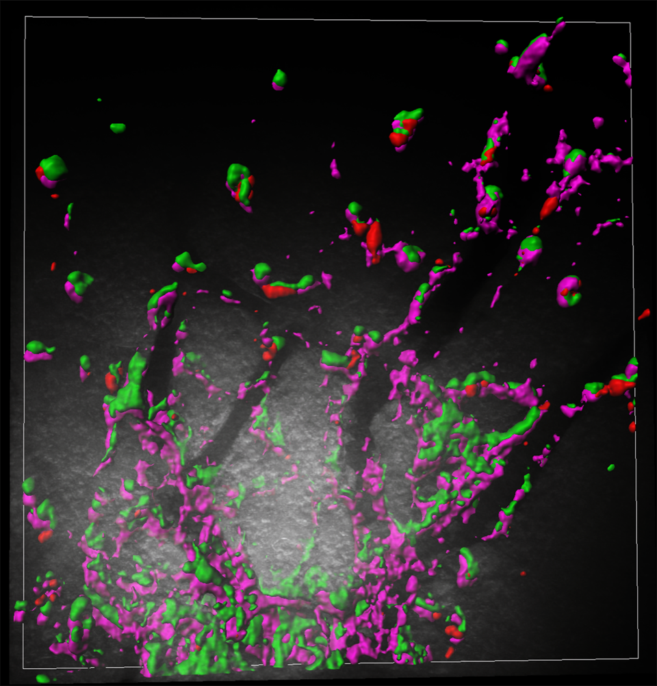
The genes in question, known as ‘retrotransposon Gag-like’ 5 and 6 (Rtl5/Rtl6), are carried by almost all mammals, and are similar to genes found in retroviruses, such as HIV. The researchers were convinced that the genes must be doing something important, as despite coming from viruses, these inherited viral genes have been preserved in the mammalian genome for at least the last 120 million years. To work out what these genes are doing the scientists needed to know where they are active, so they looked for RTL5/6 proteins, which are only produced when genes are switched on. They discovered that Rtl5 and Rtl6 are switched on in the brain in cells called microglia, which act as the ‘first responders’ to infection. Dr Kaneko-Ishino said, “we never expected that Rtl6 and Rtl5 would function in microglia when we started this work 15 years ago, and even when we knew that Rtl6 was a microglial gene we didn’t understand its significance. Our ‘eureka moment’ came during a dissection when Dr Ishino was carefully removing a mouse brain. We realised that if instead we damaged the brain, we could activate RTL6”.
The RTL6 proteins, shown in green, guard the mouse brain capillaries (the branch-like structures in black) against ‘infection’ by clustering around the magenta-coloured bacterial mimic.
The team set up fake infections in mice brains to test how the microglia producing RTL5 or RTL6 would respond to either bacteria or viruses. They found that microglia containing RTL6 protein responded to the bacteria-like mimic, whereas the microglia with RTL5 reacted to the simulated viral infection. In addition, when the researchers removed the Rtl6 gene, they found that the mice could not eliminate the fake bacterial infections, while the mice without Rtl5 could not clear the viral mimics, meaning that together Rtl5 and Rtl6 protect the brain against two of the most common types of infection.
These results provide the first example of viral-derived genes that have been re-purposed to protect mammalian brains against infection. The idea that viruses have had such a positive impact on our lives may be surprising, but examples like Rtl5 and Rtl6 demonstrate that viral invaders can, in the long run, benefit their host. According to Dr Ishino, “virus-acquired genes are essential parts of our genome, playing various – but essential – roles in mammalian and human development. We think it is possible to extend this idea to primate- and human-specific acquired genes from retroviruses to help us understand human evolution”.
To find out more about this story check out our interview with authors Masahito Irie, Fumitoshi Ishino and Tomoko Kaneko-Ishino, for our ‘The people behind the papers’ series.
With the calendar about to click over from September to October, we are bringing you one last post highlighting some of our archive content that we hope will help make your academic year a good one! In this post, we look at getting organised, both in the lab and with your data analysis.
The topic of this post was prompted by a tweet from Teresa Rayon asking for advice on lab inventory management software, so we’ve included it below so you can see the replies that Teresa received.
#NewPI question. Can people recommend Lab-inventory management software? *Free *Web-based *Easy import/export database
@Quartzy seems a good option, any opinions for/against?
After the lab is organised and you are approaching your first experiments, you need to think about how you are going to record all your data and metadata. We have an article on The Pros and Cons of having an Electronic Lab Notebook (ELN) here on the Node. The article also includes a link to five popular ELNs, updated in 2020.
We have collected below, a series of ‘how to’ guides from Joachim Goedhart, Helena Jambor, Jonas Hartmann and Steph Nowotarski covering organising, visualising and analysing data.
Once you have followed all the tips below, you are ready to present your data to the community. Helena tells us how to make a graphical abstract and how to win a poster prize (or how to make an impactful poster!)
If you have a ‘how to’ guide you would like to share, please get in touch or feel free to post it directly onto the Node. Details of how to register with the Node can be found here. You can contact us at thenode@biologists.com
Helena, Joachim and Jonas offer guides into presenting and analysing microscopy data. You can find more information on image analysis on our sister site FocalPlane, including Andrey Andreev’s recent post on presentation and analysis of calcium imaging data.
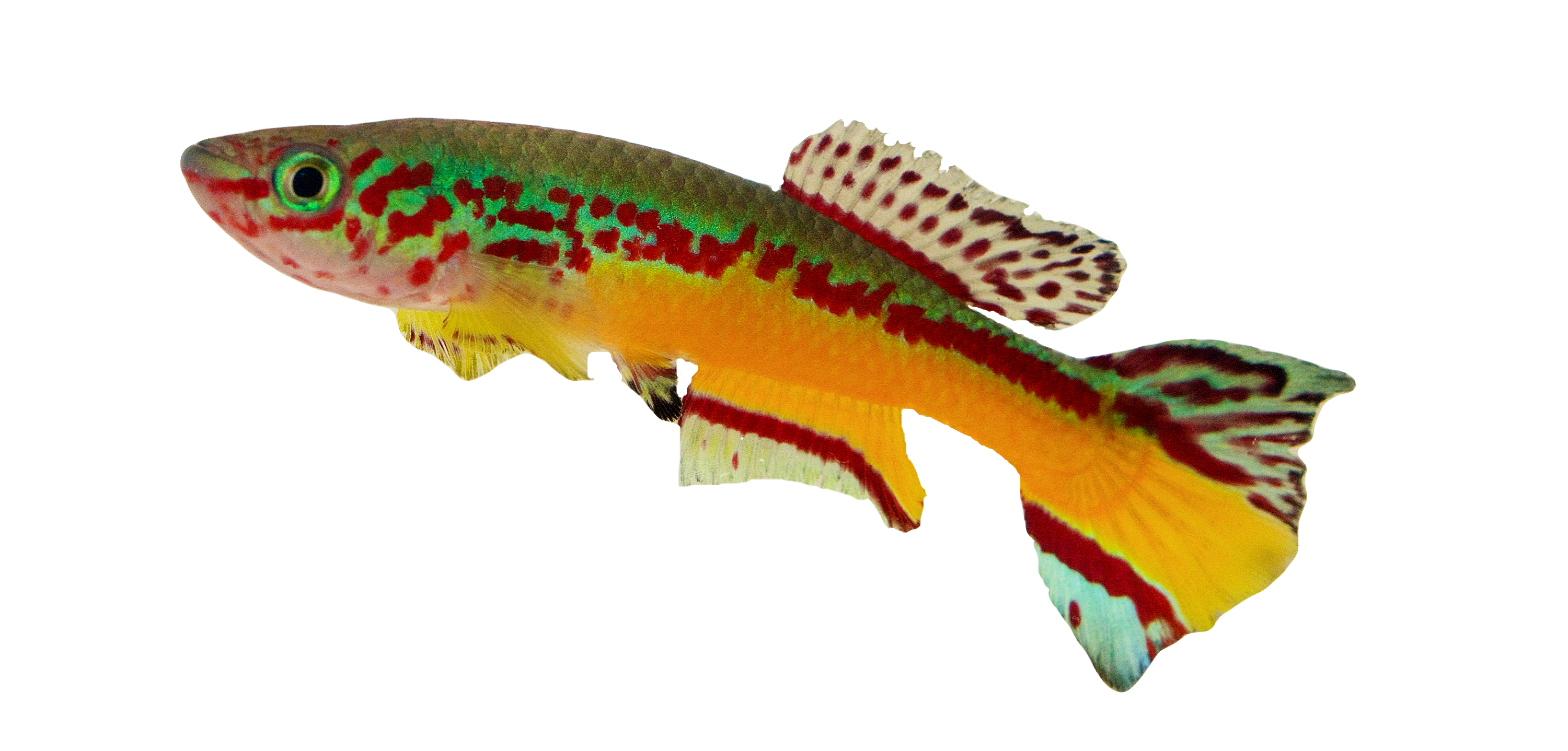
My name is Andrew Thompson, and I am an assistant professor and principal investigator of the Xtremo-Devo Lab at Western Michigan University. In our lab, we use small, colorful, tropical killifishes (Fig.1) to study how organisms adapt to extreme environments and undergo suspended animation. Killifishes include freshwater fishes in the group of Aplocheiloidei (Cyprinodontiformes) and live in small freshwater pools and streams in the tropics of South and Central America, Africa, Madagascar, India, and Southeast Asia. In fact, it is thought that “killi” is derived from the archaic Dutch word “kilde” meaning small stream or puddle.
Figure 1. A male Fundulopanchax amieti, a killifish native to Cameroon. Photo by A. Thompson.
A Seasonal Life Cycle
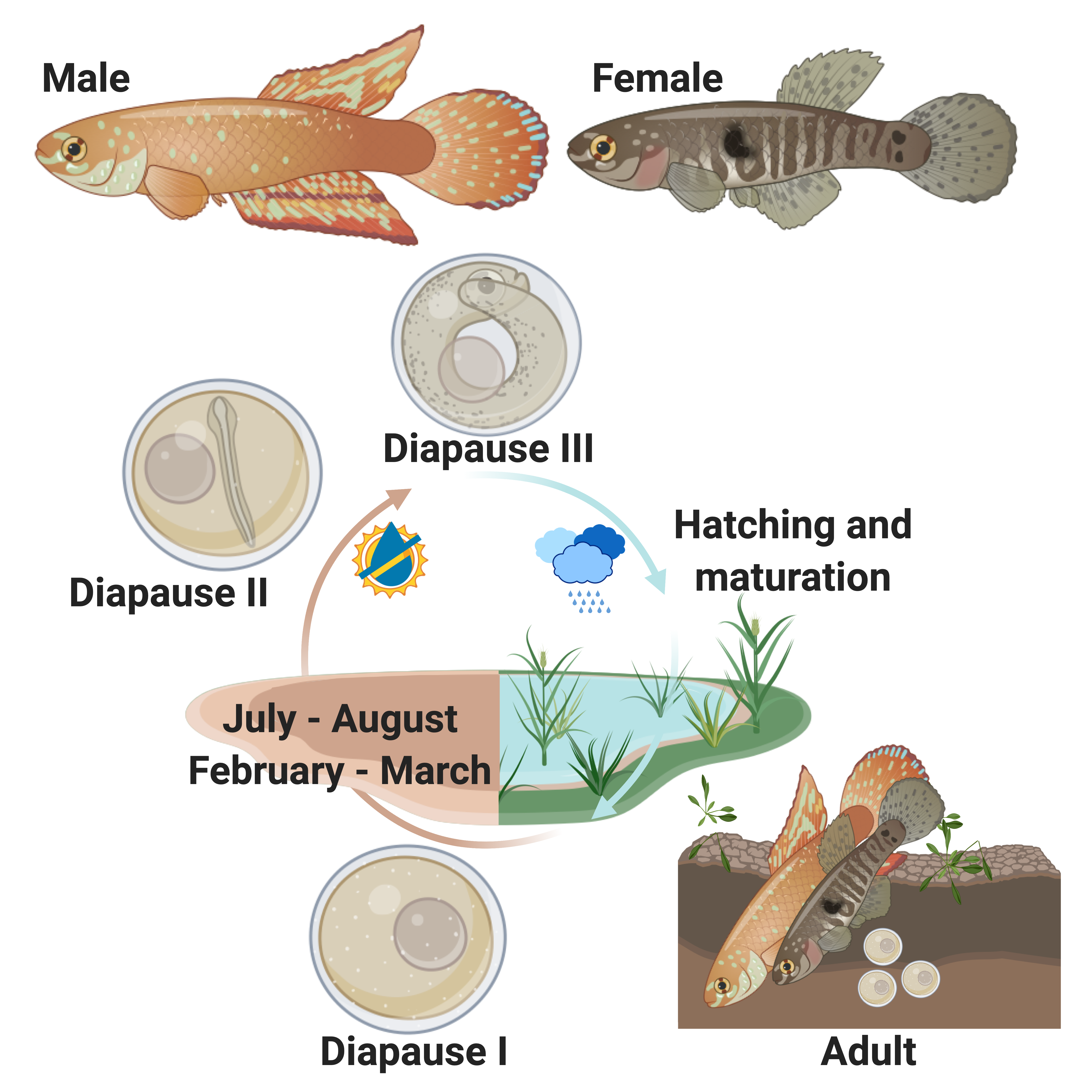
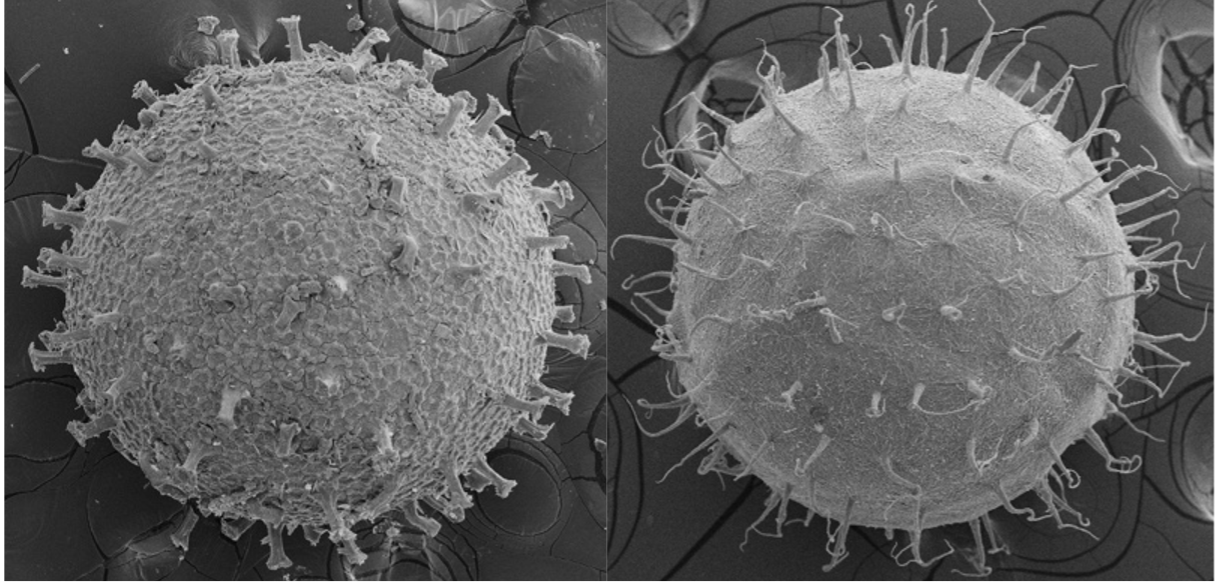
Some killifish species in Africa and South America live in pools that seasonally flood in a wet season and dry out in a dry season (Fig. 2). This can happen once a year, twice a year, or less predictably depending on weather and climate patterns (Costa 2002; Furness 2015). Unfortunately, for these killifish, the adults living in seasonal or annual pools are doomed to die in the dry season and the desiccation of their habitat. But as Dr. Ian Malcom from Jurassic Park might say, “life, uh, finds a way.” And killifish did find a way. Around the time dinosaurs were going extinct, Aplocheiloid Killifish were beginning to diversify (Thompson et al. 2021). They evolved superpowers that allowed some species to survive in these temporary habitats. More specifically, these “fish-out-of-water” bury their eggs in the soil that have tough specialized egg envelopes (Fig. 3) that keep water in and prevent the embryos from drying out during the dry season. These eggs can be covered in some amazing structures like filaments to bind them to the substrate, and even mushroom-like (Fig 3A) or corkscrew-like projections (Fig. 3B, Thompson et al. 2017a).
Figure 2. The life cycle of a seasonal killifish, the Rio Pearlfish, Nematolebias whitei (Thompson et al. 2022, Made with BioRender).
Figure 3. Scanning electron micrograph of annual killifish eggs: A.Notholebias fractifasciatus with mushroom-like projections. B. Plesiolebias aruana with corckscrew-like projections. Images by A. Thompson and C. Stone.
Dormancy and Diapause
But what about timing? These annual or seasonal killifishes cannot just grow and hatch like most fish when they get to a certain time in development. If they grow too fast or hatch at the wrong time, they will die upon hatching if the pools have not refilled or already dried up. Annual killifishes have a strategy that is unique among vertebrates in that they can arrest their development up to three different times as embryos buried in the soil (Fig. 2, Wourms 1972a; Wourms 1972b; Wourms 1972c). This arrested development is known as diapause, and few vertebrates are capable of this type of dormancy. During diapause, growth stops, cells stop dividing, and metabolic rate is greatly reduced. Killifish diapause can occur at three specific embryonic stages and can be skipped depending on environmental conditions such as temperature (Podrabsky et al. 2010). The first diapause occurs very early after fertilization when the embryo consists of stem cells (Fig.2, Wourms 1972b; Wourms 1972a). The second diapause stage occurs when the embryo is starting to develop organs (Fig. 2, Wourms 1972a; Wourms 1972c). The third diapause is used to delay hatching and begins after organs have fully developed (post organogenesis, Fig. 2, Wourms, 1972a, 1972c). Interestingly, non-annual killifishes can also delay hatching, especially if they are incubated out of water, but this might not represent a dormant or diapause phenotoype (Wourms 1972c; Varela-Lasheras and van Dooren 2014; Furness 2015; Thompson et al. 2017b).
Environmentally-Cued Hatching
Annual killifishes are the only vertebrates to stop their development after organogenesis, and this unique type of suspended animation could inform research on how vertebrate animals survive stasis with fully-developed, complex organ systems. Humans have long fantasized about “hypersleep”, a key plot point in science fiction films used to travel into deep space, save energy, stop aging, and survive in harsh environments, but killifish have been practicing this technique for millions of years. Killifish are able to use Diapause III to control hatching, remaining dormant at this stage until their habitat floods, triggering them to hatch. Hatching is a process that most animals and all vertebrates must complete as part of their development. Aquatic vertebrates like fish and frogs hatch by secreting enzymes that break down the egg envelope or zona pellucida and allow the transition from an embryo to a free-living larvae (Urch and Hedrick 1981; Yamagami 1981; Yasumasu et al. 1992; Cohen et al. 2018). While you may think of human hatching as a birthday, humans and other mammalian embryos hatch much earlier in development, during the blastocyst stages when the embryo and/or the uterus secrete enzymes that break down the zona pellucida so implantation can occur (Yamazaki et al. 1994; O’Sullivan et al. 2002; Syrkasheva et al. 2017; Leonavicius et al. 2018).
Eco-Evo-Devo
Our lab wants to figure out how animals integrate genetic and environmental cues to control progression of development in the face of changing environments. Killifish offer a unique opportunity as a research model to study suspended animation and the environmental control of hatching in a vertebrate system. While we understand the adaptive nature of dormancy and the enzymatic component of hatching, the genetic regulation of these developmental transitions or lack thereof via diapause, are relatively unknown. The study of evolutionary developmental biology or “Evo-Devo” has provided unprecedented insights into how myriad organisms turn genotype in to phenotype, but our killifish model system allows for an “Eco-Evo-Devo” approach, exploring the integration of environmental cues with the underlying genetic regulatory signals to create phenotype.
The Rio Pearlfish
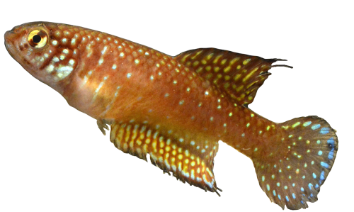
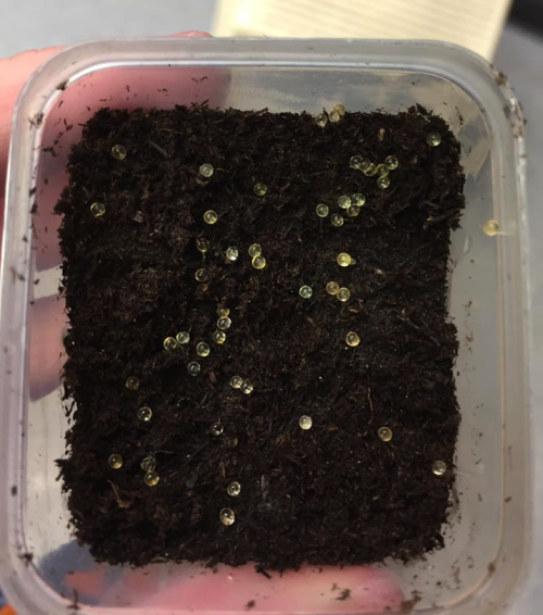
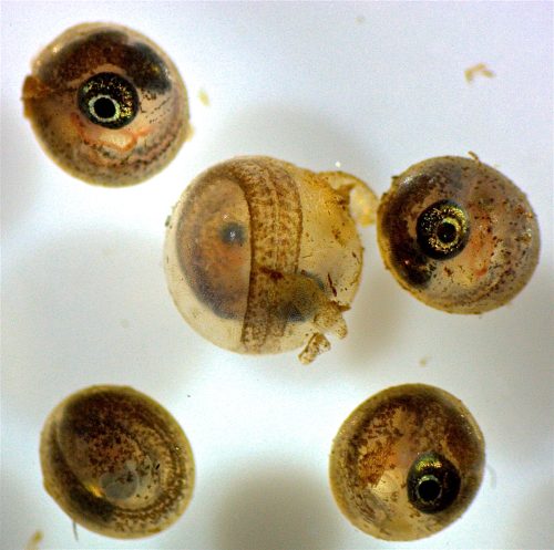
We have been developing the Rio Pearlfish (Fig. 4) as a tractable annual killifish model to study both the suspended animation and environmental control of hatching (Thompson and Ortí 2016; Thompson et al. 2022). Rio Pearlfish are quite happy in small containers that mimic their small puddles in the wild. They breed very easily in the lab in sand (Video 1) and eggs can be sifted out and used for studies that manipulate both the genome and environment of developing and diapausing eggs. For example, eggs can be incubated in humid environments outside of water, and we often place them in small plastic containers on top of moist peat moss to keep them damp and mimic seasonal desiccation (Fig. 5) Once our Pearlfish reach diapause III, we simply add water to halt dormancy, induce hatching, and begin the next generation (Fig. 6 ). Rio Pearlfish evolved a seasonal life history and the associated diapauses independently from other annual killifish species in Africa and South America (Thompson et al., 2021). We have sequenced the genome of the Rio Pearlfish, identified the hatching gland (Thompson et al. 2022), and characterized changes in gene expression between diapause III and hatched fish, many of which are convergent during dormancy across metazoans (Thompson and Ortí 2016). Thus, the Rio Pearlfish will be an invaluable laboratory model in our lab as we explore convergent adaptations to extreme environments..
Figure 4. The Rio Pearlfish, Nematolebias whitei, a bi-annual killifish native to Rio de Janeiro Brazil. Photo by A. Thompson
Video 1. Males and female Rio Pearlfish bury eggs in the substrate when spawning to protect embryos from desiccation. Males are larger and more colorful than females. Video by A. Thompson and M. Davoll.
Figure 5. Killifish eggs can be collected from the substrate and incubated out of water in small plastic containers containing peat moss. Photo by A. Thompson.
Figure 6. Rio Pearlfish, in diapause III, ready to hatch in water. The embryo in the center is starting to hatch as hatching enzymes have been secreted and started to swell and break down the egg envelope. Photo by A. Thompson, H. Wojtas, and M. Davoll.
A Future of Killifish
Overall, we aim to use our investigations into diapause and environmentally-cued hatching in the Rio Pearlfish and other killifishes to learn more about how animals adapt to stressful environments in a changing world. This is especially important in the light of pervasive human-induced environmental change. We also aim to strengthen the use of killifishes as biomedical models since they can slow growth, development, and metabolic rate. Perhaps, someday, killifish suspended animation could inform research on human diseases involving growth retardation, metabolic disorders, or abnormal cellular division.
We are currently recuiting Ph.D. and Master’s students, so if you are interested in joining our team or hearing more about our research, please contact us and check out our website here.
References:
Cohen, K. L., Piacentino, M. L., & Warkentin, K. M. (2019). Two types of hatching gland cells facilitate escape-hatching at different developmental stages in red-eyed treefrogs, Agalychnis callidryas (Anura: Phyllomedusidae). Biological Journal of the Linnean Society, 126(4), 751-767.
Costa, W. J. E. M. (2002). The neotropical seasonal fish genus Nematolebias (Cyprinodontiformes: Rivulidae: Cynolebiatinae): taxonomic revision with description of a new species. Ichthyological Exploration of Freshwaters, 13(1), 41-52.
Furness, A. I. (2016). The evolution of an annual life cycle in killifish: adaptation to ephemeral aquatic environments through embryonic diapause. Biological Reviews, 91(3), 796-812.
Leonavicius, K., Royer, C., Preece, C., Davies, B., Biggins, J. S., & Srinivas, S. (2018). Mechanics of mouse blastocyst hatching revealed by a hydrogel-based microdeformation assay. Proceedings of the National Academy of Sciences, 115(41), 10375-10380.
O’Sullivan, C. M., Liu, S. Y., Karpinka, J. B., & Rancourt, D. E. (2002). Embryonic hatching enzyme strypsin/ISP1 is expressed with ISP2 in endometrial glands during implantation. Molecular Reproduction and Development: Incorporating Gamete Research, 62(3), 328-334.
Podrabsky, J. E., Garrett, I. D., & Kohl, Z. F. (2010). Alternative developmental pathways associated with diapause regulated by temperature and maternal influences in embryos of the annual killifish Austrofundulus limnaeus. Journal of Experimental Biology, 213(19), 3280-3288.
Shafei, R. A., Syrkasheva, A. G., Romanov, A. Y., Makarova, N. P., Dolgushina, N. V., & Semenova, M. L. (2017). Blastocyst hatching in humans. Russian Journal of Developmental Biology, 48(1), 5-15.
Thompson, A. W., Black, A. C., Huang, Y., Shi, Q., Furness, A. I., Braasch, I., … & Ortí, G. (2021). Deterministic shifts in molecular evolution correlate with convergence to annualism in killifishes. BioRxiv.
Thompson, A. W., Furness, A. I., Stone, C., Rade, C. M., & Ortí, G. (2017). Microanatomical diversification of the zona pellucida in aplochelioid killifishes. Journal of Fish Biology, 91(1), 126-143.
Thompson, A. W., Hayes, A., Podrabsky, J. E., & Ortí, G. (2017). Gene expression during delayed hatching in fish-out-of-water. Ecological Genetics and Genomics, 3, 52-59.
Thompson, A. W., & Ortí, G. (2016). Annual killifish transcriptomics and candidate genes for metazoan diapause. Molecular biology and evolution, 33(9), 2391-2395.
Thompson, A. W., Wojtas, H., Davoll, M., & Braasch, I. (2022). Genome of the Rio Pearlfish (Nematolebias whitei), a bi-annual killifish model for Eco-Evo-Devo in extreme environments. G3, 12(4), jkac045.
Urch, U. A., & Hedrick, J. L. (1981). Isolation and characterization of the hatching enzyme from the amphibian, Xenopus laevis. Archives of Biochemistry and Biophysics, 206(2), 424-431.
Varela-Lasheras, I., & Van Dooren, T. J. (2014). Desiccation plasticity in the embryonic life histories of non-annual rivulid species. EvoDevo, 5(1), 1-11.
Wourms, J. P. (1972). Developmental biology of annual fishes. I. Stages in the normal development of Austrofundulus myersi Dahl. Journal of Experimental Zoology, 182(2), 143-167.
Wourms, J. P. (1972). The developmental biology of annual fishes. II. Naturally occurring dispersion and reaggregation of blastomeres during the development of annual fish eggs. Journal of Experimental Zoology, 182(2), 169-200.
Wourms, J. P. (1972). The developmental biology of annual fishes. III. Pre‐embryonic and embryonic diapause of variable duration in the eggs of annual fishes. Journal of Experimental Zoology, 182(3), 389-414.
Yamagami, K. (1981). Mechanisms of hatching in fish: secretion of hatching enzyme and enzymatic choriolysis. American Zoologist, 21(2), 459-471.
Yamazaki, K., Suzuki, R., Hojo, E., Kondo, S., Kato, Y., Kamioka, K., … & Sawada, H. (1994). Trypsin‐like Hatching Enzyme of Mouse Blastocysts: Evidence for Its Participation in Hatching Process before Zona Shedding of Embryos 6: (embryo/hatching enzyme/protease/trypsin/strypsin). Development, growth & differentiation, 36(2), 149-154.
Yasumasu, S., Yamada, K., Akasaka, K., Mitsunaga, K., Iuchi, I., Shimada, H., & Yamagami, K. (1992). Isolation of cDNAs for LCE and HCE, two constituent proteases of the hatching enzyme of Oryzias latipes, and concurrent expression of their mRNAs during development. Developmental biology, 153(2), 250-258.
Fully-funded PhD and Postdoc positions are available in The Zaidel-Bar Lab to study the regulation of the cytoskeleton during cell and tissue morphogenesis. We invite highly motivated students to apply.
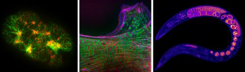
The lab takes a multi-scale approach from single proteins to the organism and system level, using a variety of cutting-edge technologies, most notably advanced microscopy, both in organoids and in the nematode C. elegans. Its goal is to understand the molecular mechanisms cells and tissues employ to change shape, migrate, sense, and generate mechanical forces that are essential for embryonic development and go awry in many diseases.
The lab welcomes outstanding international students with a degree in life sciences or in quantitative sciences, with a passion for research and interest in the cytoskeleton. The lab provides full fellowships for all students and housing on campus is an option. Lectures and courses are in English. Ph.D. studies usually last 4 years. Postdocs will be hired on a yearly contract up to 5 years.
The Zaidel-Bar lab is located in Tel-Aviv University, which is the leading interdisciplinary research and teaching university in Israel. Importantly, the scientific environment is dynamic and collaborative, with state of the art facilities, and students will have numerous opportunities to meet, learn from, and collaborate with excellent scientists.
Tel-Aviv is the cultural and commercial heart of Israel. Situated on a beautiful coast of the Mediterranean Sea, it is a fun, young city that never sleeps, with great food and weather.
To apply: send your CV (including transcripts) and a cover letter detailing your research experience and interest to: zaidelbar@tauex.tau.ac.il. Review of applications will begin immediately, and positions will remain open until filled.
 (No Ratings Yet)
 (1 votes)