Two Postdoctoral Research Associate positions (PDRA) are available in the Spagnoli lab. in the Centre for Stem Cell & Regenerative Medicine at King’s College London. Our team uses interdisciplinary approaches to study pancreatic development and stem cells. The candidates will join a Wellcome Trust-funded research programme aiming at studying the pancreatic tissue microenvironment in all its complexity using cutting-edge models.
Single-cell sequencing has unveiled a high degree of cellular heterogeneity within the pancreatic microenvironment and has opened the way for a systematic study of intercellular interactions. We seek to spatially reconstruct the organisation of functional niches in the pancreas and study how they induce distinct pancreatic differentiation programmes using mouse models and human pluripotent stem cells. This will set the stage for manipulating combinatorial 3D organ niches towards engineering pancreatic cells for regenerative medicine.
The research programme will require expertise in transcriptomics, high-resolution imaging, stem cell culture, human tissue, mouse genetics, computational analytical methods. Applicants should have a recent Ph.D. degree or have submitted his/her Ph.D. thesis. We wish to appoint one PDRA with background in development and stem cell biology and one PDRA with computational background interested in single-cell omics, spatial transcriptomics and image analysis.
The Spagnoli lab. is a young dynamic team, member of the outstanding Centre for Stem Cell & Regenerative Medicine at King’s College London. This is a world-class research environment with all facilities essential for this ambitious research programme.
Interested candidates should get in touch with Francesca Spagnoli (francesca.spagnoli@kcl.ac.uk) for further information. Please send your CV, research interests, and names and contact information of three references.
Positions will be available starting in October 2021.
More information on the group, publications and research topics in the group can be found in the laboratory website: https://www.spagnolilab.org/
In the latest episode of Genetics Unzipped we’re delving into the science behind so-called ‘genetic superheroes’, and explaining why you might have hidden powers within your genes. Despite the name, these superheroes don’t have the ability to shoot webs from their fingers or save the universe, but something with a lot more real world relevance to human health.
Instead, these people have a much more down to earth ability: carrying genetic alterations that should make them seriously ill, yet they are apparently healthy.
We take a closer look at the search for genetic superheroes, the science behind their secret powers, and what their existence means for our understanding of genetics.
If you enjoy the show, please do rate and review on Apple podcasts and help to spread the word on social media. And you can always send feedback and suggestions for future episodes and guests to podcast@geneticsunzipped.com Follow us on Twitter – @geneticsunzip
The HOUART lab is seeking to appoint three postdoctoral research associates to join a 5-year Wellcome Trust-funded research programme in developmental neurobiology. The programme aims to understand the role of axonal splicing factors and intron retaining transcripts in neuronal maturation and degeneration. Our nervous system crucially relies on local fast decisions taken long distance from the neuronal cell body. The mechanisms by which these decisions are spatio-temporally controlled remain obscure. We found that axonal splicing factors and intron-retaining mRNAs are key players in the process. We seek to understand the fundamental molecular and cellular roles they play locally in the dynamic control of neuronal connectivity, using animal models (zebrafish) and cell culture approaches.
The research programme will require expertise in zebrafishgenetics, transcriptomics, proteomics, high-resolution (live and fixed) molecular imaging, and sophisticated computational analytical methods. We wish to appoint one scientist with expertise in cutting-edge high-resolution/molecular imaging technologies; one with strong zebrafish neurobiology background and one with very strong protein/RNA biology experience.
Prof. Corinne Houart is a member and Deputy Head of the outstanding Centre for Developmental Neurobiology and MRC Centre for NeuroDevelopmental Disorders at King’s College London. Her lab is affiliated member of the Francis Crick Institute. This rich research environment provides world-class research facilities to the post holders.
Deadline for application 31st March 2021. For further information, please contact Prof. Corinne Houart, corinne.houart@kcl.ac.uk
The Ward Lab (https://www.ward-lab.org/) at the University of Texas Medical Branch is seeking a Postdoctoral Fellow to lead core projects focused on cardiovascular functional genomics.
The goal of the Ward Lab is to dissect the global role of regulatory elements in directing gene expression in healthy, stressed and disease states in cardiovascular disease-relevant cell types. We use a variety of population and evolutionary genomics approaches and induced pluripotent stem cell-based tools to tackle this problem.
Projects are available in several areas including:
-gene regulatory dynamics during differentiation to cardiovascular cell types
-gene regulatory processes in response to disease-relevant perturbations
-inter-individual variation in response to disease-relevant perturbations
We are looking for a highly motivated, enthusiastic individual to join our growing team. Candidates should have received their Ph.D. within the last year in Molecular Biology, Cell Biology, Evolutionary Biology, Cancer Biology, Genetics, Systems Biology, Computational Biology, or a related field. They should have excellent communication skills and a good track record of productivity, including a first-author paper.
The University of Texas Medical Branch, located on the island of Galveston, is a member of the University of Texas System, the Texas Medical Center (the largest medical center in the world based in Houston -approximately 50 miles away), and the Gulf Coast Consortia in Quantitative Biomedical Sciences. This environment provides a vibrant research community. There are excellent Core facilities on campus including Next Generation Sequencing, Flow cytometry and Proteomics.
Please apply by sending a cover letter, C.V., and contact information for three references via email to Dr. Michelle Ward (miward {@} utmb {.} edu). Review of applications will begin immediately and continue until the position is filled. Informal enquiries are welcome.
UTMB Health strives to provide equal opportunity employment without regard to race, color, national origin, sex, age, religion, disability, sexual orientation, gender identity or expression, genetic information or veteran status. As a VEVRAA Federal Contractor, UTMB Health takes affirmative action to hire and advance women, minorities, protected veterans and individuals with disabilities.
A Postdoctoral Researcher position is available in the La Manno lab at the École Polytechnique fédérale de Lausanne (EPFL) in Switzerland. We are looking for an ambitious candidate with a Ph.D. in the developmental biology of the nervous system or pathologies thereof, interested in applying cutting-edge methods in spatial transcriptomics and single-cell biology to understand prenatal brain development.
The research project offered will be focused on the spatio-temporal organization of neural progenitor cells across the developing nervous systems in health and disease. The project, funded by SNF, is low-risk and high-impact.
The successful candidate will analyze the spatial distribution and abundance of different newly discovered radial glial subpopulations in both normal and perturbed murine brain. Cutting-edge spatial transcriptomics technologies will be applied to obtain a comprehensive description of the spatiotemporal patterning of the ventricular zones across different brain regions.
The study involves a cell-type centric comparison between normal development and clinically relevant perturbations associated with congenital malformations and mental disabilities. The high-throughput measurements will identify key system-level parameters affected and if they are conditional to particular cell types.
The overarching goal is understanding the functional implications of the distribution and abundance of radial glial subtypes and the modalities teratogens and metabolic alterations can disrupt it.
Candidate Qualifications
An advanced degree of experience with mouse work and, in particular, with in-utero manipulations is required. Furthermore, some degree of experience with histology preparation and imaging of the developing brain is expected. In particular, having used single-molecule fluorescent in situ hybridization will be considered a significant plus.
A keen interest in image analysis or programming is a plus; however, no significant previous experience is required. The candidate will have the possibility to learn the bioinformatics skills required for a fulfilling analysis of the generated data.
Ideally, there will be a track record of peer-reviewed publications. Good written and oral skills and the ability to work collaboratively in a team are expected.
About us
The Laboratory of Neurodevelopmental Systems Biology is part of the Brain Mind Institute at the Swiss Federal Institute of Technology Lausanne (EPFL). EPFL is one of the top-ranking universities in the world, and its research environment is characterized by its multi-disciplinarity, bridging neuroscience, computation, and engineering.
The long-term goal of the La Manno lab is the description and modeling of the different cellular states appearing during neurodevelopment, their diversification and fate commitment. Using single-cell genomics and spatial transcriptomics tools, we aim to answer key questions in developmental biology, neuroscience, and pathology.
We have been analyzing single-cell genomics data since its early times (Islam et al., Nature Methods 2013). We have developed RNA velocity, a new analysis framework that allows the inference of lineage relationships from scRNA-seq data (La Manno et al., Nature 2018).
We contributed discovered new radial-glial populations in the human midbrain (La Manno et al., Cell 2016), and more recently released a single-cell atlas of the entire prenatal nervous system development (La Manno et al., Biorxiv 2020)
What we offer
We offer a well-funded, low-risk high-gain project to be performed in a friendly and constructive work environment. We will provide you with the freedom to be a creative, independent scientist. We strive to ensure a good work/life balance and flexible working hours.
The successful candidate will have the opportunity to interact and participate in the scientific activities of the EPFL School of Life Sciences. The laboratory is located in Lausanne with state-of-the-art facilities and a vibrant interdisciplinary research community.
EPFL offers an English-speaking work environment and competitive salaries and benefits. The position is fully funded for four years (renewed yearly). Remuneration is determined in accordance with the EPFL “Scientific collaborator” directive (a minimum salary of 82,000 Swiss francs, and adapted depending on years of experience).
EPFL is an equal opportunity employer and a family-friendly university. We strive to increase diversity and strongly encourage minorities to apply.
Application
Please send your request as a single PDF file – including a CV, a complete list of publications, a statement of research interests, and the contact information of at least two reference persons – to nsbl.openings@epfl.ch
We are looking forward to receiving your application.
Applications are invited for two postdoctoral fellows in the Brückner lab at The University of California San Francisco, Broad Center of Regeneration Medicine and Stem Cell Research, Department of Cell and Tissue Biology.
The Brückner lab investigates mechanisms how organ development and transdifferentiation are regulated by neuronal input and environmental conditions, using a model of blood cell development in Drosophila melanogaster (Corcoran et al. bioRxiv 2020; Makhijani et al. Nature Comms 2017; Makhijani et al. Development 2011). Transdifferentiation generates specialized cell types independent of stem or progenitor cells. Despite the high interest in this unique phenomenon, it is largely unknown how transdifferentiation is regulated in vivo.
We offer two federally funded projects that focus on the genetic dissection of molecular mechanisms in the neuronal regulation of blood cell development, signaling, and transdifferentiation. In collaboration with the Perrimon lab at Harvard Medical School, we use single cell/ single nucleus RNA sequencing (snucRNAseq) to identify transcriptional mechanisms how activated neurons control blood cells and potentially other target tissues. This research is expected to reveal fundamental principles how environmental cues, through activation of sensory neurons, regulate transdifferentiation and other cell behaviors in organ development. It will suggest similar principles in other species and organ systems where environmental sensors and tissue precursors coincide, including systems of blood, skin, lung, and digestive system.
Positions will be available starting in April and July 2021, respectively.
Candidates must hold a recent PhD, MD or MD/PhD degree, or anticipate such degree in the near future. Underrepresented and diversity candidates are in particular encouraged to apply. The ideal candidate is a talented and creative scientist with a background in Drosophila genetics. General skills in molecular and cell biology are expected. Prior experience in bioinformatics would be an advantage but is not required. We are looking for motivated candidates who have good communication skills and are reliable, hardworking and fun, and have prior research publication(s). We anticipate transition to future independent funding.
UCSF and the Broad Center offer a vibrant scientific environment and state of the art facilities. The Brückner lab provides infrastructure, a collegial environment, training and opportunities to work in teams. The Perrimon lab at Harvard Medical School/ Howard Hughes Medical Institute provides collaboration and offers training on the single cell aspect of the project.
COVID-19: As of March 2021, UCSF is offering COVID vaccinations to all education-related personnel including postdocs. UCSF offers free asymptomatic COVID testing to employees at all times. The Brückner lab observes safe practices including mask wearing, social distancing, reduced lab occupancy, and remote meetings where possible.
Please email your CV, research interests, and names and contact information of three references to katja.brueckner@ucsf.edu
We are seeking to recruit a talented and motivated Postdoctoral Research Scientist to investigate the gene regulatory control of human pluripotent states. This position is within Peter Rugg-Gunn’s team in the Epigenetics Programme at the Babraham Institute, Cambridge, UK.
The central aim of this three-year project is to investigate new regulators of human naïve cell reprogramming that we have recently identified, and to develop a mechanistic understanding of how they function. The job holder will engineer human pluripotent cell lines with inducible degradation systems targeting the identified regulators and will use these cell lines to investigate the molecular and cellular defects that arise following protein degradation. The job holder will also use CUT&Tag methods to identify genome-wide occupancy of the proteins of interest. We have particular expertise in reprogramming and capacitation transitions, developmental cell models including gastruloids, gene targeting, and in relevant assays such as proteomics and single cell transcriptomics / epigenomics. The overall significance of this work will be to establish exciting new links between gene regulatory mechanisms and the control of pluripotency during human development. We anticipate that modulating the identified pathways will improve the generation of naïve cells and open up new ways to deliver cell types with useful translational properties.
The ideal candidate will be interested in stem cell and developmental biology, particularly in the gene regulatory mechanisms that underpin lineage specification and reprogramming. The Epigenetics Programme provides a highly collaborative and thriving research environment with particular strengths in stem cell, developmental and ageing biology. We have access to onsite state-of-the-art facilities run by dedicated staff, including High-Throughput Sequencing, Bioinformatics, Imaging and Gene Targeting. We have close links to Cambridge University through affiliations with the Stem Cell Institute, the Centre for Trophoblast Research, the Epigenetics Club, and with the many departments and companies that we work with.
Multiple positions are available in the Stottmann lab as we relocate to the Institute for Genomic Medicine at Nationwide Children’s Hospital and Ohio State University. We study the genetic basis of structural birth defects. Projects usually focus on novel genes and mutations identified through human whole genome sequencing. We use sequencing analysis and a range of molecular embryological tools including genome editing in animal models and in vitro studies. Candidates will develop a robust research program in close consultation with the PI. We prefer applicants with multiple first-author publications and experience in mouse genetics, molecular biology and/or embryology. We also look for applicants with experience with iPSC culture. More information is at our current web page at Cincinnati Children’s:
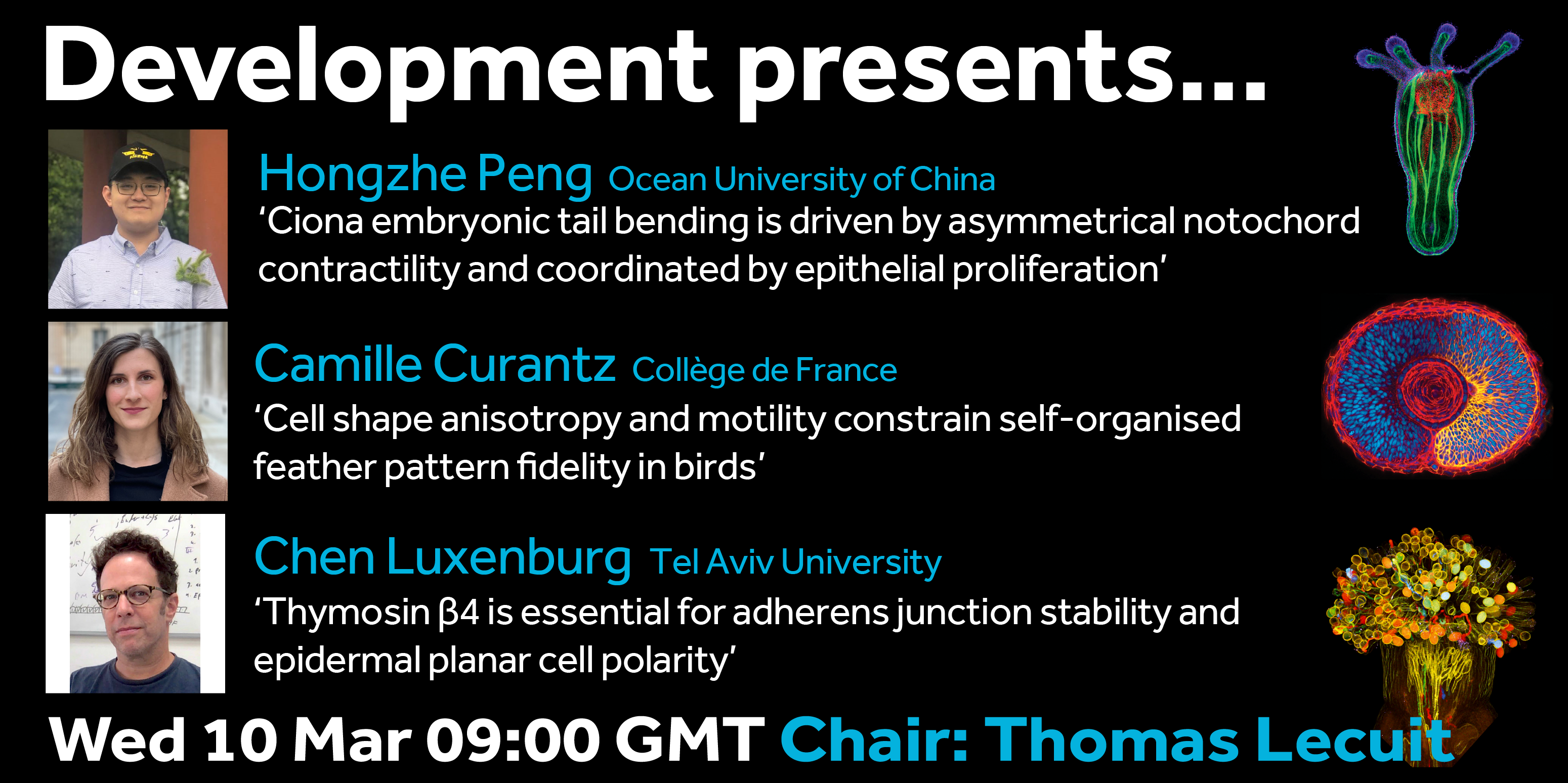
The next webinar in our Development presents… series will be chaired by Development editor, Thomas Lecuit (Institut de Biologie du Développment de Marseille). Thomas has brought together three talks on the topic of mechanics and morphogenesis.
The webinar will be held in Remo, our browser-based conferencing platform – after the talks you’ll have the chance to meet the speakers and other participants at virtual conference tables. If you can’t make it on the day, talks will be available to watch for a couple of weeks after the event; details will be posted on the Node or you can sign up to our mailing list for email alerts.
For more information about what to expect in Remo, go to
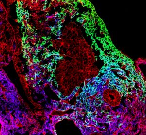
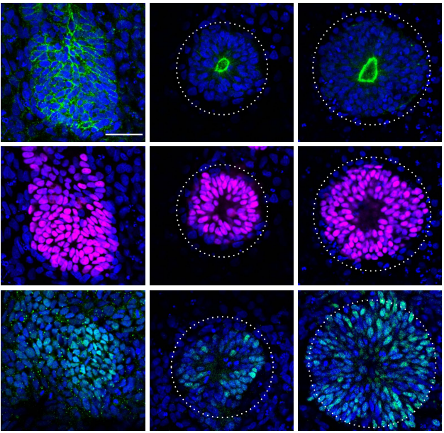
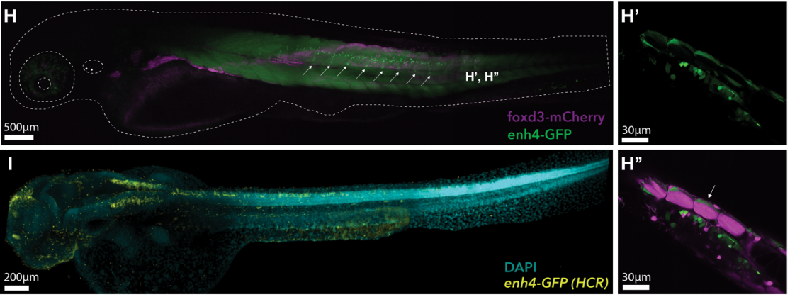
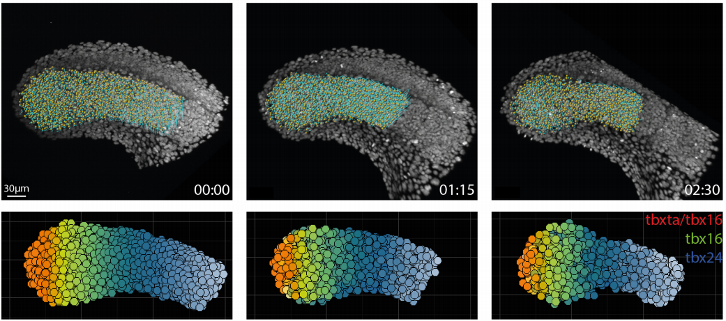
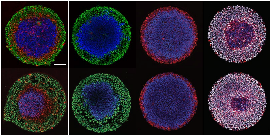
Hand2 delineates mesothelium progenitors and is reactivated in mesothelioma
Karin D. Prummel, Helena L. Crowell, Susan Nieuwenhuize, Eline C. Brombacher, Stephan Daetwyler, Charlotte Soneson, Jelena Kresoja-Rakic, Manuel Ronner, Agnese Kocere, Alexander Ernst, Zahra Labbaf, David E. Clouthier, Anthony B. Firulli, Héctor Sánchez-Iranzo, Sundar R. Naganathan, Rebecca O’Rourke, Erez Raz, Nadia Mercader, Alexa Burger, Emanuela Felley-Bosco, Jan Huisken, Mark D. Robinson, Christian Mosimann
Long noncoding RNA VENTHEART is required for cardiomyocyte specification and function
Albert Dashi, Wilson L.W. Tan, Chukwuemeka George Anene-Nzelu, Bangfen Pan, Autio Matias Ilmari, Zenia Tiang, Robin J.G. Hartman, Justus Stenzig, Heming Wei, Chen Gao Bin, Matthew Andrew Ackers-Johnson, Bing Lim, Anna Walentinsson, Vidhya Vardharajan Iyer, Malin K.B. Jonsson, Roger S. Foo
Dichotomous regulation of lysosomes by MYC and TFEB controls hematopoietic stem cell fate
Laura García-Prat, Kerstin B. Kaufmann, Florin Schneiter, Veronique Voisin, Alex Murison, Jocelyn Chen, Michelle Chan-Seng-Yue, Olga I. Gan, Jessica L. McLeod, Sabrina A. Smith, Michelle C. Shoong, Darrien Paris, Kristele Pan, Andy G.X. Zeng, Gabriela Krivdova, Kinam Gupta, Shin-Ichiro Takayanagi, Elvin Wagenblast, Weijia Wang, Mathieu Lupien, Timm Schroeder, Stephanie Z. Xie, John E. Dick
Drosophila functional screening of de novo variants in autism uncovers deleterious variants and facilitates discovery of rare neurodevelopmental diseases
Paul C Marcogliese, Samantha L Deal, Jonathan Andrews, J Michael Harnish, V Hemanjani Bhavana, Hillary K Graves, Sharayu Jangam, Xi Luo, Ning Liu, Danqing Bei, Yu-Hsin Chao, Brooke Hull, Pei-Tseng Lee, Hongling Pan, Colleen M Longley, Hsiao-Tuan Chao, Hyunglok Chung, Nele A Haelterman, Oguz Kanca, Sathiya N Manivannan, Linda Z Rossetti, Amanda Gerard, Eva Maria Christina Schwaibold, Renzo Guerrini, Annalisa Vetro, Eleina England, Chaya N Murali, Tahsin Stefan Barakat, Marieke F van Dooren, Martina Wilke, Marjon van Slegtenhorst, Gaetan Lesca, Isabelle Sabatier, Nicolas Chatron, Catherine A Brownstein, Jill A Madden, Pankaj B Agrawal, Roberto Keller, Lisa Pavinato, Alfredo Brusco, Jill A Rosenfeld, Ronit Marom, Michael F Wangler, Shinya Yamamoto
Glutamatergic dysfunction precedes neuron loss in cerebral organoids with MAPT mutation
Kathryn R. Bowles, M. Catarina Silva, Kristen Whitney, Taylor Bertucci, Jacob C. Garza, Nathan C. Boles, Kevin H. Strang, Sidhartha Mahali, Jacob A. Marsh, Cynthia Chen, Derian A. Pugh, Yiyuan Liu, Joshua E. Berlind, Jesse D. Lai, Susan K. Goderie, Rebecca Chowdhury, Steven Lotz, Keith Lane, Khadijah Onanuga, Celeste M. Karch, Justin K. Ichida, John F. Crary, Stephen J. Haggarty, Alison M. Goate, Sally Temple
Developmental and behavioral phenotypes in a new mouse model of DDX3X syndrome
Andrea Boitnott, Dévina C Ung, Marta Garcia-Forn, Kristi Niblo, Danielle Mendonca, Michael Flores, Sylvia Maxwell, Jacob Ellegood, Lily R Qiu, Dorothy E Grice, Jason P Lerch, Mladen-Roko Rasin, Joseph D Buxbaum, Elodie Drapeau, Silvia De Rubeis
A Human Multi-Lineage Hepatic Organoid Model for Liver Fibrosis
Yuan Guan, Annika Enejder, Meiyue Wang, Zhuoqing Fang, Lu Cui, Shih-Yu Chen, Jingxiao Wang, Yalun Tan, Manhong Wu, Xinyu Chen, Patrik K. Johansson, Issra Osman, Koshi Kunimoto, Pierre Russo, Sarah C. Heilshorn, Gary Peltz
The making of cauliflowers: the story of unsuccessful flowers
Eugenio Azpeitia, Gabrielle Tichtinsky, Marie Le Masson, Antonio Serrano-Mislata, Veronica Gregis, Carlos Gimenez, Nathanaёl Prunet, Jérémy Lucas, Etienne Farcot, Martin M. Kater, Desmond Bradley, Francisco Madueño, Christophe Godin, Francois Parcy
Apple ripening is controlled by a NAC transcription factor
Zoë Migicovsky, Trevor H. Yeats, Sophie Watts, Jun Song, Charles F. Forney, Karen Burgher-MacLellan, Daryl J. Somers, Yihi Gong, Zhaoqi Zhang, Julia Vrebalov, James G. Giovannoni, Jocelyn K. C. Rose, Sean Myles
Gene loss during the transition to multicellularity
Berenice Jiménez-Marín, Jessica B. Rakijas, Antariksh Tyagi, Aakash Pandey, Erik R. Hanschen, Jaden Anderson, Matthew G. Heffel, Thomas G. Platt, Bradley J. S. C. Olson
A novel adhesive complex at the base of intestinal microvilli
Christian Hartmann, Eva-Maria Thüring, Birgitta E. Michels, Denise Pajonczyk, Sophia Leußink, Lilo Greune, Frauke Brinkmann, Mark Glaesner-Ebnet, Eva Wardelmann, Thomas Zobel, M. Alexander Schmidt, Volker Gerke, Klaus Ebnet
 (No Ratings Yet)


 (1 votes)