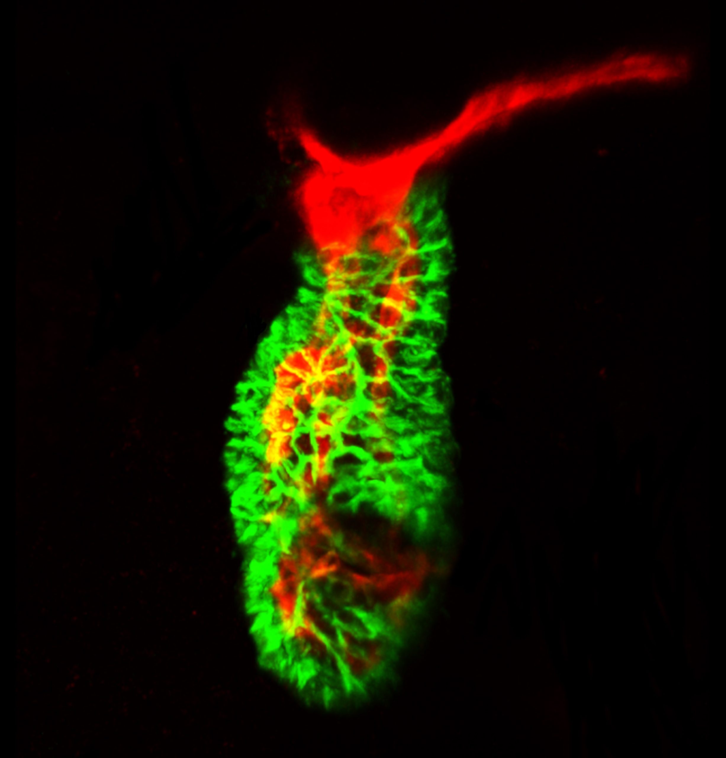
I started off as quite little—just one cell, in fact. No heart, no brain, no blood flowed in me and yet, somehow I found the motivation in me to divide. One me made two and two mes made four, till many tiny mes made me a full-fledged embryo with a heart full of hope. As I grew older, my heart grew with me. My body and heart needed to synchronize their growths, you see, because if my heart grew slower, then it would pump less blood into my system, and my other organs that needed blood to grow would not be able to do so properly. This posed a challenge for me– how do I make sure that my body knows my vessels are growing, and my vessels know my heart is growing? Please don’t tell me that’s yet another thing my brain has to manage. Wait, do I even have a brain yet!?
Anyway, perhaps it’s easiest if I use some intrinsic property of my blood, the fluid that connects my body and vessels and heart, to transport this important message. This way, as my heart beats faster and more blood flows through my system, my vessels will take the hint and grow accordingly. Going back to connections, let me first focus on my outflow tract, which connects my heart to my aortic vasculature. Perhaps if I tell my outflow tract to widen as blood flow and contractility increase, it will maintain my blood pressure while also allowing information in blood to pass through to my other systems. Ugh, so how I do that?
Hmm. What if the cells in the inner endothelial lining of my outflow tract respond to blood flow and contractility by dividing? What if they accumulate cells from the neighboring vessels, in conjunction with neighboring vessels accumulating them from more distal vessels? Maybe I’ll do both—yes, that sounds like a good strategy. But how do I regulate this growth? Maybe I should have many opposing signals, reacting to multiple inputs, towards the unified goal of getting the outflow tract to a particular lumen size at a particular developmental stage. I’m feeling lazy, I’ll just repurpose my good ol’ TGFb pathway to do this—put some receptors in my endothelial cells and have my blood carry physical and molecular cues to activate them.
Great, I think I have a functioning cardiovascular system! And it’s caught in this self-regulating loop where function informs form, which feeds back to function. I feel so smart! Kudos to me figuring this out over evolution. Well, I guess I’ll give an additional kudos to this grad student who figured it out in 5 years.
We are looking forward to an exciting UK SouthWest Zebrafish Meeting at the 11 Sep 2020!
If you have not already, register and also submit your abstracts for either talks or posters. Talks and posters will be given to students, early career researchers and technicians. Our deadline for abstract submission is Friday 24th July, so submit your abstracts on our website: https://swzm2020.wixsite.com/swzm2020/registration. On the website you will find the online registration form, as well as the abstract form to fill out and upload.
Virtual Meeting:
We have been working behind the scenes to build our virtual meeting! We are planning to hold the meeting via a central hub on Padlet.com. This means we can host an online poster exhibition, secure to all delegates, where you will be able to browse and read posters, leave comments and interact with presenters and crucially vote for ‘best poster’! Prizes will be awarded for best talks and posters! We will also use Zoom to host our series of talks, break-out sessions, talks from sponsors, as well as an online ‘Zebrafish Zoom Café’ for breaks and general discussion. More details to come!
New Sponsors:
Good news! We can now confirm our meeting will be free from any registration fees for delegates. We can now confirm we have been kindly sponsored by The Company of Biologists, through a Scientific Meeting Grant, so we can waive registration fees. We can also announce we are kindly sponsored by Tecniplast and DanioLabs.
Special Issue:
We would like to announce that a Special Issue of ‘Histochemistry and Cell Biology’ focussing on ‘In vivo cell biology in zebrafish’ will accompany the meeting. Please email us for further details. We are looking forward to hearing from you with research!
Please pass this information on to anyone you think may be interested and feel free to tweet about the event using #SWZM20 and follow us on Twitter @Swzm20!
Looking forward to hearing from you all,
Steffen, Lucy, Holly, Michael, Josh, Chengting & Yosuke
(South West Zebrafish Organising Committee)
Maud Slye. Photo by Helen Balfour Morrison, Library of Congress – Public Domain
In this episode of Genetics Unzipped, Kat Arney tells the stories of two women – one a scientist fascinated by dancing mice, the other a seamstress with a deadly family legacy – who made significant contributions to our understanding of cancer as a disease driven by genetic changes, paving the way for lifesaving screening programmes for families.
Kat’s upcoming new book Rebel Cell: Cancer, Evolution and the Science of Life, explores what we’ve learned so far about where cancer comes from, where it’s going, and how we might finally beat it. It’s coming out in the UK on the 6th of August and in the US on the 29th September – and is available now to pre-order from Amazon or your retailer of choice.
While she was researching the book, Kat came across the stories of two remarkable women who both made significant contributions to our fundamental understanding of cancer, but who have tended to be overlooked in many tellings of the history of cancer research. Here are their stories.
Born in Minnesota in 1879, Maud Slye was a cancer pathologist who dedicated her career to studying patterns of cancer inheritance in more than 150,000 mice. But as well as being a dedicated scientist (as well as a part-time poet), her ideas about eugenics brought controversy.
Even so, Slye’s work earned her a gold medal from the American Medical Society in 1914 and from the American Radiological Association in 1922. She was also awarded the Ricketts Prize from the University of Chicago in 1915 and an honorary doctorate from Brown University in 1937. She was even nominated for a Nobel prize in 1923.
Running parallel to Slye’s work in mice was the research carried out by Aldred Warthin, a doctor working at the University of Michigan in Ann Arbor. One day in 1895, a chance meeting between Warthin and a local seamstress, Pauline Gross, set the two of them off on a 25-year-long quest to understand why so many members of Pauline’s family had died from cancer at a young age.
Pauline spent years compiling detailed family histories, enabling Warthin to trace the pattern of inheritance through Family G, as it became known. Like Slye, Warthin was a fan of eugenic ideas, and his work fell out of favour after his death. Pauline’s detailed genealogy lay undisturbed in a closet in the university until the 1960s, when American doctor Henry Lynch and social worker Anne Krush rediscovered her work and continued extending and investigating Family G. Today, members of Family G – and others around the world carrying dangerous variants in mismatch repair genes – can undergo genetic testing, with a range of preventative and screening options available.
The story of Pauline and Family G, and the impact that their genetic legacy has had on the family down the generations, is beautifully told in the book Daughter of Family G, a memoir by Ami McKay.
If you enjoy the show, please do rate and review on Apple podcasts and help to spread the word on social media. And you can always send feedback and suggestions for future episodes and guests to podcast@geneticsunzipped.com Follow us on Twitter – @geneticsunzip
By Héloïse Dufour, Shigeyuki Koshikawa and Cédric Finet
In this post we will discuss our recent paper entitled “Temporal flexibility of gene regulatory network underlies a novel wing pattern in flies” [1]. We initiated the present project in Sean Carroll’s lab where the pigmentation in drosophilids was used as a model to study evolutionary genetics. When we started back in 2010, we knew that the black dots on Drosophila guttifera wings result from the co-option of the protein Wingless during evolution [2]. However, D. guttifera does represent a particular case: the black dots on the wing are associated with campaniform sensilla, which is not the case for other wing colour patterns in drosophilids. The most vivid examples of wing pigmentation patterns are found among the Hawaiian flies, from simple pigmentation around the veins to complex black and white patterns [3], but we were aware that the Hawaiian flies are hardly tractable in the lab. In the meantime, we came across the species Samoaia leonensis which was maintained at the National Drosophila Species Stock Center. We decided to investigate the making of the wing colour pattern in this species.
The genus Samoaia: a case study of pigmentation and its evolution
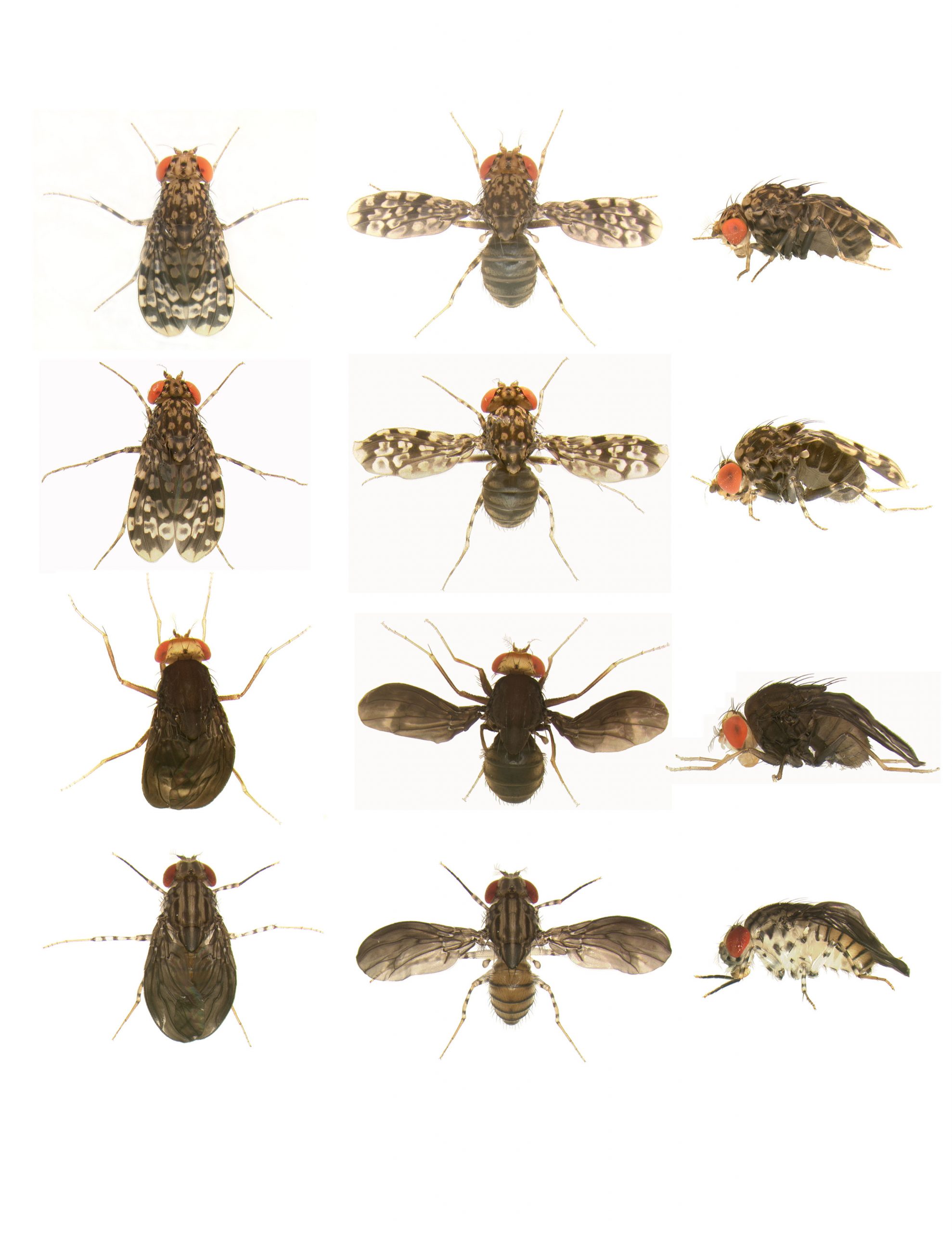
Samoaia is a small genus of seven described species endemic to the Samoan Islands in the central South Pacific. For example, the main model of our study S. leonensis exhibits a beautiful and complex black and white spot pattern on its wings (Figure 1). This species is a perfect case of pattern matching, with white spots not only covering the black wings, but the legs, the head, the abdomen, and the notum as well. [For etymology lovers, it is tempting to see in leonensis a reference to the lion, then to the jaguar and its spotted fur. Well, entomology systematics can be sometimes down-to-earth: the species S. leonensis was simply discovered around Leone on Tutuila Island.]
The entire genus Samoaia is an excellent model to study the evolution of pigmentation, especially the modularity of pigmentation. The species S. attenuata has indeed white spots on the legs only (Figure 1), suggesting that the whole body ‘camouflage’ of S. leonensis might result from the stepwise gain of spots in different organs during the course of evolution.
Figure 1. From top to bottom: Samoaia leonensis, Samoaiaocellaris, Samoaia hirta, Samoaiaattenuata. Image: courtesy of Héloïse Dufour.
Key finding nº 1
We showed that the co-option of the transcription factor Engrailed underlies the making of the white wing spots in S. leonensis wings. Whereas the expression of Engrailed is restricted to the posterior wing compartment in early pupal stages, its expression becomes spotty in both anterior and posterior compartments in later stages. This result is a big surprise for Drosophila developmental biologists for whom engrailed is the posterior identity gene by definition. Would it mean that there is a critical point beyond which engrailed is no longer required for AP patterning?
We tested this hypothesis in D. melanogaster. We silenced or overexpressed engrailed specifically in the wing at different time points. We were able to precisely define three different time windows during which (i) disturbing engrailed expression results in severe morphological defects, (ii) disturbing engrailed results in minor vein defects, (iii) disturbing engrailed expression does not lead to visible phenotype. We argue that the co-option of a given gene is possible beyond its corresponding critical point.
Key finding nº 2
We found that the co-option of Engrailed in the S. leonensis wing is partially independent from the other genes of the AP specification network. For instance, hedgehog and engrailed are not co-expressed in the anlagen of the future white spots, whereas these two genes are co-expressed in the posterior wing disc. Would it mean that the interactions between players of the AP specification network are labile over development? Is it a specificity of S. leonensis late development?
Again, we tested this hypothesis in D. melanogaster. In the D. melanogaster wing disc, Engrailed acts in coordination with other players of the anterior-posterior (AP) specification network. For example, Engrailed both activates the expression of Hedgehog and represses Cubitus interruptus in the posterior compartment. We found that the AP network is only partially maintained in late pupal stages in D. melanogaster. For instance, the depletion of engrailed transcripts in the developing late pupal wing no longer causes the expansion of Cubitus interruptus expression into the posterior compartment. Our results suggest that the temporal flexibility of regulatory gene networks might facilitate gene co-option during evolution.
Key finding nº 3
We further investigated the function of several genes of the AP specification network over wing development in D. melanogaster. Except for cubitus interruptus and hedgehog that behave in a similar way, we found that every single gene of the gene regulatory network has its own functional time window. This property might facilitate the co-option of a single gene independently from the whole gene network. We followed the same approach for supplementary genes, and found that wingless and Distal-less have a critical time point particularly early. Wingless and Distal-less have been co-opted to generate wing pigmentation in D. guttifera [2] and D. biarmipes [4], respectively. We propose that genes with an early critical point might be more easily co-opted during evolution.
A perfect model but…
The only fly in the ointment is the suitability of Samoaia flies for functional developmental biology. Let us now share a little bit of the unsaid B-side of the project. We injected S. leonensis embryos for two years and finally obtained a single transgenic line that turned to be useless on its own. The members of the Carroll lab wondered how it was possible to accumulate so many impediments. The “curse of Samoa has struck again” became quickly the new leitmotiv in the lab. It may be useful for the community to know a few of the technical challenges. First, the chorion of S. leonensis embryo is very thick and hard. After the first attempts, the injection post was scattered with dozens of broken needles like the French lines after the battle of Waterloo. We therefore dechorionated the embryos by bleaching prior to microinjection, and let them hatch in halocarbon oil to prevent them from desiccation. The larvae looked pretty healthy but 90% of them died within the next few days. After one year of laborious work we finally got transgenics carrying the transgene UAS-engrailed. Believe us, GFP never appeared so beautiful and ecstatic. We were halfway and Christmas was almost there. Such a gift! For us the coming new year meant more and more injections. Again, we encountered numerous issues and got a single transgenic larva carrying the transgene nab-Gal4. The larva died, bringing away our last hope of seeing a pigmentation phenotype in S. leonensis.
Fly wing coloration: future directions
The Samoan Islands and their colorful flies would have been a real change of scene. But the call of the genetics is too strong, we need tractable species… Back to the real world, we wonder what mystery is still unsolved in the pigmentation patterns of flies [5]. Many genes are probably involved in the pattern formation, and many of them still await discovery [6]. We have to clarify how the coordinated regulation of many genes was acquired to make novel pigmentation patterns, and how pre-existing gene regulatory networks were (or not) modified and recruited. In addition to the on/off regulation of gene expression, the transport of signaling molecules, hormones and precursors of pigments could be also key factors for pigmentation evolution. Besides pigmentation, the structural coloration of membranous wings region has been proposed to be play a role in visual communication. Little is known about how the wing structural coloration is formed during development and how it evolved. Fly wing coloration has many chapters, some still being written as time flies…
Location: Highfield Campus
Salary: £30,942 to £36,914 per annum Full Time – Fixed Term until 31/01/2022
Closing Date: Thursday 25 June 2020
Interview Date: Wednesday 01 July 2020
Reference: 1266420BJ
A Research Fellow position is available in Dr. Salah Elias laboratory at the School of Biological Science (SoBS) – University of Southampton (UoS), to study the mechanisms of asymmetric cell division during mammary gland development and homeostasis. The position is available until 31/01/2022 in the first instance.
The Project
Our lab focusses on studying the mechanisms that regulate mammary stem cell fate and dynamics in normal development and breast cancer. This exciting project is a collaboration between our group and Dr. Philip Greulich group based at the School of Mathematics at UoS. It will employ combined in vivo lineage tracing, quantitative three-dimensional (3D) high-resolution imaging and next generation sequencing as well as mathematical/computational modelling to identify novel mechanisms that control mitotic spindle orientation in mammary stem cells; and determine how these mechanisms influence cell fate outcomes.
The Successful Candidate
We are looking for a creative, ambitious and skilled Postdoctoral Researcher Scientist willing to challenge an innovative project by adopting a pro-active attitude and an analytical approach, with a strong interest in interdisciplinary collaboration.
You will be responsible for the development of the wet-lab part of the project, which includes experimental design, data collection and interpretation. You are also expected to contribute to new ideas for research projects, develop ideas for writing grant proposals, prepare scientific reports, write up results for publication in international peer-reviewed journals.
You will hold a PhD* or equivalent professional qualifications and experience in stem cell and/or developmental biology (or related field). A strong evidence of proficiency in cell biology and quantitative advanced microscopy in vivo is necessary. Experience in Next Generation Sequencing is desirable.
The Environment
There is a strong interdisciplinary research focus at SoBS bringing together researchers from Biological and Medical Sciences, Computer Sciences, Physics and Mathematical Sciences with experimental work housed in a £45 million building that encompasses cutting-edge research infrastructures. You will have full access to all resources and undertake appropriate training in the use of the equipment of our state-of-the-art core facilities to accomplish your studies. You will thrive within a unique international, stimulating and challenging environment.
For informal enquiries, please contact Dr Salah Elias S.K.Elias@soton.ac.uk and/or Dr. Philip Greulich P.S.Greulich@soton.ac.uk
Given COVID-19 outbreak, virtual interviews will be held using Microsoft Teams or Skype.
Equal Opportunities
SoBS holds an Athena SWAN Silver Award, demonstrating commitment to equal opportunities and gender balance in the workplace.
Application Procedure
You should submit your completed application form online at www.jobs.soton.ac.uk. Please include (1) a cover letter outlining your scientific interests, describing how you meet the requirements of the position, and an outline of future goals; (2) a curriculum vitae, (3) contact information for at least two references.
If you need any assistance, please contact Hannah Farrance (Recruitment Team) on recruitment@southampton.ac.uk . Please quote vacancy reference number 1266420BJ on all correspondence.
*Applications will be considered from candidates who are working towards or nearing completion of a relevant PhD qualification. The title of Research Fellow will be applied upon successful completion of the PhD. Prior to the qualification being awarded the title of Senior Research Assistant will be given.
The Company of Biologists Workshops provide leading experts and early-career researchers from a diverse range of scientific backgrounds with a stimulating environment for the cross-fertilisation of interdisciplinary ideas. The programmes are carefully developed and are intended to champion the novel techniques and innovations that will underpin important scientific advances.
While the 2019 Workshops have been postponed due to the COVID-19 pandemic, we hope to be running our scheduled events from January 2021 onwards. We’re also looking ahead to 2022: we are currently seeking Workshop proposals.
Workshop proposals should take into account the following:
The focus should be on cutting-edge scientific research in topic areas that are novel and not covered by traditional conferences.
Proposals that concentrate on emerging or cross-disciplinary themes are particularly encouraged.
Organisers should be experts in their field, or have sufficient standing to attract world-class speakers and attendees.
Particularly strong postdoc and early-career researcher applications will be considered.
An indication of how the Workshop will contribute to establishing new collaborations or research directions should be provided.
Each Workshop will consist of 30 delegates including 20 speakers and 10 early-career researchers (places to be applied for). Proposers should ensure maximum diversity in the proposed attendee list.
The next deadline for applications is 31 July 2020 – apply here.
The aim of the studentship is to examine how lymphatic vessels contribute to diabetic kidney disease and their potential as a therapeutic target. The student will (i) use three-dimensional imaging technologies to study the lymphatics of diabetic kidneys down to the detail of single cells; (ii) isolate cells from diabetic kidneys and perform single cell RNA sequencing to see how gene expression changes in lymphatics on a cell-by-cell basis, and how lymphatics might be interacting with other cells in the diseased kidney and (iii) target kidney lymphatics using gene therapy; delivering VEGF-C, a lymphatic growth factor, to the diabetic kidney. The project will utilise multiple techniques ranging from animal husbandry, three-dimensional imaging technologies, flow cytometry, single cell RNA-sequencing and gene therapy strategies.
Applicants should have, or expect to receive an upper second-class Bachelor’s degree and a Master’s degree (or equivalent work experience) in a relevant discipline or an overseas qualification of an equivalent standard. The student will receive a starting stipend of £19,000 per annum (including London weighting) as well as the cost of tuition fees for UK/EU students (applicants from non-EU countries can apply but will have to personally fund the difference between the home/EU rate and the overseas rate).
To apply, please send a current CV including the contact details of two professional referees as well as a cover letter to ich.dbc.admin@ucl.ac.uk.
Enquiries regarding the post can be made to Dr David Long (d.long@ucl.ac.uk).
Deadline for receipt of applications: 19th June 2020, 5pm
The Alenius lab has a fully funded postdoc position opening at at Umeå university, Sweden. Our group combines Drosophila and mouse genetics to investigate the fundamental mechanisms that control neuronal activity. We are shifting gears for this project and the focus is to identify hormones that regulate taste and olfaction in Drosophila.
We seek an enthusiastic, highly motivated candidate with a strong background in either Drosophila molecular biology or cell signalling. The position involves exploring candidate hormones from a screen using state of the art Drosophila genomic tools, behaviour analyses and biochemistry. Thus, additional skills in Drosophila neuroscience, imaging, and behavior analysis are considered a plus.
Candidates are encouraged to send applications (cover letter, CV, and contact information of 3 references) to the link below. Application deadline is June 23, 2020. Reviewing of applications will start immediately until the position is filled. Openings are available immediately. If you have questions do not hesitate to write: mattias.alenius@umu.se
A Press Release from the University of Pennsylvania School of Medicine – see the Development paper by Lisa Vrooman, Marisa Bartolomei and colleagues here.
An experimental study from researchers in the Perelman School of Medicine at the University of Pennsylvania links a specific procedure – embryo culture – that is part of the assisted reproduction process (ART) to placental abnormalities, risk for preeclampsia, and abnormal fetal growth. The team, led by Marisa Bartolemei, PhD, a professor of Cell and Developmental Biology, published their findings today in Development.
Millions of births across the world have occurred with the aid of ART, and while its use continues to rise globally, this revolution in human reproduction does come with some problems, the underlying cause of these issues remain unclear.
“The question has always been, is increased risk a function of infertility or is it due to these procedures, because you’re doing all these manipulations outside the normal environment,” Bartolomei said.
Bartolomei and colleagues used a mouse model to study the effects of four individual ART procedures – hormone stimulation, in vitro fertilization (IVF), embryo culture and embryo transfer – on placental development and fetal growth. All four procedures led to reduced fetal weight at mid-gestation, and at late gestation groups utilizing embryo culture still had reduced fetal weight, larger placentas, and altered placental cell composition. The full IVF procedure led to an increased risk of preeclampsia, and the embryo culture procedure, a necessary component of IVF, was associated with defective methylation of placental DNA, which has the potential to result in abnormalities in the placenta and possible adverse effects on the fetus.
“With the ART process, there’s hormone stimulation to produce eggs, the actual IVF, the embryo culture, and the embryo transfer procedure – there’s a lot going on,” said Lisa Vrooman, PhD, a postdoctoral fellow of Cell and Developmental Biology and first author on the paper. “In the mouse model, we were able to pull apart those four different procedures and look at how they individually contribute to placental development. We also looked at different time points – one close to placental formation, a mid-point, and then at term – to try to understand how placental development may be altered at these different time points.”
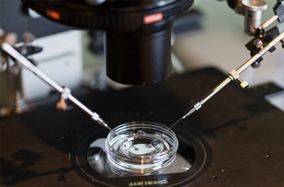
ART procedures in mice cause placental abnormalities unrelated to underlying infertility. Researchers found that the embryo culture procedure – where the fertilization of the egg with the sperm takes place in a medium meant to replicate the essential nutrients found in the oviduct and is placed in an incubator meant to mimic the womb – had the strongest effects on abnormalities and adverse outcomes.
“ART is more art than science”
“ART is more art than science,” Bartolomei said. “We don’t really know exactly what’s going on in the human body. Reproductive endocrinologists are looking at whether or not the embryo in the embryo culture developed in what we think is the right amount of time, with the right number of cells for the stage it’s in, and so on. The embryo sits in culture for a week, as opposed to oviducts in the mother’s body and the embryo culture is an attempt to simulate that environment.”
The authors conclude that efforts should be focused on optimizing embryo culture to ensure healthy outcomes for mothers and offspring.
This study is part of a larger effort to investigate infertility and reproduction at Penn through the National Centers for Translational Research in Fertility and Reproduction. Bartolomei’s research team partners with Christos Coutifaris, MD, PhD, Professor of Obstetrics and Gynecology, Monica Mainigi, MD, Assistant Professor of Obstetrics and Gynecology, and Jeremy Wang, MD, PhD, Professor of Developmental Biology at the School of Veterinary Medicine.
“This collaboration between clinical and basic researchers is designed to look at causes of infertility and when you have infertility using assisted reproduction, what are features that can be optimized for healthier outcomes – that’s the goal of this work,” said Bartolomei.
###
Co-authors include Eric A. Rhon-Calderon, Olivia Y. Chao, Duy K. Nguyen, Laren Narapareddy, Asha K. Dahiya, Mary E. Putt, and Richard M. Schultz. Funding for this study came from National Centers for Translational Research in Reproduction and Infertility (HD068157), The Lalor Foundation, Ruth L. Kirschstein National Service Award (HD089623), Roy & Diana Vagelos Scholars Program, and the National Institute of Nursing Research (T32NR007100).
Today let’s delve into a curious case involving induced pluripotent stem cells (iPSCs) and leukemic stem cells (LSCs). Most blood cells derived from iPSCs are unable to engraft in immunodeficient mice. However, Wesely and colleagues observed an exceptionally high engraftment efficiency of cell lines derived from an individual affected by acute myeloid leukemia (AML). In particular, in vitro they observed typical round cells together with cobblestone-like, firmly adherent cells. The latter displayed markers of immaturity, a more quiescent cell cycle, but foremost they were responsible for the successful engraftment in immunodeficient mice. Collectively, these properties prompted the authors to identify the cells as induced Leukemic Stem Cells (iLSCs). They proved that the iLSCs can become the round cells, but not vice versa, suggesting a stem cell nature. In addition, the adherent phenotype allowed the easy separation of the two populations. In-depth transcriptomic analysis, both at single-cell and population level, was coupled with the study of chromatin accessibility. The iLSCs displayed a molecular resemblance to the leukemic stem cells isolated from AML patients, based on a 42-genes signature. Finally, the authors identified the transcription factor RUNX1 as critical for iLSC phenotype maintenance, as it is involved in the expression of 16 of the 42 genes in the LSC signature. In conclusion, this work describes for the first time the derivation of LSCs from iPSCs. This could be a fantastic tool for the study of the cancer stem cell theory, as those rare cells are not prospectively isolated, but only studied after transplantation. Since these iLSCs were derived from a single patient, it will be interesting to isolate them from other AML-iPSCs lines.
Wesely et al. “Acute Myeloid Leukemia iPSCs Reveal a Role for RUNX1 in the Maintenance of Human Leukemia Stem Cells”
 (9 votes)

 (No Ratings Yet)
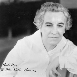
 (2 votes)