The Regeneration Next Initiative (RNI) was established by the Duke University School of Medicine to enhance discovery and applications in the broad field of tissue regeneration. RNI is pleased to announce a new 10-week summer research program designed to give undergraduate students hands-on experience in graduate level biomedical research in the area of tissue regeneration. We welcome applicants who are seriously considering joining a graduate program after completing their undergraduate degree.
Regeneration Next Summer Fellows will be selected through a competitive process and matched with a faculty mentor in tissue regeneration/ tissue engineering. The RNI Summer Research program will operate simultaneously with Duke’s Summer Research Opportunities Program (SROP).
RNI Summer Fellows will participate in professional development and community gatherings organized by SROP. This will maximize the RNI Fellows’ research experience and networking opportunities. RNI Fellows will be able to attend seminars, participate in professional development workshops, and present their work at the end of their training.
Students will receive shared on-campus apartment accommodation, travel assistance, and a competitive stipend of $4,000.
Important Dates:
Application period: November 4, 2019 – February 7, 2020.
May 26, 2020 – Program Begins
August 1, 2020 – Program Ends
To be eligible, students must be enrolled in an accredited undergraduate program in the United States at the time of application. We welcome international students but students must already have a valid student visa.
The application and supporting materials must be received by 5pm, 7 February 2020. Before starting your application please have the following ready (You will not be able to save an incomplete application and continue at a later time; we suggest that you write your essay responses in Microsoft Word or another word processing program)
(1) Essay answers to the following:
– Why are you interested in participating in the RNI Summer Research Fellows program? How would participating in the program align with your career goals? Limit your response to 600 words or less.
– Describe any past research experiences. Include the institution where you conducted the research, the context (research lab or through coursework), your research mentor’s name, and a brief description of the project. Also, state the skills that you learned during the experience and how the skills have helped you to grow as a scholar and researcher. Please limit your response to 600 words or less.
– Please describe your current research interest(s). If there are particular departments or faculty at Duke that you are interested in, please state that here. Please limit your response to 400 words or less.
(2) A transcript (pdf) (If you have multiple transcripts, you must compile them into one pdf document)
We’re excited to announce the launch of theNode Network, a global directory of developmental and stem cell biologists. The Node Network is designed to help those organising conferences, assembling committees, seeking speakers for seminar series, looking for referees and so on to identify individuals who might not otherwise come to mind.
The Network is entirely inclusive – any member of the developmental and stem cell biology community, at any stage of their career, can join. It is also designed to help promote diversity in our field – as well as providing information on scientific field, model organism, place of work and career stage, members can also voluntarily provide details on aspects of diversity such as gender, race/ethnicity, LGBTQ+ identity and disability status. You are welcome to give as much or as little information as you like. We hope the Network will help promote diversity at our conferences and make ‘manels’ a thing of the past, but it can also be used solely in terms of science.
The idea was first suggested to us by James Briscoe, Development’s Editor-in-Chief (check out his editorial, written with Executive Editor Katherine Brown, announcing the launch and discussing other aspects of inclusion and diversity). We were also influenced by initiatives like Anne’s List, DiversifyEEB and Diversify PlantSci – directories of women (or women plus under-represented minority) scientists which aim to help people looking to fill various roles (reviewer, speaker, panel member, etc) to diversify their pool.
After discussions with Development’s editors and board, we next considered whether the directory should be focused on diversity, or should be totally inclusive (but usable with diversity in mind). We also had to consider how we would display sensitive information in line with GDPR regulations – we consulted our in house GDPR and legal experts, and asked the Information Commissioner’s Office for advice. We also consulted the Wellcome Trust Diversity and Inclusion team for advice on terminology regarding diversity information.
But what we really needed to know was what you the community thought about the idea. Last summer we ran an extensive survey which got hundreds of responses, and were reassured (and delighted) to find that 99% of respondents would be interested in accessing such a directory, and 97% would consider adding themselves to it. The majority of respondents were in favour of something inclusive for the entire developmental biology and stem cell field, rather than something specifically labelled as a diversity directory – although there was still strong support for including diversity information. Survey respondents also shared ideas about how to develop the directory, and noted potential uses we hadn’t thought of (like collaboration and recruitment).
So then we built it (helped as ever by our IT Consultants), and it’s now live for you to use. Of course, it will only be helpful to the field if people use it, so please consider registering to access the Network, and if you’re happy with the idea of being contacted as a reviewer, speaker, panel member, collaborator etc., consider becoming a member too. You can also circulate this flyer (PDF link) to your lab, your peers, your departments and your societies!
For more information about how to use the Network, go to the homepage and follow the links. You can also read our information page and FAQs. You can get to the Network from elsewhere in the Node via the top menu bar.
Established by the British Society for Developmental Biology in 2014, The Gurdon/The Company of Biologists Summer Studentship scheme provides financial support to allow highly motivated undergraduate students an opportunity to engage in practical research during their summer vacation. Each year, ten successful applicants spend eight weeks in the research laboratories of their choices, and the feedback we receive is outstanding. You can read accounts from previous years here. If you’re interested in applying or hosting a student in 2020, applications need to be in by the end of March.
Our ninth report from the class of 2019 comes from Vlad Arimia (UCL) who studied chick neural induction in Claudio Stern’s lab in UCL.
It makes me nervous: identifying inductive signals
Developmental biologists have sought the mechanisms regulating neural induction ever since Spemann and Mangold demonstrated that grafts of the dorsal blastopore lip can induce ectopic neural axes (Spemann & Mangold, 1924). Almost a century later, neural induction still has its mysteries, but perhaps not for long.
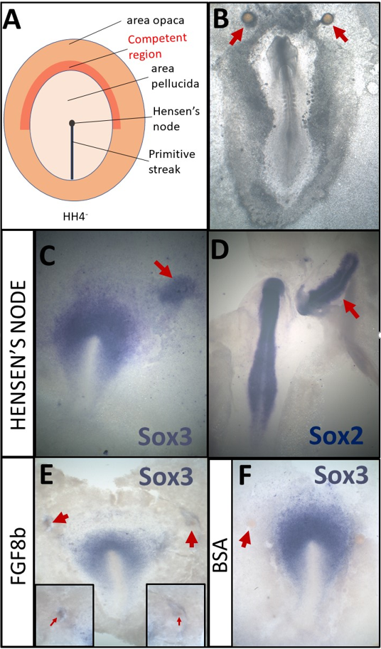
The Stern lab now mixes classical embryology with transcriptomics to re-evaluate neural induction in the modern molecular era. Grafts of Hensen’s node to a region of competent area opaca (Fig. 1A) can induce ectopic neural tubes from ectoderm that does not normally contribute to the nervous system. Combining this assay for ectopic neural induction with RNA-seq at various time points, they have identified all genes that change in response to a node graft up to the time when neural plate formation begins (Trevers et al., 2018). Additionally, many secreted signalling factors have been identified from the transcriptomes of key signalling tissue. These screens have provided a wealth of new tools that have started to shed new light on the complex nature of neural inducing signals and how ectodermal cells respond to them.
Figure 1 – Overview of grafts performed according to the neural induction assay A- Schematic of a HH4- chick embryo. The organizer, Hensen’s node, is the tip of the primitive streak B- An embryo which was cultured overnight with grafts of heparin-acrylic beads (arrows) C- Sox3 in situ hybridisation of an embryo which had been cultured for 5h with a node graft D- Sox2 in situ hybridisation of an embryo which had been cultured overnight with a node graft. E- Beads soaked in FGF8b can induce expression of Sox3 after 5h. F- Beads soaked in 0.1% BSA do not induce Sox3 after 5h
My project focused on two secreted molecules that had not yet been explored in the context of neural induction. The aim was to test if either could induce similar transcriptional changes in responding ectodermal tissue as those seen in the neural induction assay. Excitingly, one of these molecules is novel not only in the context of neural induction but also in terms of its possible role in embryonic development altogether.
Under the watchful eye of Katherine Trevers (a postdoc in the lab and a great mentor) I learnt how to culture chick embryos (New, 1955; Stern &Ireland, 1981) and mastered the micromanipulations necessary for my project (Fig. 1B-F).
These consisted of grafting beads soaked overnight in the putative inducer to the competent region of the area opaca (Fig1A, B) and incubating them for 5h. Then, I would carefully remove the bead and dissect the (tiny!) region of tissue directly beneath it which had been exposed to the putative inducer. Dissected pieces of tissue were stored at -80°C until I had collected 24 of them – a much bigger challenge than I had anticipated! After considerable perseverance I collected all the necessary samples, which were then analysed by NanoString nCounter technology. This technique uses fluorescently barcoded probes to count the number of mRNA molecules in a sample and the Stern lab has a large probe set that includes all the transcription factors whose expression changes in response to neural induction (more than 200!), as well as many controls.
It was tremendously exciting to quantify the expression of these genes in response to our putative inducers and compare them to a vehicle control. Especially when our analysis revealed that both molecules induced interesting transcriptional responses- one upregulating and the other downregulating different subsets of genes. These results are strong indicators that the molecules I was testing may contribute to neural induction. However, these preliminary results need to be validated by checking the transcriptional responses identified through methods such as in situ hybridisation. Also, loss-of-function experiments would further confirm our observations. All in all, I am happy that in a relatively short time I was able to learn enough embryology as to get to tangible results!
In addition to the experiments described above, I also had the opportunity to repeat some classical experiments myself. By grafting Hensen’s node, I confirmed that 5h of signals can induce the neural marker Sox3 (Fig. 1C) while Sox2 can be induced after an overnight culture (Fig. 1D). I also repeated experiments demonstrating that beads soaked in FGF8b (Fig. 1E, F) can induce early markers, such as Sox3 (Streit et al., 2000). Thus, I have not only made a small contribution towards understanding the molecular basis of neural induction, but also repeated some of the key experiments that have been the fundament of research in this field.
Although challenging, I found these embryology techniques extremely satisfying and enjoyable. So much so, that I miss being at the bench surrounded by all the amazing members of the Stern lab (part of them in Figure 2). Whenever Nidia (our lovely lab manager), asked how I was, I would reply that I was having fun. And indeed, I had a wonderful summer in Claudio Stern’s lab at UCL as part of the neural induction subgroup.
Thank you!
References
NEW, D. A. T. (1955). A new technique for the cultivation of the chick embryo in vitro. J. Embryol. Exp. Morphol., 3, 326-331.
Spemann, H., & Mangold, H. (1924). Über induktion von Embryonalanlagen durch Implantation artfremder Organisatoren. Archiv für mikroskopische Anatomie und Entwicklungsmechanik, 100(3-4), 599-638.
STERN, C. D. & IRELAND, G. W. (1981). An integrated experimental study of endoderm formation in avian embryos. Anatomy and Embryology, 163, 245-263.
Streit, A., Berliner, A. J., Papanayotou, C., Sirulnik, A., & Stern, C. D. (2000). Initiation of neural induction by FGF signalling before gastrulation. Nature, 406(6791), 74–78. https://doi.org/10.1038/35017617
Trevers, K. E., Prajapati, R. S., Hintze, M., Stower, M. J., Strobl, A. C., Tambalo, M., Ranganathan, R., Moncaut, N., Khan, M. A. F., Stern, C. D., Streit, A. (2018). Neural induction by the node and placode induction by head mesoderm share an initial state resembling neural plate border and ES cells. Proceedings of the National Academy of Sciences, 115(2), 355 LP – 360. https://doi.org/10.1073/pnas.1719674115
We are seeking outstanding postdoctoral candidates to join the Perry lab at the University of California, San Diego. Our group uses genetic and genomic approaches to study the development and evolution of neural systems. We use the insect visual system as a model to understand how the genome encodes the complexity of the brain and nervous system. We are interested in the mechanisms that generate the exquisite diversity of ways in which animals perceive and interact with the world.
We are specifically seeking independent, passionate, and highly motivated applicants for a postdoctoral position to study the evolution and development of butterfly color vision, with a focus on understanding the specific genetic changes that produce a more complex retinal mosaic. Butterflies have doubled the number of R7 photoreceptors in their retinas, allowing for an increased number of color comparisons (see Perry et al. Nature 2016). We use CRISPR to test gene function directly in developing butterfly retinas. A second part of this project will be aimed at understanding how the brain interprets this additional input and the role of developmental plasticity. A portion of the work will involve making Drosophila retinas more like butterfly retinas, and then using sophisticated genetic tools in Drosophila to understand the impact on downstream neural circuits. A Ph.D. in the biological sciences with at least three years of laboratory research experience in molecular or developmental biology is required. Experience with Drosophila or other genetic model systems is preferred but not required.
This is a renewable two-year position with full benefits, which will be extended as needed upon good performance of the candidate. Salary will be competitive and dependent on the level of experience of the candidate. Applicants should email a CV and a description of research interests to Prof. Perry (mwperry@ucsd.edu), along with contact information for three references. Applications submitted by February 15th, 2020 will receive priority consideration, but the position will remain open until filled. Start date is flexible.
It is an incredibly exciting time to be a developmental biologist as new tools such as CRISPR and single cell sequencing allow us to move beyond model systems in order to ask targeted questions about the mechanisms that adapt animals to their unique environments. Apply and join the adventure!
We are seeking outstanding postdoctoral candidates to join the Perry lab at the University of California, San Diego. Our group uses genetic and genomic approaches to study the development and evolution of neural systems. We use the insect visual system as a model to understand how the genome encodes the complexity of the brain and nervous system. We are interested in the mechanisms that generate the exquisite diversity of ways in which animals perceive and interact with the world.
We are specifically seeking independent, passionate, and highly motivated applicants for a postdoctoral position to study the evolution of novel neural types. We identified a range of examples of where novel types of neurons have appeared in the visual systems of different insect species (see Perry, Konstantinides et al. 2017 Annual Reviews Genetics) and are interested in 1) the genetic changes that produce a new type of cell and 2) how novel neurons plug in to existing circuitry in a way that is useful for the animal. In a potential initial project, the successful candidate would focus on a fly visual system innovation known as the “Love Spot”. The Love Spot (LS) is a male specific modification of the dorsal-frontal eye found in over fifteen families of Diptera. It provides high sensitivity for the detection and pursuit of conspecific females. More information can be found in Perry and Desplan 2016 Current Biology “Love Spots”. The successful candidate will dissect cell-type specific gene regulatory networks that work together to produce this sexually dimorphic complex feature using the house fly Musca domestica as a model.
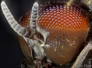
Bonus points if you can identify this bug and whether it has a love spot.
A Ph.D. in the biological sciences with at least three years of laboratory research experience in molecular or developmental biology is required. Experience with Drosophila or other genetic model systems is preferred but not required. scSeq experience (e.g. 10x Genomics) is desirable. This is a renewable two-year position with full benefits, which will be extended as needed upon performance of the candidate. Salary will be competitive and dependent on the level of experience of the candidate. Applicants should email a CV and a description of research interests to Prof. Perry (mwperry@ucsd.edu), along with contact information for three references. Applications submitted by February 15th, 2020 will receive priority consideration, but the position will remain open until filled. Start date is flexible.
It is an incredibly exciting time to be a developmental biologist as new tools such as CRISPR and single cell sequencing allow us to move beyond model systems in order to ask targeted questions about the mechanisms that adapt animals to their unique environments. Apply and join the adventure!
Established by the British Society for Developmental Biology in 2014, The Gurdon/The Company of Biologists Summer Studentship scheme provides financial support to allow highly motivated undergraduate students an opportunity to engage in practical research during their summer vacation. Each year, ten successful applicants spend eight weeks in the research laboratories of their choices, and the feedback we receive is outstanding. You can read accounts from previous years here. If you’re interested in applying or hosting a student in 2020, applications need to be in by the end of March.
Our eighth report from the class of 2019 comes from Réiltín Ní Theimhneáin (NUI Galway) who studiedcnidarian reprogramming inUri Frank’s lab at NUI Galway.
Slightly different to many of the fortunate students supported by the BSDB, I do not come from an especially strong scientific background. Approaching my final year of undergraduate medicine, I wanted to spend the summer studying something not offered by the traditional medical electives. I wanted a better appreciation for the more fundamental cellular processes that underly developmental biology. I wanted some sort of foundation to work from, so I could better understand some of the science that may eventually be translated to medicine in the future. I was also curious, how would I fare out of my natural habitat of wards and surgical theatres; would I take to laboratory work, or spend my 8-weeks fumbling with protocols and pipettes?
I began my internship at Prof Uri Frank’s laboratory in the Biomedical Sciences building at NUI Galway. Under the supervision of postdoctoral researcher, Dr Miguel Salinas-Saavedra, I was given a crash course in laboratory techniques, methods to correctly follow protocols and the various rules surrounding the culture and manipulation of Hydractinia. During my internship I was expected to develop my practical skills and have an appreciation and understanding of the role of cellular senescence in the reprogramming of Hydractinia.
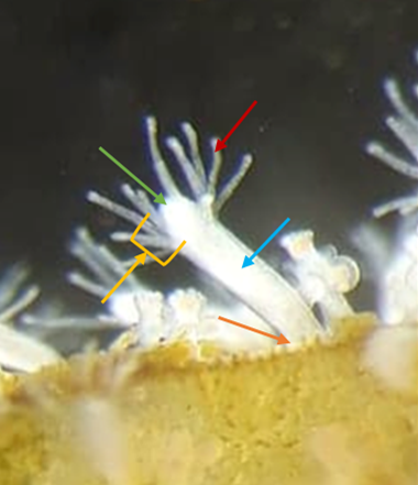
Hydractinia, or ‘tiny jellyfish’ as my family dubbed them, are a cnidarian model organism that has been employed in development, regeneration, and allorecognition studies. Roughly 8 mm in length Hydractinia are naturally found on the shells of hermit crabs. Prof Frank’s laboratory is the only one in Ireland to study these animals, and one of less than 10 laboratories worldwide. Similar to the freshwater cnidarian Hydra, Hydractinia have remarkable regenerative capacities. Figure 1 shows a photo of an anotated feeding polyp.
Figure 1 – Photograph of a Hydractinia feeding polyp. Structures highlighted are the hypostome (green), tentacles (red), Body (blue), stolon (orange) and head (yellow arrow with bracket).
Hydractinia possess a population of adult stem cells, known as interstitial cells or i-cells. These cells are highly proliferative and normally replace cells during tissue homeostasis and regeneration1. I-cells are anatomically restricted to certain areas in the adult; they are excluded from the intact head (but can migrate to it upon injury). Interestingly, isolated heads that lack i-cells, can nevertheless regenerate a fully functional animal that includes germline competent i-cells.
The mechanisms of conversion from terminally differentiated head cells to stem cells (i.e. i-cells) is currently unknown. This idea fascinated me. The concept also represented an opportunity to study the mechanisms that can destabilize the fate of animal cells in a regenerative, non-malignant context. Preliminary work done in the Prof Frank’s laboratory suggests cellular senescence might be a potential trigger that induces dedifferentiation in neighbouring cells. This information lead to lots of reading; I needed a better understanding cellular senescence and cell cycle limitations before I started practical experiments!
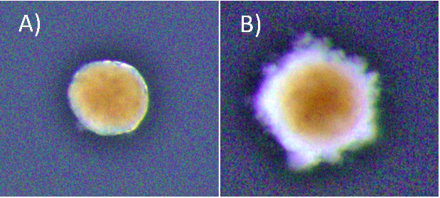
At the start of my internship I tried a number of different experiments centred around cellular senescence in Hydractinia. Using the fluorescent probe SPiDER-ßGal to detect β-galactosidase activity, we sought to detect potentially senescent cells (a hallmark of senescence is high β-galactosidase activity that can be detected using SPiDER-βGal). Having detected β-galactosidase activity, Dr Salinas-Saavedra sought to optimise a method of performing an in vivo time-lapse on the confocal microscope. I meanwhile conducted some visual morphological studies in which I cut hypostomes and kept them in sea water with 5% cell culture medium, changed at regular intervals, to see the time frame associated with complete regeneration. The ultimate goal was to form fully regenerated polyps from cut hypostomes and go on to prove that these polyps were fully functional and could reproduce sexually. As the time passed we decided that the concentration of the media was too low, and the regime was changed to 10% medium for 1h changed daily. Unfortunately, by day 45 all samples had expired. Fig 2 shows an example of a healthy and an expired sample. Due to my limited time at the lab, optimising the protocol proved to be an impossible task but the experiment will be replicated by the lab, perhaps with shorter daily medium exposure.
Figure 2 – Both photographs are of a hypostome cut from a feeding polyp, kept them in sea water and exposed to 10% medium for 1h changed daily. (A) An example a healthy sample of a hypostome at day 43 post-cut. (B) An expired sample of hypostome at day 45 post-cut. * Brightness in the photos was increased +40% for viewing purposes.
We also wanted to form a timeline to establish when cell replication occurs in hypostomes of Hydractinia at various days post cutting. To do this I conducted a double staining experiment in which I stained for both EdU and Piwi1 protein, using hypostomes at days 0-7 post-cutting along with full polyps to be used as controls. EdU is used to assay DNA synthesis in tissues and to detect cells which have undergone DNA synthesis during time of incubation. Unfortunately, EdU staining proved to be difficult in my particular case. Initial experiments failed to stain, despite positive results for the antibody and DAPI staining; the latter stains DNA. We hypothesised that I had cut the control polyps too high from the stolon (above the proliferative zone) to see any EdU staining or the EdU protocol simply had not worked. Additional issues presented with poor staining due to tissue contraction.
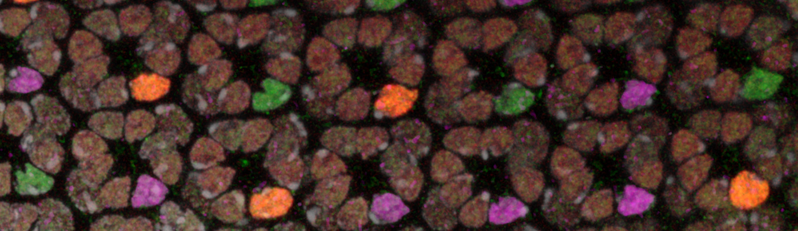
To try and mitigate this problem I used a new EdU and antibody staining protocol. For this I used feeding polyps cut at the stolon. To combat the contraction issue, I added MgCl2 for 15 minutes incubation prior to fixation to one sample, and sea water to the other as a control. Figure 3 shows an example of a feeding polyp from either sample group stained with DAPI, EdU and Piwi1 viewed under a confocal microscope. Interestingly, although very little change was seen in terms of tissue contraction, EdU staining appeared much stronger in the control sample in comparison to the MgCl2 exposed sample. It would have been interesting to see whether a longer time in MgCl2 would yield a more notable difference in tissue contraction.
Figure 3: Fluorescent staining comparing hydractinia polyps incubated in MgCl2 prior to fixation on left, to control on right. Blue stain – DAPI; green stain – EdU; red stain; Piwi1.
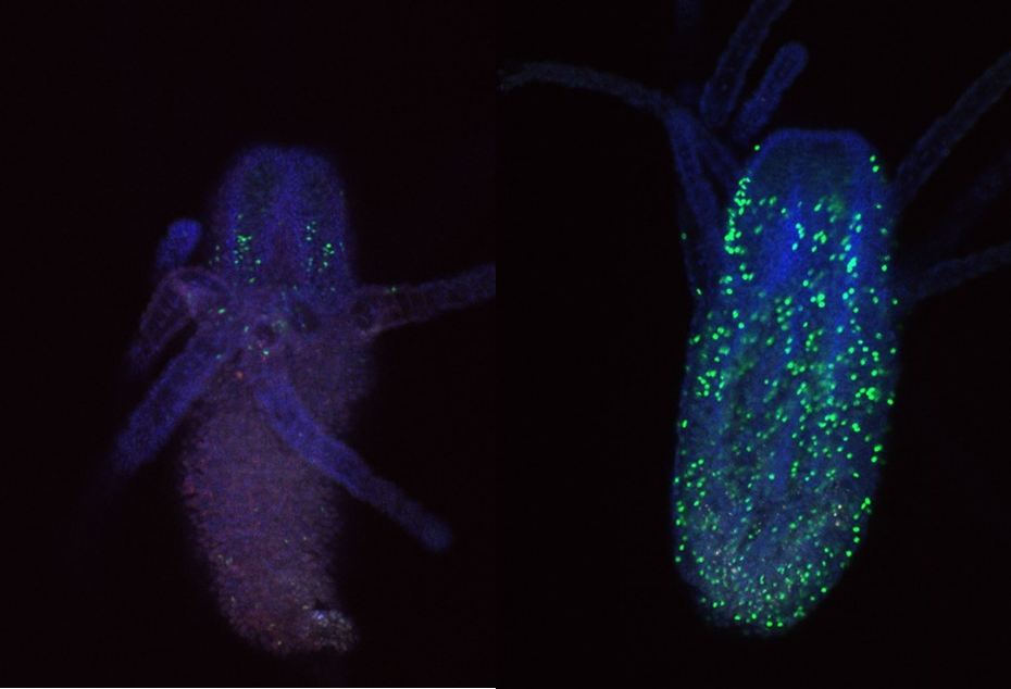
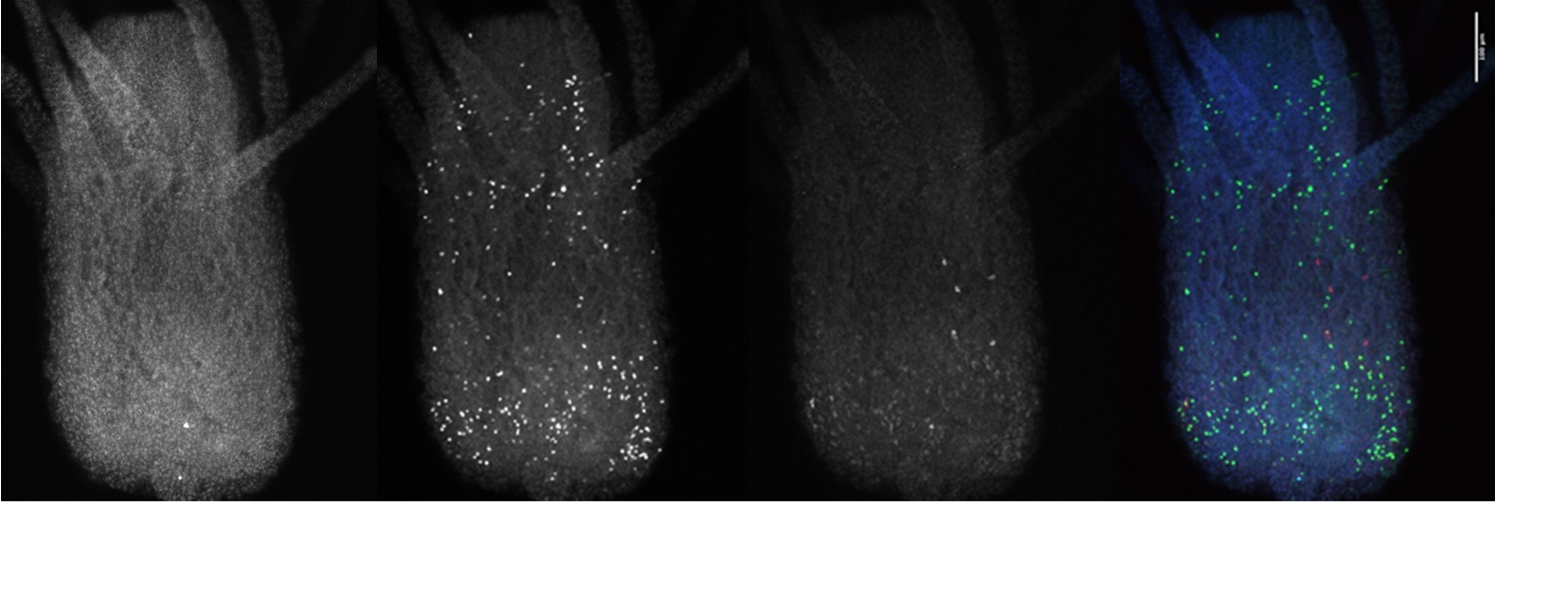
I also looked closly at the Piwi1 staining. PIWI proteins are a germline marker in hydractinia3. Staining for PIWI1 in these polyps enabled me to look at the i-cells; checking their quantity and location. It was apparent that some polyps had been cut too high as the expected i-band was not present. Figure 4 is an example of the seperated strands of DAPI, EdU, Piwi1 with the far right coloured image ultimatly compiled from each layer overlapping. Given the limited data, more experiments need to be conducted to draw any strong conclusions. Ultimately, I gained very real insight into the challenges faced daily be researchers and have a new appreciation for the resilience of those conducting experiments.
Figure 4: Hydractinia polyp stained with DAPI, EdU, Piwi1 with image compiled of the three strands (far right) viewed under confocal microscope.
The weeks felt very short as I approached the end of my internship. I would like to thank Prof Uri Frank and all of my laboratory colleagues for their endless support and incredible patience. With the support of the BSDB Grant I was able to work as part of a cutting-edge research team, perform independent experiments, participate in weekly meetings and discuss concepts with experienced researchers. I learnt specific methods and better understood protocols. The transferable laboratory skills that I have developed will certainly help me to secure a research position after graduation, and hopefully enrich my medical career. I wish the lab all the best in their endeavours to understand the fundamental questions in regenerative biology.
References
Plickert, Günter, et al. (2012)“Hydractinia, a Pioneering Model for Stem Cell Biology and Reprogramming Somatic Cells to Pluripotency.” The International Journal of Developmental Biology, vol. 56, no. 6-7–8, , pp. 519–534, www.ijdb.ehu.es/web/paper.php?doi=123502gp, 10.1387/ijdb.123502gp. Accessed 10 July 2019.
Gahan, James et al. (2016). The interstitial stem cells in Hydractinia and their role in regeneration. Current opinion in genetics & development. 40. 10.1016/j.gde.2016.06.006.
Plickert, Günter, et al. “Hydractinia, a Pioneering Model for Stem Cell Biology and Reprogramming Somatic Cells to Pluripotency.” The International Journal of Developmental Biology, vol. 56, no. 6–8, 2012, pp. 519–34, www.ncbi.nlm.nih.gov/pubmed/22689372, 10.1387/ijdb.123502gp. Accessed 10 July 2019.
Established by the British Society for Developmental Biology in 2014, The Gurdon/The Company of Biologists Summer Studentship scheme provides financial support to allow highly motivated undergraduate students an opportunity to engage in practical research during their summer vacation. Each year, ten successful applicants spend eight weeks in the research laboratories of their choices, and the feedback we receive is outstanding. You can read accounts from previous years here. If you’re interested in applying or hosting a student in 2020, applications need to be in by the end of March.
My interest in research and the lack of lab experience I had increased my thirst for a laboratory based summer studentship. Successfully this summer, I was awarded the BSDB Gurdon Studentship to work in Dr David Long’s lab at UCL Great Ormond Street Institute of Child Health. As I truly believe that research and medicine go hand-in-hand, I saw this as a first step towards a career as a clinician-scientist.
As an undergraduate medical student, I had zero lab experience and everything I learnt was new to me. Day to day, I was supervised in the lab by an MB/PhD student and previous Gurdon Studentship awardee, Daniyal Jafree. I was surprised that from the very first day, I started getting my hands on. It took me a good 30 seconds to pipette up 1 ml of liquid; I became quicker as weeks went by. It was even a struggle at the beginning to get my brain thinking of volume in terms of microlitres, rather than millilitres!
I had the privilege to work on a topic that has only received attention in recent years; understanding the role of macrophages during kidney development. For a very long time macrophages were only thought of as part of the immune system and that they all originated from monocytes. But this is not the complete story because the macrophage subpopulation is heterogeneous (they don’t just originate from monocytes) and very little is known about the different populations and their functions.
Of all the techniques I learnt in the lab, one of my favourite techniques turned out to be the most challenging. I learnt how to dissect mouse embryonic kidneys under a microscope. I started by trying every tool imaginable, ranging from syringe needles to scalpels to sharp or blunt forceps and scissors. After a few unsuccessful attempts (mistaking the stomach for the kidney!) I learnt how to spot the kidney and eventually became proficient enough to help the research team in other dissections for some big experiments. I also tried my hand at flow cytometry to discriminate different macrophage populations within the developing kidney; another technique which I became independent with.
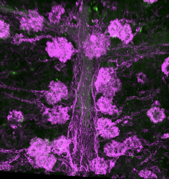
Perhaps the coolest of the techniques I got to try was imaging. By treating mouse embryonic kidneys with a cocktail of chemicals, adding specific and optically clearing the kidneys (making them transparent!) I could take three-dimensional pictures of entire mouse embryonic kidneys using confocal imaging. Each experiment took up to a week, and it was occasionally frustrating as things didn’t always go to plan. For example, after a big experiment that I was really excited to see the images for, I was heartbroken to look under the microscope and see a big bubble in the middle of the kidney, making it almost impossible to get any useful images. This experience taught me that sometimes lab research can be frustrating and unpredictable. However, sometimes the imaging came out quite well, and I captured some really detailed pictures of the blood vasculature in developing mouse kidneys (Picture 1). Using this technique, I found a really unexpected phenotype in one of the lab’s mouse models, something that is apparently still puzzling the lab to this day! Apart from learning laboratory techniques and analysis, I also had the opportunity to present at one of Dr Long’s bimonthly lab meetings. Scary at first, but looking back, it was an invaluable experience. I got really useful feedback that I look forward to trying out in my future presentations.
Picture 1: A chain of mouse kidney glomeruli at E18.5. The kidneys were stained for a blood vascular marker, optically cleared and imaged using a confocal microscope.
I was very lucky to have worked in Dr Long’s lab. Dr Long always found the time in between his busy schedule to check up on my progress, and to answer any questions that I had. I don’t think I have ever experienced eight weeks fly by that fast, and this is largely thanks to Daniyal and everyone in the lab, all who created such friendly and supportive environment. Towards the end of my placement, we even paid a trip to the bowling lanes! (Picture 2). Overall, this BSDB studentship has given me an insight into what working in a scientific laboratory is really like, and a taster of what to expect in the career of a research scientist. I highly recommend any undergraduate student, especially those with little or no lab experience, to take up this fantastic opportunity.
Picture 2: Me (last one on the right side of table) along with Dr Long’s group having dinner before bowling!
NSF and AHA funded post-doctoral position available in the Davidson lab at Swarthmore College.
Searching for a post-doc who is passionate about both teaching and research. We are studying the interplay between division and inductive signaling. In particular, we are exploring how signaling receptors are trafficked in dividing cells to generate asymmetric induction of a cardiac progenitor lineage. We are also using comparative genomics to explore the evolutionary constraints that shape gene regulatory networks. We study these questions in the invertebrate chordate, Ciona robusta. Ciona embryos consists of extremely low cell numbers allowing high resolution analysis of intra-cellular dynamics in intact embryos. The ease of generating transgenic Ciona embryos make this an excellent model organism for undergraduate research. We have recently initiated a collaboration with Danelle Devenport at Princeton focused on similar processes in cultured mouse epithelial cells. This is a great opportunity for a post-doc with an interest in undergraduate teaching and research. My former post-doc recently started a tenure-track position at a small liberal arts college and it was clear that the combination of a strong research record along with a demonstrated commitment to undergraduate mentoring and teaching made her a strong candidate for this very competitive career track.
Applicants should have a PhD (or be close to completing one) in a relevant subject area. Excellent communication skills and a commitment to undergraduate mentoring are essential.
To apply or if you have questions about the position – please send your CV and a cover letter describing your interest to Bradley Davidson at bdavids1@swarthmore.edu. I will be in touch with instructions for submitting a formal application.
Established by the British Society for Developmental Biology in 2014, The Gurdon/The Company of Biologists Summer Studentship scheme provides financial support to allow highly motivated undergraduate students an opportunity to engage in practical research during their summer vacation. Each year, ten successful applicants spend eight weeks in the research laboratories of their choices, and the feedback we receive is outstanding. You can read accounts from previous years here. If you’re interested in applying or hosting a student in 2020, applications need to be in by the end of March.
Our sixth report from the class of 2019 comes from Matyas Bubna (QMUL) who studied frog neural crest in Roberto Mayor’s lab at UCL.
Visualising neural crest induction, migration and differentiation in Xenopus
Throughout my undergraduate studies I have become increasingly captivated by the intricacy and elegance of animal development. Especially interesting to me is how processes such as morphogenesis, tissue patterning or cell migration, which can appear incomprehensible, emerge from relatively simple interactions at the molecular level. Observing how a single cell transforms into a complex organism is a unique and thought-provoking experience. I am grateful to the BSDB for the studentship allowing me to explore this field. I would strongly recommend the Gurdon studentship to anyone interested in topics ranging from evolution, epigenetics, cell signalling, cancer, to stem cells and regeneration – all of these processes may be elucidated by taking a developmental point of view.
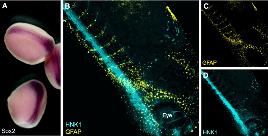
During the summer studentship I have learnt to work with Xenopus laevis embryos and keep track of their development. The aim of my project was to optimise techniques for visualising the neural crest with other tissues relevant to its induction, migration and differentiation. To do this I used two well established techniques in developmental biology: in situ hybridisation and immunofluorescence (see Figure 1).
Figure 1: A) Colorimetric in situ hybridisation with neural plate marker Sox2 in late neurula stage Xenopus embryos. B-D) Whole-mount fluorescent immunostaining for neuronal marker HNK1 and glial marker GFAP in a tailbud (Neiuwkoop and Faber stage 31) embryo.
Why study neural crest cells?
The neural crest is a fascinating population of cells unique to vertebrates, which is induced at the neural plate border and following neurulation delaminates and migrates away from the neural tube. A subset of these cells migrates into head regions where it gives rise to a variety of tissues including bones, cartilage, as well as neurons. This cranial neural crest also migrates into the branchial (or pharyngeal) arches and contributes in a major part to the craniofacial skeleton. This has undergone major changes during vertebrate evolution. For instance, some of the jaw bones present in our common ancestors with reptiles have given rise to the middle ear bones of mammals (Santagati & Rijli, 2003). A possible explanation for this versatility of the neural crest is that its cells retain multipotency for longer than the three embryonic germ layers, prompting some to consider it a ‘fourth germ layer’ (Simoes-Costa & Bronner, 2015).
The diversity of neural crest-derived cells, which also includes melanocytes, Schwann cells, meninges or the cornea, makes it an important model of differentiation with a potential for therapeutic applications. It is also a great model for studying cell migration (Szabó & Mayor, 2018) and by proxy epithelial to mesenchymal transition in cancer metastasis. Recent work even attributed the ability to remove cellular debris from early neural tube to migrating neural crest cells, potentially through a macrophage-like mechanism (Zhu, et al., 2019).
Going further with established techniques
I have been testing and optimising techniques to visualise proteins and gene expression that may help elucidate how the neural crest gets induced and how it migrates through the embryo. For example, I have image the neural crest with nearby mesoderm, which is required during induction, using a combination of fluorescent in situ hybridisation (FISH) and immunostaining. In order to visualise expression of two different genes within the embryo a double in situ hybridisation (ISH) can be used. This is especially important in Xenopus, as no antibody is known to efficiently and exclusively label the neural crest. Although colorimetric ISH is easier and does not require clearing of the tissues, it doesn’t enable exploring the 3D structure by imaging the whole embryo at once. I have used confocal and multiphoton microscopy to analyse the embryos (see Figure 2). Unfortunately, it seems that using two RNA probes at the same time reduces signal intensity and although I have tried to optimise the signal amplification reaction and bleaching to neutralise endogenous peroxidase activity, I have not been able to reduce the background.
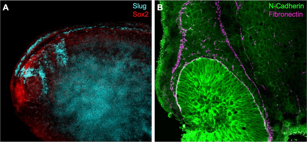
Figure 2: A) Fluorescent in situ hybridisation using RNA probes for Slug (neural crest) and Sox2 (neural plate) in a whole-mount stage 22 Xenopus embryo. B) Dissected embryo immunostaining shows N-Cadherin expression in migratory neural crest. Fibronectin outlines the streams of cranial neural crest.
One possibility is to delimit the neural crest using an antibody for fibronectin, an extracellular matrix component that encourages neural crest migration. Using double immunostaining (Figure 2B) I was able to confirm migratory neural crest cells express N-Cadherin near the optic vesicle as fibronectin outlines the neural crest streams (Scarpa, et al., 2015). Towards the end of my summer project, I have also manipulated cell contractility using drug treatments to observe changes in neural crest cell behaviour using several antibodies.
I thank Prof Roberto Mayor for supervising this summer project and Dr Adam Shellard for teaching me methods used in the lab. I have learnt a lot about how research is done and presented my results at a lab meeting in the final week and used this experience to transition from undergraduate study into my PhD.
References
Santagati, F. & Rijli, F. M., 2003. Cranial neural crest and the building of the vertebrate head. Nature Reviews Neuroscience, Issue 4, p. 806–818.
Scarpa, E. et al., 2015. Cadherin Switch during EMT in Neural Crest Cells Leads to Contact Inhibition of Locomotion via Repolarization of Forces. 34(4), pp. 421-434.
Simoes-Costa, M. & Bronner, M. E., 2015. Establishing neural crest identity: a gene regulatory recipe. Development, Volume 142, pp. 242-257.
Szabó, A. & Mayor, R., 2018. Mechanisms of Neural Crest Migration. Annual Review of Genetics , Volume 52, pp. 43-63.
Zhu, Y. et al., 2019. Migratory Neural Crest Cells Phagocytose Dead Cells in the Developing Nervous System. Cell, 179(1), pp. 74-89.
Established by the British Society for Developmental Biology in 2014, The Gurdon/The Company of Biologists Summer Studentship scheme provides financial support to allow highly motivated undergraduate students an opportunity to engage in practical research during their summer vacation. Each year, ten successful applicants spend eight weeks in the research laboratories of their choices, and the feedback we receive is outstanding. You can read accounts from previous years here. If you’re interested in applying or hosting a student in 2020, applications need to be in by the end of March.
Exploring the genetic control of microRNAs in Drosophila melanogaster
This summer, thanks to the BSDB Gurdon Studentship, I was able to work closely with a PhD student in the lab of Sarah Newbury at the Brighton and Sussex Medical School. Under their supervision I was able to get an applied, practical approach to lab work, much different from that experienced during my undergraduate course.
My project involved looking at the genetic control of microRNAs in Drosophila melanogaster, otherwise known as the common fruit fly. MicroRNAs are small-noncoding RNAs that participate in RNA silencing and regulation of gene expression. miRNAs are able to base-pair with their complementary target mRNA, and through this they are able to silence them, either by their subsequent degradation or prevention of their translation.
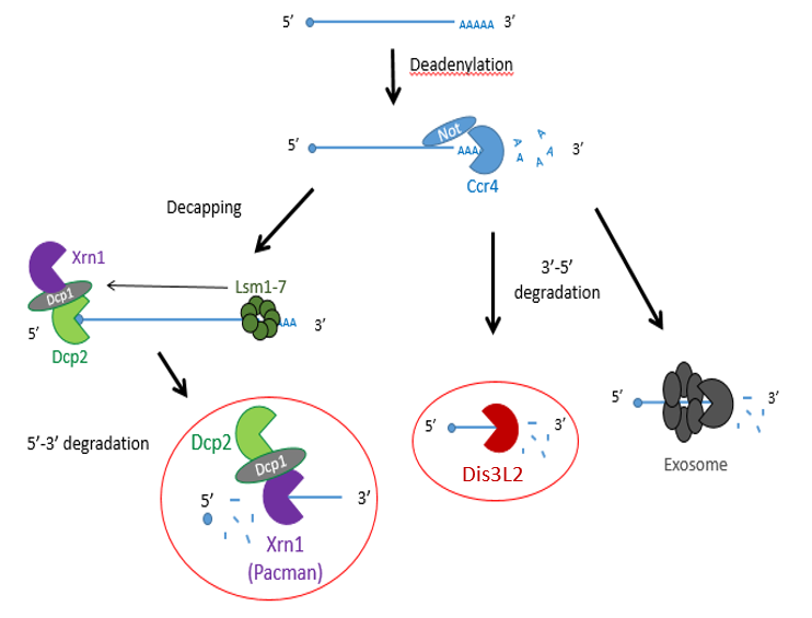
Previous studies have shown that exoribonucleases, known as Pacman (XRN1) and Dis3L2 degrade different microRNAs through the mRNA decay pathway, shown in Figure 1.
Figure 1 – The mechanisms of mRNA decay. Most mRNAs undergo decay through the deadenylation dependent pathway shown above. The poly(A) tail is first removed by a deadenylase (shown here as Ccr4-Not) and then proceeds down 3 possible routes: decapping by enzymes (shown as Dcp1/2) , followed by 5’ → 3’ decay by XRN1 (Pacman in Drosophila) or 3’ → 5’ decay by Dis3L2 or the exosome.
The project made use of wing imaginal discs, which are highly proliferative organs found in Drosophila that will eventually develop into the adult fly’s wings. Prior work in the lab had found that there were significant differences between the size of these discs in both pacman and dis3L2 mutants, with dis3L2 mutants having much larger discs (Towler et al. 2016), and pacman mutants having smaller ones (Waldron et al. 2015). Both exoribonucleases are conserved to humans and their defects have shown to be significant in human disease as well. For example, mutations of DIS3L2 in humans have been linked to Perlman syndrome, an overgrowth syndrome which presents as organomegaly and is associated to a high risk of developing Wilm’s tumours (Astuti et al. 2012). Together this suggested that there could be an involvement of these exoribonucleases in the control and regulation of cell proliferation and apoptosis within these discs, potentially through their degradation activity. Therefore, the aim of my project was to study the difference between levels of specific miRNAs (which were extracted from these WIDs) in both pacman and dis3L2 mutants, compared to their respective control wildtype.
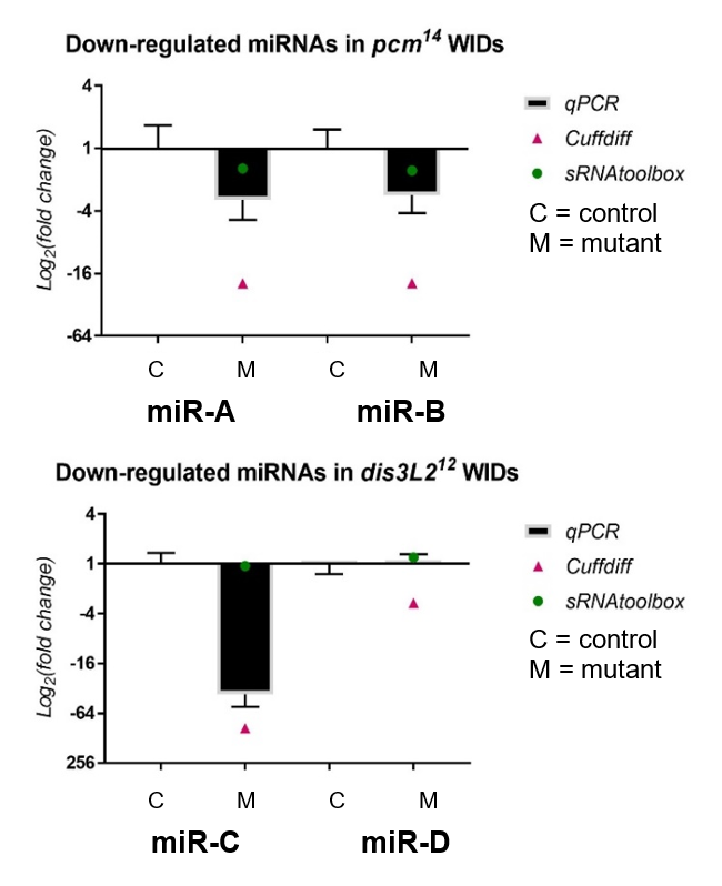
My results, shown in Figure 2, were obtained through the use of qPCR (quantitative or real-time PCR). Also shown on Figure 2 are two other datasets from previous high-throughput RNA sequencing experiments, known as Cuffdiff and sRNAtoolbox.
Figure 2 – The fold change of miRNAs downregulated in both dis3L2 and pacman (pcm) mutants normalized to their controls, obtained through my qPCR and previous RNA-seq methods.
Looking at my data, the pacman mutants showed that there was a negative fold change in the levels of two miRNAs, miR-A and miR-B when normalized to their wildtype’s levels. This indicated that the two miRNAs are downreguated in the pacman mutants. This confirmed the results obtained from the RNA sequencing, as they both showed that the miRNAs were downregulated as well.
Results from the dis3L2 mutants were less definite. For one of the miRNAs, miR-C, there was a negative fold change when normalized to the wildtype’s level. Again, this confirmed results obtained from the RNA-seq, but my data, and data from Cuffdiff, suggested a much stronger downregulation than that from sRNAtoolbox. For the other miRNA, miR-D, qPCR showed that there was no significant fold change in the mutants. This agrees with results from one of the RNA-seq, but not the other. Data from Cuffdiff showed a slight negative fold change of miR-D, suggesting it is downregulated in the mutants. However, my data, and that from sRNAtoolbox, suggested there is no change between the wildtypes and mutants.
Further areas of my project involved looking at the difference in volume of the abdomen and ovaries in flies with a defect in the gene of a TUTase known as Tailor, to see whether it was involved in controlling their size. TUTases, or Terminal Uridylyl Transferases, are enzymes that uridylate miRNAs, potentially serving as a signal for exoribonuclease-mediated degradation such as Dis3L2. This involved me freezing flies in liquid nitrogen, then photographing and dissecting out their ovaries. The limited number of ovaries measured suggested that there were no significant differences in the sizes between the wildtype and mutants.
In addition to this, I also looked at the differences in sizes of wings in Tailor mutant and double mutants of Tailor and Dis3L2 compared to wild types. This included dissecting wings and mounting them on a slide then taking measurements using computer software and a microscope. We hypothesised that the wings from the Tailor mutants would be the same size as the wildtype but the Tailor/Dis3L2 double mutants would have larger wings than the wildtype. My results here were too limited to draw a reliable conclusion but indicated there was no difference in wing sizes between the mutants and wildtype.
Overall, my time in the lab has proved highly valuable and interesting. Learning about the techniques in lectures and actually performing them yourself in a lab are completely different experiences and I am really grateful. I feel it was really important for me to experience the fly work alongside the molecular work as this has helped me to familiarize myself with numerous practises and therefore determine what is most interesting and suitable for me in the future. But not only did I learn how to perform these techniques, I was also introduced into the world of Drosophila, ranging from learning about their genome, recognising their phenotypes, and performing my own genetic crosses to learning their general upkeep and how to do simple things such as egglays or anesthetizing the flies. This experience for me has really increased my interest in developmental biology. Being able to produce my own data and learning to work independently in a lab have also strengthened my desire to do a PhD and I’m very appreciative that I got to experience something not many undergraduate students will. On top of all this, being inside a lab with such a friendly and welcoming group of people has made it all that more enjoyable.
A huge thank you to everyone in the lab for their support and assistance, and to the BSDB for making this opportunity possible through the Gurdon Studentship.
Sources
Towler BP, Jones CI, Harper KL, Waldron JA, Newbury SF. 2016. A novel role for the 3′-5′ exoribonuclease Dis3L2 in controlling cell proliferation and tissue growth. RNA biology 13: 1286-1299.
Waldron JA, Jones CI, Towler BP, Pashler AL, Grima DP, Hebbes S, Crossman SH, Zabolotskaya MV, Newbury SF. 2015. Xrn1/Pacman affects apoptosis and regulates expression of hid and reaper. Biology open 4: 649-660.
Astuti D, Morris MR, Cooper WN, Staals RH, Wake NC, Fews GA, Gill H, Gentle D, Shuib S, Ricketts CJ et al. 2012. Germline mutations in DIS3L2 cause the Perlman syndrome of overgrowth and Wilms tumor susceptibility. Nature genetics 44: 277-284.
 (No Ratings Yet)
 (5 votes)