Postdoctoral position available to study mechanisms of segmentation in the invertebrate chordate amphioxus
Three-year NSF-supported postdoctoral position is available at University of California San Diego, Department of Scripps Institution of Oceanography, La Jolla, CA, USA to study the evolution of segmentation in chordates. This is a multi-level study focused on somite formation in the basal chordate amphioxus. The study involves dissecting the genetic mechanism of somite segmentation in embryos and larvae of the basal chordate, amphioxus. The effects of perturbing gene function on the mechanics of somite segmentation will be monitored at the single-cell level by 3-D reconstructions with serial blockface scanning electron microscopy. RNA-Seq will address questions of gene hierarchy. Applicants with a background in evo/devo and/or developmental mechanisms of amphioxus are preferred. A PhD is required. Interested applicants please submit a curriculum vitae, names, addresses and e-mail addresses for three references to Dr. Linda Z. Holland, Marine Biology Research Division, Scripps Institution of Oceanography, University of California San Diego, La Jolla CA, 92093-0202 (tel 858-534-5607; fax 858-534-7313; email lzholland@ucsd.edu). UCSD is an equal opportunity employer.
We have an open position immediately available for a postdoc to study the transcriptional regulation of polarized axon outgrowth in the simple embryos of the tunicate Ciona, taking advantage of their simplicity at the genetic and cellular levels. The larval central nervous system (CNS) of Ciona is a miniaturized but typically chordate CNS containing only 177 neurons, and represents only the second complete “connectome” ever mapped. The tractability and low-cost of Ciona embryos make them especially suited for candidates who plan on starting a research program at primarily undergraduate institutions. In fact, part of the proposed project includes outreach initiatives aimed at involving both undergraduates and high school students in applying cutting-edge techniques like CRISPR/Cas9, RNAseq, and optogenetics to addressing fundamental questions in chordate neurodevelopment.
a) Diagram of the Ciona larval nervous system. b) Larva showing cholinergic CNS neurons labeled by electroporation of VAChT reporter plasmid. c) CNS development in Ciona.
In this specific project, we are trying to dissect a regulatory network for the intrinsic control of neuronal polarization and polarized axon outgrowth in the descending decussating neurons (ddNs), a single left/right pair of neurons proposed to be homologous to vertebrate Mauthner cells. This builds on our recent papers, and capitalizes on the lab’s contributions to developing and adapting the latest methods for CRISPR/Cas9-mediated, tissue-specific gene knockouts.
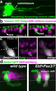
a) Pair of ddNs labeled by Dmbx reporter plasmid. b) Fixed series images of MG cells labeled with fluorescent Golgi apparatus (green) and histone (pink) reporters, showing Golgi position inversion. c) ddN cytosol labeled with CFP (white) showing axon projecting medially. d) Pax3/7 overexpression in the MG results in ectopic ddNs that project across the midline.
NSF funding is available starting immediately, with the possibility of extension pending further funding support. Our lab (www.tunicates.org) is at the Georgia Institute of Technology (Georgia Tech), located near downtown Atlanta, GA, USA, a dynamic, multicultural city boasting a vibrant neurobiology and biomedical research community fostered by Georgia Tech together with nearby Emory University and Georgia State University.
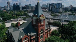
Georgia Tech campus overlooking the Atlanta skyline and Krone Building where our lab is located.
To inquire, please contact alberto.stolfi@biosci.gatech.edu
CENTURI seeks to attract outstanding computer scientists, physicists, or mathematicians with a theoretical and/or computational biology project.
The selected candidates, who are expected to bridge biology and other disciplines will be affiliated to two institutes and will be offered competitive funding.
The questions that are being addressed by this interdisciplinary research initiative include:
How is information encoded in living systems?
How do genes control cell fates and functions?
How do signalling dynamics impact on cells?
How do mechanics influence cell and tissue dynamics?
How do networks form, function collectively and evolve?
The selected candidates, who are expected to bridge biology and other disciplines will be affiliated to two institutes and will be offered competitive funding.
Applicants should send a curriculum vitae with a complete list of publications, a 2-page summary of research achievements and projects in English, and arrange for 2-3 letters of recommendation to be sent to PIcall@centuri-livingsystems.org, before February 28th, 2020.
Short-listed candidates will be invited to submit a full proposal by April 22nd, 2020, and for an interview between May 25th and May 27th, 2020. CENTURI promotes equal opportunities for employment.
CENTURI brings together leading institutes in biology, physics, mathematics, computer science and engineering to decipher the complexity and dynamics of living systems. CENTURI offers an exceptional international environment for the development of interdisciplinary projects, in developmental biology, immunology and neurosciences.
CENTURI is mainly located on the Luminy campus of Aix-Marseille University and is affiliated to Aix- Marseille University, CNRS, INSERM and École Centrale Marseille.
Established by the British Society for Developmental Biology in 2014, The Gurdon/The Company of Biologists Summer Studentship scheme provides financial support to allow highly motivated undergraduate students an opportunity to engage in practical research during their summer vacation. Each year, ten successful applicants spend eight weeks in the research laboratories of their choices, and the feedback we receive is outstanding. You can read accounts from previous years here. If you’re interested in applying or hosting a student in 2020, applications need to be in by the end of March.
Our first report from the class of 2019 comes from Franklin Lo who studies at the University of Edinburgh. His Gurdon studentship took him to…John Gurdon’s lab at the Gurdon Institute, where he was supervised by Sir John, Jerome Jullien and Khayam Javed.
I am Franklin Lo, a 4th year undergraduate student at the University of Edinburgh. My summer internship was carried out in Professor Sir John Gurdon’s lab at Gurdon Institute of the University of Cambridge and I was supervised by Professor Sir John Gurdon, Dr Jerome Jullien and Khayam Javed.
The major focus of my internship was to understand the functions of oocyte-specific B4 linker histone during nuclear reprogramming (NT). This maternal factor is abundant in oocytes and eggs, and present also in early embryo until the mid-blastula transition. The injection of somatic cell nuclei into Xenopus oocytes has been shown to reverse the restriction of some gene expressions and induce expression of pluripotency genes including POU5F1 and SOX2. This cell division independent process utilises components in the germinal vesicle (GV) of oocytes, and mechanism of reprogramming in NT is thought to be identical to the reprogramming of sperm genome by eggs after fertilisation. Therefore, NT is an excellent approach to gain further insights on the mechanism of nuclear reprogramming in eggs.
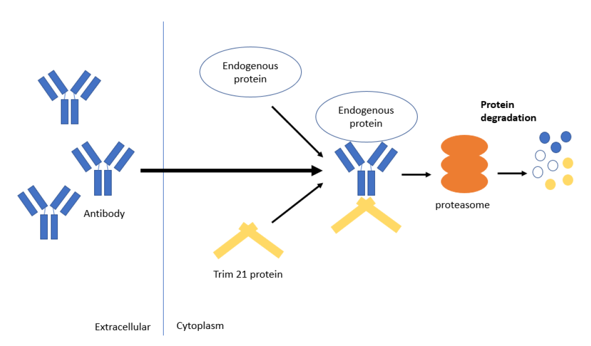
B4 is an important component of reprogramming by NT since it substitutes somatic histones in injected somatic cell genome and drives the transcription of pluripotency genes. A recently developed novel antibody-based methodology called Trim-Away enables targeting and degradation of specific proteins in Xenopus oocytes including B4 linker histone without any genetic manipulations. The procedure involves the injection of Trim 21 mRNA or protein into the cytoplasm or GV of oocytes, respectively, followed by GV injection of antibody specific to a maternal factor. The rationale of Trim-Away is that the constant region of antibody binding to its target protein interact with Trim 21 protein, which then recruits ubiquitin proteasome that degrades the protein-antibody-Trim21 complex (Fig. 1). By using this technique, we have targeted B4 linker histone to study the effect of its degradation on the efficacy of reprogramming after NT.
Figure 1. Mechanism of protein degradation by Trim-Away. The sophisticated characteristic of this method is its capability to knock down endogenous proteins such as maternal factors efficiently, which is not possible with techniques like gene editing and RNA interference
To perform these experiments, my first few weeks of the internship was dedicated on practising DNA/mRNA injection into the GV and cytoplasm of oocytes using Drummond microinjector under a light microscope. GV injection was especially a tough challenge for me since GV is hidden just underneath the pigmented region of Xenopus oocytes. A precise positioning of glass needle against oocytes and controlling the depth to penetrate are of utmost importance for successful GV injection. While my success rate began to hover around 50-60% after few weeks of practice, everyone else can aim the GV at approx. 90% efficiency! Such accuracy is important to run experiments smoothly. Nonetheless, I was compensating my inefficiency of GV injection by both increasing the number of oocytes I inject and co-injecting DNA encoding fluorescent protein which gives an indication of successful injection. Trim-Away is a very useful technique and future attempts may involve targeting different maternal factors to study the mechanism of nuclear reprogramming.
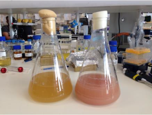
I was also responsible for generating both Trim 21 fused with mCherry and B4 linker histone proteins using bacteria. It started off with cloning of these genes into an appropriate plasmid vectors and transforming it into a BL21 bacteria for protein expression. Protein purification worked but the yield was extremely lower than our prediction. After numerous attempts, we managed to produce a large quantity of these proteins using a strain of bacteria called Rosetta. I still remember the joy of seeing pink bacterial culture as Rosetta strain was highly expressing Trim 21- mCherry (Fig. 2). Unfortunately, my time in the lab was limited and I could not purify these proteins.
Figure 2. Protein expression using Rosetta strain. Trim21-mCherry production (right) turns bacterial culture medium pink while wild type B4 protein (left) production did not change the colour of culture medium.
Having gained experiences to work with oocytes, I also began to work with Xenopus embryos which is made by fertilising eggs with sperms extracted from testis. Oocytes and eggs are naturally protected by a jelly-like coating which impede penetration of resulting embryos with a microinjector. But unlike oocytes, de-jellying process of embryos are difficult, and I often killed the embryos by de-jellying excessively. The unusual hot weather in Cambridge also deteriorated the egg quality and heightened the difficulty of working with embryos; researchers working with Xenopus often avoid performing important experiments during the summer. The time frame for injection is also shorter in embryos since they divide and develop, hence time window for injection is shorter than oocytes which are static. Despite encountering these challenges, I still found experiments with embryos very exciting as mRNA injected can affect embryo development and produce unique phenotypes.
Despite being a Nobel laureate, professor Gurdon is always humble and active as a researcher – he is very focused and still loves to perform experiments. Everyone, including myself, gains strong motivations to work hard from seeing his working style. His talkative and approachable personality makes him a great researcher even more; he often taught me procedures to work with oocytes and invited me to his tea-time for further discussions.
I was also surprised to see that everyone in the lab is very enthusiastic and keen about learning new information even from outside of their field of interest. This nature must be acting as a strong foundation for them to come up with creative ideas. The experience I had at Gurdon lab was stimulating and fantastic, I very much appreciate mentorship, patience and kindness from Professor Gurdon, Jerome and Khayam. Working closely with several supervisors enabled me to participate on multiple projects and gain a variety of invaluable experiences.
I would also like to convey my gratitude to other lab members namely, Nigel Garrett, Can Aztekin, Dr Eva Hörmanseder, Dr Ming-Hsuan Wen, Dr Chris Penfold and Dilly Bradford for their immense support and mentorship during the internship.
Finally, I would like to thank BSDB for providing me this opportunity and I strongly recommend students who wish to do a lab internship to apply for this studentship. It offers students to gain hand-on experiences in an exciting lab!
Animal cytokinesis is driven by an actomyosin ring that assembles at the cell equator and constricts to physically separate the two daughters. Although myosin is known to be essential for cytokinesis in multiple systems, whether this requirement reflects its motor or actin crosslinking activities has recently been a matter of contention. A new paper in Development now addresses this problem using the first divisions of the Caenorhabditis elegans embryo as a model. We caught up with the paper’s three first authors Daniel Osório, Elaine Chan and Joana Saramago, and their supervisor Ana Carvalho, Principal Investigator at the University of Porto’s i3S consortium, to find out more about the story.
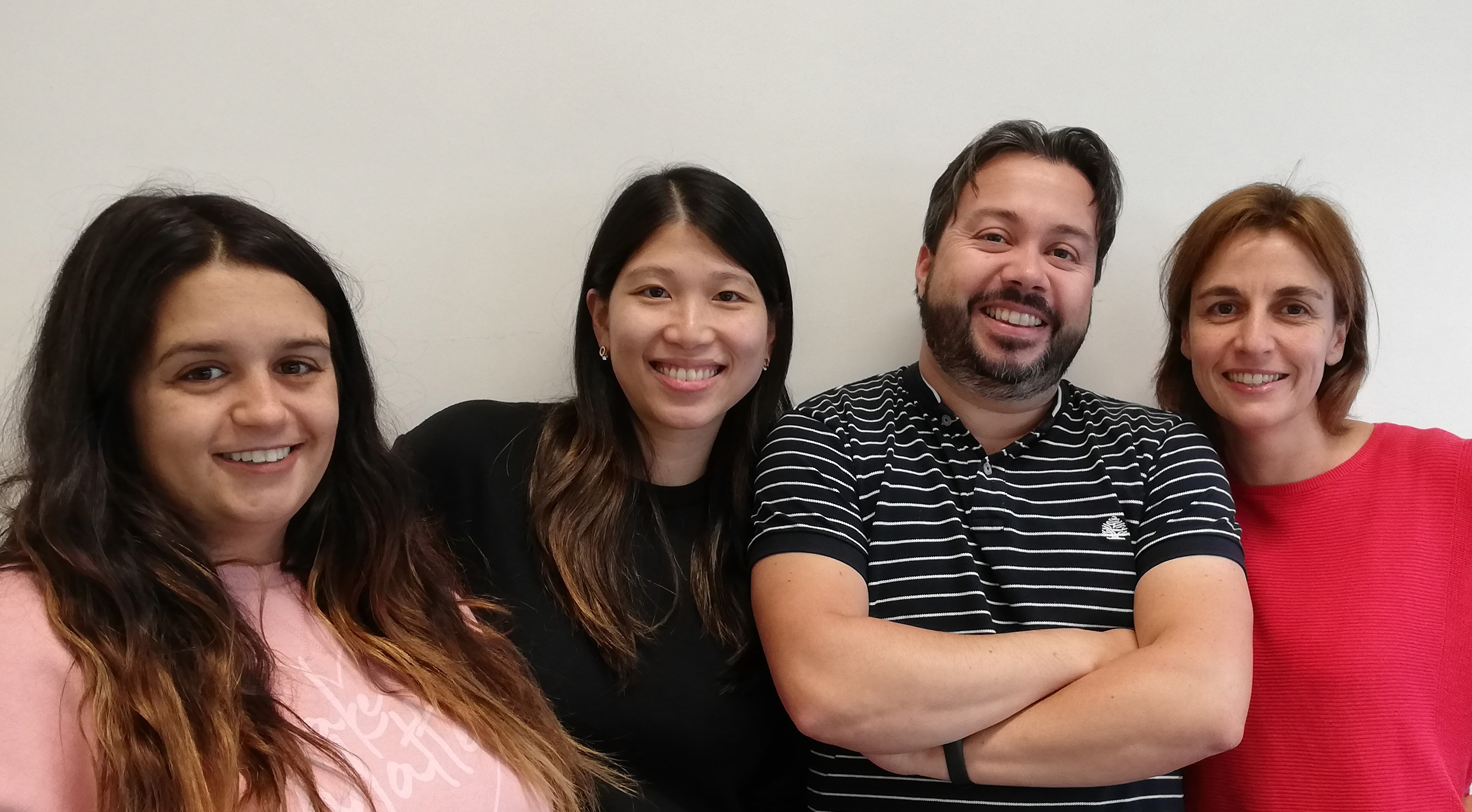
Joana, Elaine, Daniel and Ana (L to R)
Ana, can you give us your scientific biography?
AC I graduated in biochemistry from the Faculty of Sciences of the University of Porto and did my doctoral studies on mitosis with Professor Bill Earnshaw at the Wellcome Trust Centre for Cell Biology in Edinburgh, UK. Then I moved to San Diego, USA, to do my postdoctoral work on cytokinesis in Professor Karen Oegema’s group at the Ludwig Institute for Cancer Research. In 2012, I moved to IBMC, the Institute for Molecular and Cell Biology in Porto, where I launched my independent research group. IBMC is currently part of the i3S, Institute for Research and Innovation in Health, a large consortium of three research institutes headed by the University of Porto.
Daniel, Elaine and Joana: how did you come to work in the Carvalho lab, and what drives your research today?
DO When I finished my PhD with Edgar Gomes in Paris working on the cell biology of nuclear positioning mediated by actin-binding proteins, I was looking to return to Portugal and my soon-to-be wife, having been away for almost 7 years. I found that the Carvalho lab had been recently established and had an opening in a C. elegans project to work with actomyosin and cytokinesis. In 2009 I had been one of the lucky ones selected for the physiology course of the Marine Biological Laboratory in Woods Hole, and during that course I got my first contact with C. elegans by interacting with the Oegema-Desai labs where Ana did her post-doc. Although we did not meet at the time I was very impressed by the model and its possibilities, so I guess the universe aligned later on and I was selected by Ana to take on this ambitious project.
EC In 2012, I got married to my Portuguese husband. After marriage, we decided to move from San Diego to Portugal, but at that time finding a research job in Portugal was not easy because of the economic crisis. But I did not give up my research dream. I contacted Ana because I really liked her research projects studying cell division in C. elegans, which was similar to my PhD background on bacterial cell division. After meeting her, we realised that we had both worked in the same university in San Diego before moving to Portugal, just in different buildings. We never had a chance to meet on the UCSD campus but we got our chance in Portugal! Ana and I have been working on actomyosin projects together for almost 7 years.
JS In 2013, I started my Master’s thesis and I knew that I wanted to do cell biology, and I started to search for an interesting lab. I visited a few labs, but when I saw a beautiful C. elegans embryo undergoing cytokinesis on Ana’s computer I knew that I had found what I was looking for. I also loved the idea of doing live microscopy, which was a dream of mine since I was a little girl: to see what our eyes can’t see. I liked it so much that I stayed to do my PhD after my Master’s.
Why has myosin’s precise role in cytokinesis been controversial?
DO Well, first and foremost because if you deplete or inactivate myosin, cytokinesis fails in most situations and systems. This only allows for the conclusion that myosin is required for the process, not that myosin motor activity is required. Second, I think it has been controversial because we tend to take the concept of conservation of processes too far: we want to assume that cytokinesis in a small yeast cell works exactly like it does in adhered cells or in a C. elegans embryo where divisions are so fast. Although the players and mechanisms are clearly conserved and a lot has been learned from studies in all systems from yeast to mammalian cells, we should accept that there may be system-specific variations. These should not be looked at as controversies, but may in fact reflect differences between the systems. Finally, the lack of good structural and quantitative information on the actomyosin organisation of the contractile ring in animal cells has limited the interpretation of phenotypes. Recent work using both electron and super-resolution microscopy are improving knowledge in the field.
Can you give us the key results of the paper in a paragraph?
DO, EC, JS & AC The motor activity of non-muscle myosin II is essential for cytokinesis and contractile ring contractility in C. elegans embryos, and the motor activity of muscle myosin II is required for contraction but dispensable for actin organisation in the body muscle of C. elegans adults.
C. elegans one-cell embryo undergoing cytokinesis. The embryo expresses non-muscle myosin II (NMY-2) fused to GFP. Three stills corresponding to three consecutive time points during cytokinetic furrow ingression have been overlaid (first time point in yellow, second in cyan and third in magenta).
Why do you think myosin motor mutants have a slower rate of contractile ring constriction?
DO Although several mechanisms may be involved, our work suggests that actin filament sliding through myosin motor activity is indeed important for constriction of the contractile ring. This is in agreement with previous proposals and with the organisation of the actomyosin network observed in recent electron microscopy and super-resolution studies. Therefore, mutants with reduced or absent motor activity have a reduced or absent ability to slide actin filaments, thus presenting a reduced rate of ring constriction.
When doing the research, did you have any particular result or eureka moment that has stuck with you?
DO I would not say eureka moments but there were some particular events that definitely contributed to the advance of the story. One was testing the effects of myosin mutations in the muscle. Although muscle and non-muscle myosins have biochemical differences and the muscle has a different organisation when compared to what we know about the contractile ring, we wanted an in vivo strategy that could help validate in vitro results as a complementary approach. Additionally, since we were not able to use animals expressing just the motor dead mutants (as they were sterile and did not produce embryos), the use of a wild-type copy from another location in the genome that could specifically be depleted by RNAi was a seemingly simple but very important step. Finally, getting the in vitro myosin motility assay to work was important. Although it has been established for many years, it is quite a challenging and meticulous process requiring several proteins to be expressed and purified in a functional form. This was quite time-consuming and not straightforward – in a sense I consider it a eureka moment for the lab because we started from scratch.
AC Being able to generate C. elegans in which the only source of UNC-54, the major muscle myosin, is UNC-54(R710C) and observing them moving normally was an important moment. Previous work has considered the equivalent mutation in non-muscle myosin II from mammalian cells to be ‘motor-dead’ based on an in vitro motility assay. As this residue and the region where it is located is highly conserved between muscle and non-muscle myosins of several organisms, we expected to obtain animals whose muscles were unable to contract and therefore were compromised in their movement. The result we obtained emphasised the fact that results from in vitro studies should be validated in vivo, where more complex scenarios exist.
And what about the flipside: any moments of frustration or despair?
DO Well, trying to analyse the specific role of any component of the cytokinetic contractile ring can be challenging. There is a quote from Ray Rappaport that explains it better than I can: ‘When I began working on cytokinesis, I thought I was tinkering with a beautifully made Swiss watch, but what I was really working on was an old Maine fishing boat engine: overbuilt, inefficient, never-failed and repaired by simple measures’. Additionally, myosin itself is quite a large and amazing motor and when tinkering with its gears it becomes complicated to decouple the effects on its motor versus actin-binding activities. All in all, everything involving myosin biochemistry was a challenge as we did not have any expertise.
So what next for you three after this paper?
DO I’m currently trying to capitalise on the multiple myosin strains we generated to study the interplay between muscle and non-muscle myosins in different C. elegans actomyosin-dependent contexts.
JS I am in the last year of my PhD and am currently trying to understand whether different types of myosin play a role during embryonic cytokinesis in C. elegans.
EC I am currently studying how the surrounding cortex influences the contractile ring and formin regulation throughout ring constriction.
Where will this work take the Carvalho lab?
AC There are still so many fascinating questions in the field and so studying the process of cytokinesis will continue to entertain us for a while. Our team is also moving onto exploring other cellular contexts involving actomyosin contractile networks in the powerful C. elegans system.
There are still so many fascinating questions in the field
What is the current situation for developmental and cell biology in Portugal?
AC Doing developmental and cell biology in Portugal is fun. Portugal is a beautiful and pleasant place to live, and more and more laboratories work in these fields. If only funding from the national Portuguese Foundation for Science and Technology would be more reliable and research positions more stable!
DO I think there are several very good cell and developmental biology labs in Portugal. The main challenge they face is funding and work position stability. The funding calls mostly centred on the Portuguese Foundation for Science and Technology have been quite erratic and there are very few alternatives for people trying to establish their labs in a more permanent way. Additionally, the amounts funded by national projects are not so high and this is quite an expensive field, so it is almost essential to secure funding from the European Research Council and other European sources, which are extremely competitive. As for work positions after PhD completion, the country is finally moving from a scholarship-based system to work contracts for post-docs and researchers. However, the university system is currently quite locked and most institutes, whether they are affiliated with the universities or not, have very limited possibilities to hire even the most talented PIs. This reduces my hopes that the country will succeed in maintaining and increasing the current level of its scientific community.
Finally, let’s move outside the lab – what do you like to do in your spare time in Porto?
DO Porto has become quite an amazing city over the years. There is now a whole new food, wine and craft beer scene that I love to explore while rediscovering some of the old classics. I like to walk and run around the river and seaside and when I have some free time I try to go up the river to the beautiful Douro valley where part of my family comes from. Those vineyards and amazing views bring back memories of my summer holidays as a kid. As I age, I grow more fond of them and I must confess I dream about having a vineyard of my own and producing my own wine!
EC In my spare time, I like doing different outdoor activities with my family – Porto has a wonderful beach for surfing and a large city park for my kid to ride a bike and play football.
JS I live near the beach and I like to go there and play with my beautiful baby girl. I also love to be surrounded by my friends, and we do a lot of dinners in each other’s houses.
AC Going for long walks by the beach is always reinvigorating, and going out for drinks and good food with friends is the best!
Applications are invited for a postdoctoral fellow in the Brückner lab at The University of California San Francisco, Broad Center of Regeneration Medicine and Stem Cell Research.
The laboratory investigates inter-organ communication in innate immunity and hematopoiesis, focusing on the regulation of hematopoiesis by sensory inputs and signals from the nervous system, and molecular mechanisms of organismal immunity to bacterial infection (Makhijani et al. Nature Communications 2017; Sanchez Bosch et al. Developmental Cell 2019). The NIH funded project on organismal immunity will investigate a signaling relay between blood cells and the respiratory epithelia.
The ideal candidate is a talented and productive scientist with a background in developmental biology and genetics. Underrepresented minority candidates are in particular encouraged to apply. Prior work with Drosophila genetics, neurobiology or hematopoiesis/macrophage biology is desirable. General skills in molecular and cell biology, and good communication skills are expected. Candidates must hold a recent PhD, MD or MD/PhD degree, or anticipate such degree in the near future. We are looking for candidates who are motivated, hardworking, and have prior publications.
UCSF and the Broad Center offer a vibrant scientific environment and state of the art facilities. The Brückner lab provides infrastructure, a collegial environment, training and opportunities to work in teams.
Please send a CV, statement of research interests, and names and contact information of three references to:
Katja Brückner, Ph.D.
Associate Professor
Eli and Edythe Broad Center of Regeneration Medicine and Stem Cell Research
Department of Cell and Tissue Biology
University of California San Francisco
35 Medical Center Way, Room RMB 1026
San Francisco, CA 94143-0669
Tel +1 415 476 3827
Fax +1 415 476 9318
katja.brueckner@ucsf.edu http://bruecknerlab.ucsf.edu/
We are seeking to recruit a Drosophila Neuroanatomy Biocurator to join our team at the University of Cambridge. The post is suitable for those with an undergraduate degree (Research Assistant level) or post-PhD (Research Associate level).
The post holder will contribute to two Drosophila databases: Virtual Fly Brain (VFB; http://virtualflybrain.org) and FlyBase (http://flybase.org/). If you are looking for a fulfilling, fly-related career away from the bench, and enjoy the challenge of organising complex data and presenting it clearly and concisely, then this is the job for you!
Virtual Fly Brain is a resource that helps Drosophila neuroscientists map and visualize neuronal circuits, and find the experimental reagents to manipulate the circuits to reveal their role in perception, learning, memory and complex behaviours. The successful applicant will aid this process in three ways: 1) by annotating the expression of Gal4 drivers within the adult brain, using data from large scale datasets and the primary research literature; 2) assisting in the integration of connectomics data and single-cell RNA sequencing datasets; and 3) leading the effort to assess the functionality of improvements to the website, by recruiting members of the local neuroscience community and working with them and other VFB members to perform usability testing of the website. Much of the curated data will also be integrated into FlyBase, the primary community resource for Drosophila genetic, genomic and functional data.
Are you interested in the mechanisms that regulate organ development and prevent cancer formation?
We are looking for highly motivated and talented candidates to join our research team, lead by Dr. Zuzana Koledova.
We study processes, which govern mammary epithelial morphogenesis and homeostasis, and deregulation of which can lead to breast cancer. The main focus of the lab is on the role of mammary stromal cells in regulation of epithelial behaviour and the interplay of fibroblast growth factor signalling, cytokines and hormones in mammary gland morphogenesis and breast cancer. The approaches include advanced 3D organoid cultures of mouse and human primary tissue, genetic mouse models, state-of-the-art imaging techniques and mathematical modelling. The research group has extensive collaborations within the European Network of Breast Development and Cancer and beyond.
1. PhD position:
Anticipated start date:The position is available from September 2020 or upon agreement.
Requirements:
MSc. degree from biology, biochemistry or its equivalent
Interest in developmental and fundamental processes of life
High motivation and work ethics, scientific curiosity and critical thinking, self-drive
Interest in learning new methods, dexterity
Willingness to travel abroad for research internship and conferences
Tissue culture skills, imaging and/or mouse work experience are advantageous
We offer:
Interdisciplinary project on highly relevant scientific topic
Work in young, enthusiastic team with active international collaborations
Participation at international meetings, internships at collaborating laboratories
Anticipated start date: The position is available from March/April 2020 or upon agreement but no later than by 30th November 2020.
The candidate should:
be a researcher who has received a PhD or its equivalent within the last 7 years
be a researcher who has worked at least two whole years in the last three years outside the territory of the Czech Republic in the field of research with a working time of at least 0.5 full-time equivalent, or who has been PhD student (or equivalent) abroad
have a publishing record – in the last three years at least two publication outputs registered in the Thomson Reuters Web of Science, Scopus or ERIH PLUS databases and at the same time publications such as “articles”, “books”, “book chapters”, “letters” and “reviews”.
be intellectually curious, self-driven and productive, have excellent communication skills, and enjoy working in a committed team
have experience in the field of developmental or cell biology; experience in 3D cell culture techniques, live cell imaging and image analysis is highly desirable
The application should include:
a CV including a summary of education and research experience, publication activity, involvement in research grants, etc.
a scanned copy of the PhD diploma or an official letter certifying submission of a doctoral thesis for thesis defence and the planned defence date
Postdoc Position (NIH-funded): Cell & Dev Biology of Neurons – How are Dendrites Shaped? We seek a creative and productive individual who has a beginning or ongoing interest in neuron cell & dev biology. For initial studies, some familiarity with cell culture, immuno-precipitations/ running gels and microscopy would be a plus. The applicant should be finishing their PhD soon or have recently completed it. In this NIH-funded project we address the question: how are dendrite morphologies generated? The shaping of dendrites must take place properly for normal neuron connectivity to arise in the central nervous system, with alterations contributing to pathologies including mental retardation and dementia. Through the binding of novel protein-partners, we discovered that delta-catenin regulates the branching and length of dendrites. We propose to identify both the upstream pathways involved (e.g. ligands and kinases), and the downstream mediators (e.g. cytoskeletal modulators) that promote these dramatic branching versus lengthening outcomes. We will additionally examine related catenin proteins and their complexes. Laboratory members can select from a number of experimental systems including primary rat hippocampal neurons, mammalian cell lines, and frog embryos, plus undertake collaborative ventures involving other models. Pierre McCrea PhD (PI) has extensive mentorship experience. Prior trainees have gone on to successful academic as well as other rewarding scientific careers.
Environment. The Texas Medical Center in Houston houses UT MDACC and multiple other top-flight scientific institutions within walking distance (Baylor College of Medicine; UT Health Science Center; Rice University; Methodist Research Institute; TX A&M Institute of Biosciences and Technology), or within bicycling distance (U Houston). This immense depth of collaborators/ programs/ cores insures a cutting-edge research/ ideas environment. Houston boasts a diverse culture near the Gulf Coast, and affordable living in our nation’s fourth largest city. MDACC offers well-regarded professional development opportunities and benefits. This is an NIH pay-scale position.
Please send:
Cover letter or email stating your general career objectives and research interests
PhD and Postdoc positions are available in the group “Cellular sex and physiology” in the Institute of Biology Valrose – Nice (France).
We are interested in developmental, metabolic or behavioural sex differences and aim to understand how sex chromosome constitution (XX versus XY) impacts physiology across the body. We are currently identifying new genes/pathways and concepts driving sex chromosome effects using multi-scale approaches combining biochemistry, genetics and cell biology. For this, we use the fruit fly Drosophila melanogaster as a model system (see Hudry et al., 2016 Nature and Hudry et al., 2019 Cell). More information can be found on the lab webpage (http://ibv.unice.fr/research-team/hudry/).
The positions will be funded through an ERC starting grant and would ideally start in 2020. We seek highly motivated candidates with interests in cell and developmental biology. Candidates should have a good working knowledge in live imaging and image analysis or prior experience conducting behavioural experiments. Previous experience with Drosophila is a plus, but is not mandatory. We strongly value team spirit and a positive work environment.
If you are interested, please contact Bruno Hudry (applicationhudry@gmail.com) with your CV and a brief statement of your research interests, key areas of expertise and up to 3 recommendation contacts.
 (No Ratings Yet)
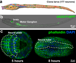
 (1 votes)