A 3-years-PhD position is available at the Chédotal Lab at the Vision Institute in Paris.
The goal of the PhD project will be to describe the organization of the developing human eye and characterize molecular and cellular mechanisms underlying human retinogenesis. We propose to revisit eye formation combining modern and innovative single-cell molecular and 3D imaging approaches on human embryo/fetal eye samples. Data from light sheet fluorescent microscopy (LSFM), spatial transcriptomics and in situ sequencing will be incorporated into one scaffold to provide a comprehensive 3D map of human retinogenesis. The project will provide significant and detailed insights into human ophthalmology, generating the first reference map of human retina development. It will further contribute to the Human Cell Atlas initiative. The thesis will be directed by Dr Alain Chédotal.
An experience in bioinformatics and transcriptomics or 3D imaging would be a plus but is not required.
Candidates should hold a diploma or a master’s degree in Neuroscience, Developmental Biology, Microscopy or Bioinformatics
They may send their application (in a PDF-format) to Dr Alain Chédotal (alain.chedotal@inserm.fr), including a CV and contact information for two references.
Welcome to our monthly trawl for developmental biology (and related) preprints.
Here’s the last cache of preprints of 2019 – happy preprinting in 2020! They were hosted on bioRxivandarXiv. Let us know if we missed anything. Use these links to get to the section you want:
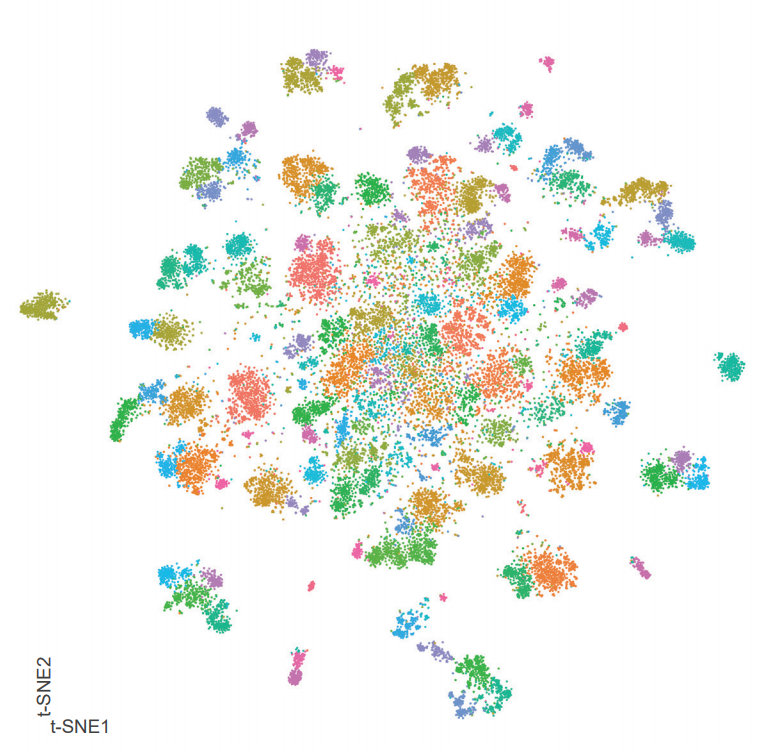
Conserved epigenetic regulatory logic infers genes governing cell identity
Woo Jun Shim, Enakshi Sinniah, Jun Xu, Burcu Vitrinel, Michael Alexanian, Gaia Andreoletti, Sophie Shen, Brad Balderson, Guangdun Peng, Naihe Jing, Yuliangzi Sun, Yash Chhabra, Yuliang Wang, Patrick P L Tam, Aaron Smith, Michael Piper, Lionel Christiaen, Quan Nguyen, Mikael Bodén, Nathan J. Palpant
Notch ligand Dll4 impairs cell recruitment into aortic clusters and limits hematopoietic stem cells
Cristina Porcheri, Ohad Golan, Fernando J. Calero-Nieto, Roshana Thambyrajah, Cristina Ruiz-Herguido, Xiaonan Wang, Francesca Catto, Yolanda Guillen, Roshani Sinha, Jessica González, Sarah J. Kinston, Samanta A. Mariani, Antonio Maglitto, Chris Vink, Elaine Dzierzak, Pierre Charbord, Bertie Göttgens, Lluis Espinosa, David Sprinzak, Anna Bigas
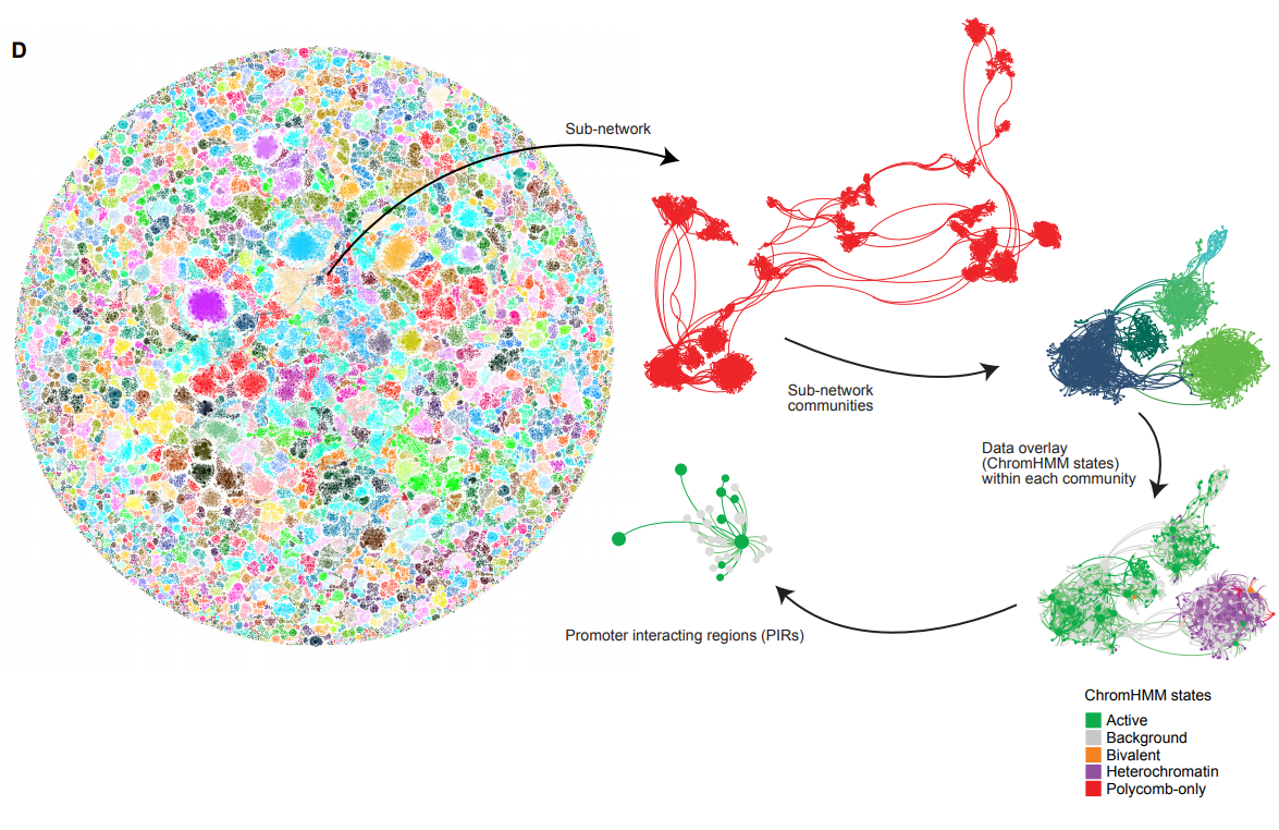
A Holistic Analysis of the Intestinal Stem Cell Niche Network
Darrick M. Hansen, Paloma Ivon Meneses Giles, Xi C. He, Shiyuan Chen, Ariel Paulson, Christopher M. Dekaney, Jennifer Wang, Deqing Hu, Aparna Venkatraman, Woosook Kim, John Kaddis, Barbara J. Olack, James C.Y. Dunn, Calvin Kuo, Susan Henning, Alan M. Hanash, Courtney W. Houchen, John Lynch, Martin G. Martin, Joyce C. Niland, Matthias Stelzner, Melissa Wong, Timothy C. Wang, Jian Yu, Kelley Yan, Linheng Li
A translational kidney organoid system bolsters human relevance of clinical development candidate
Amy Duyen Westerling-Bui, Thomas W. Soare, Srininivasan Venkatachalan, Michael DeRan, Eva Maria Fast, Alyssa B. Fanelli, Sergii Kyrychenko, Hien Hoang, Grinal M. Corriea, Wei Zhang, Maolin Yu, Matthew Daniels, Goran Malojcic, Xin-Ru Pan-Zhou, Mark W. Ledeboer, Jean-Christophe Harmange, Maheswarareddy Emani, Thomas T. Tibbitts, John F. Reilly, Peter Mundel
Characterization of SETD1A haploinsufficiency in humans and Drosophila defines a novel neurodevelopmental syndrome
Joost Kummeling, Diante E Stremmelaar, Nicholas Raun, Margot RF Reijnders, Marjolein H Willemsen, Martina Ruiterkamp-Versteeg, Marga Schepens, Calvin CO Man, Christian Gilissen, Megan T Cho, Kirsty McWalter, Margje Sinnema, James W Wheless, Marleen EH Simon, Casie A Genetti, Alicia M Casey, Paulien A Terhal, Jasper J van der Smagt, Koen L van Gassen, Pascal Joset, Angela Bahr, Katharina Steindl, Anita Rauch, Elmar Keller, Annick Raas-Rothschild, David A Koolen, Pankaj B Agrawal, Trevor L Hoffman, Nina N Powell-Hamilton, Isabelle Thiffault, Kendra Engleman, Dihong Zhou, Olaf Bodamer, Julia Hoefele, Korbinian M Riedhammer, Eva MC Schwaibold, Velibor Tasic, Dirk Schubert, Deniz Top, Rolph Pfundt, Martin R Higgs, Jamie M Kramer, Tjitske Kleefstra
In vivo mRNA structure regulates miRNA cleavage in Arabidopsis
Minglei Yang, Hugh C. Woolfenden, Yueying Zhang, Xiaofeng Fang, Qi Liu, Maria Louisa Vigh, Jitender Cheema, Xiaofei Yang, Matthew Norris, Sha Yu, Alberto Carbonell, Peter Brodersen, Jiawei Wang, Yiliang Ding
Genomic adaptations to aquatic and aerial life in mayflies and the origin of wings in insects
Isabel Almudi, Joel Vizueta, Alex de Mendoza, Chris Wyatt, Ferdinand Marletaz, Panos Firbas, Roberto Feuda, Giulio Masiero, Patricia Medina, Ana Alcaina, Fernando Cruz, Jessica Gómez-Garrido, Marta Gut, Tyler S. Alioto, Carlos Vargas-Chavez, Kristofer Davie, Bernhard Misof, Josefa González, Stein Aerts, Ryan Lister, Jordi Paps, Julio Rozas, Alejandro Sánchez-Gracia, Manuel Irimia, Ignacio Maeso, Fernando Casares
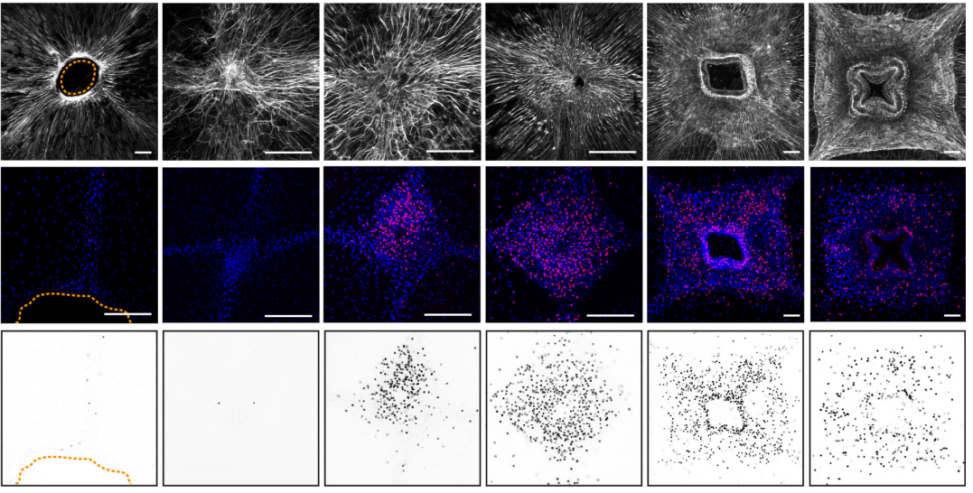
The nucleus acts as a ruler tailoring cell responses to spatial constraints
A.J. Lomakin, C.J. Cattin, D. Cuvelier, Z. Alraies, M. Molina, G. Nader, N. Srivastava, J.M. Garcia-Arcos, I.Y. Zhitnyak, A. Bhargava, M.K. Driscoll, E.S. Welf, R. Fiolka, R.J. Petrie, N. Manel, A.M. Lennon-Duménil, D.J. Müller, M. Piel
Visualizing the metazoan proliferation-differentiation decision in vivo
Abraham Q. Kohrman, Rebecca C. Adikes, Jayson J. Smith, Michael A. Q. Martinez, Taylor N. Medwig-Kinney, Nicholas J. Palmisano, Maria D. Sallee, Ononnah B. Ahmed, Nicholas Weeks, Nuri Kim, Simeiyun Liu, Wan Zhang, Ariel M. Pani, David Q. Matus
Insights from a survey-based analysis of the academic job market
Jason D. Fernandes, Sarvenaz Sarabipour, Christopher T. Smith, Natalie M. Niemi, Nafisa M. Jadavji, Ariangela J. Kozik, Alex S. Holehouse, Vikas Pejaver, Orsolya Symmons, Alexandre W. Bisson Filho, Amanda Haage
The closing date for applications is 15 February 2020
The Mass lab is looking for a postdoctoral candidate with a strong neuroscience, neurodevelopment and/or immunology background to study the role of nanoplastics in the development of neurological disorders as part of an ERC starting grant project. The position is for 2 years with a possible extension. The employment is planned to start on 1 April or upon agreement.
The goal of the NanoGlia project is to determine which type of plastic particles can pass from the mother to the developing fetus, whether this can lead to neurodevelopmental disorders and whether this is a microglia-dependent process. To achieve this, a guinea pig model is used due to its anatomical similarity to the human placenta. Further methods include histology, electrophysiology, single-cell RNA-sequencing, and flow cytometry.
About us
Our lab is located in the LIMES Institute, which is highly interdisciplinary, addresses basic research questions in developmental biology, immunology, genetics, and biochemistry and thus provides all the basic equipment required for the project. For more specific methodology we are closely collaborating with groups at the DZNE, Life&Brain and the Medical Faculty.
Qualifications
The candidate is required to hold a PhD degree in neuroscience/developmental biology or similar topics
Postdoctoral experience in the same areas is an advantage
We are looking for candidates with hands-on experience or an interest in expanding their knowledge in molecular biology, electrophysiology, high-dimensional flow cytometry, confocal microscopy, and bioinformatic analyses (particularly scRNA-seq)
Experience with guinea pigs as a model and background in R/Python is a plus
Finally, we are looking for applicants with a good track record of peer-reviewed scientific publications and teamwork
We offer
A thriving academic environment
A professional career development program
Participation in the university-wide pension system (VBL)
Access to extensive university sports program
A salary based on the TV-L scale (E13, 100%)
The university is committed to diversity and equal opportunity. It is certified as a family-friendly university. Applications from suitable candidates with a certified disability or equivalent status are particularly welcome.
For further information, please contact Professor Elvira Mass by email:
Please send your complete application as 1 combined PDF file including cover letter, CV, list of references (full address, incl. email and phone number), and list of publications to
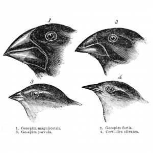
Charles Darwin’s famous finches from the Galapagos Islands
In this episode of Genetics Unzipped Kat Arney explores the myths and misconceptions behind two of the most iconic images in evolutionary biology: the much-parodied March of Progress – a series of human ancestors walking across the page, portraying the inexorable journey from monkey to man – and the famous finches of the Galapagos islands, which are supposedly the inspiration for Charles Darwin’s theory of natural selection.
Where did these infamous images come from, and do they really show what everyone seems to think they do?
If you enjoy the show, please do rate and review and spread the word. And you can always send feedback and suggestions for future episodes and guests to podcast@geneticsunzipped.com Follow us on Twitter – @geneticsunzip
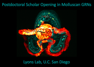
The Lyons Lab at Scripps Institution of Oceanography (a department at U.C. San Diego) is recruiting a full-time Postdoctoral Scholar to support research projects funded by an NIH MIRA award. The Lyons Lab (www.lyonslab.org) focuses on cell type differentiation and morphogenesis with a particular interest in how these processes evolve. The postdoc will contribute to our lab’s goal to build the first comprehensive developmental gene regulatory networks (GRN) controlling early events in molluscan development (e.g. germ-layer segregation, organizer signaling, and gastrulation). GRN analysis (both experimental and synthetic) will be carried out in the marine slipper snail Crepidula. The postdoc can take advantage of a growing tool kit for functional genomics in Crepidula, as well as a new marine transgenics facility for maintenance of stable lines. In collaboration with Dr. Lyons the postdoc will be responsible for leading an independent project of mutual interest that aligns with the postdoc’s career goals.
The postdoc will join a motivated group of students, staff, and postdocs who are broadly interested in cell and developmental biology of marine organisms. Additionally, the Lyons Lab offers a broad range of other systems for comparative developmental studies among molluscs and echinoderms, and provides a highly interdisciplinary and collaborative environment within our group, and with other labs at the Scripps Institution of Oceanography (https://scripps.ucsd.edu/), with U.C. San Diego’s main campus (https://ucsd.edu/), and with other institutions in the greater San Diego Area.
Qualifications:
We are seeking talented applicants who have a Ph.D. degree (or are close to earning one), and training in molecular biology, cell biology developmental biology, bioinformatics, or a related field. Candidates with expertise in cis-regulatory element analysis, network inference, and bioinformatics are encouraged to apply.
Strong experience and interest in one or more of the following areas is preferred, but not required:
Genomics: Preparation of samples and analysis of data for RNA-seq, ATAC-seq, scRNA-seq, BAC libraries, etc.
CRISPR/Cas9: Design and delivery (microinjection, electroporation) of CRISPR/Cas9 components for induction of DNA double-strand breaks.
Transgenesis: Generation of transient and stable transgenes via transposons, CRISPR/Cas9 genome editing, etc.
To Apply:
Submit the following items, as a single PDF, to Dr. Lyons (d1lyons@ucsd.edu):
1) Cover letter explaining your interest in the position and qualifications
2) CV
3) Statement of research/career goals
4) Names and contact information for at least three references
Review of applications will begin immediately and continue until position is filled.
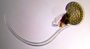
We are seeking EVODEVO POSTDOC candidates to apply any of the 3 currently open calls to join our lab in BARCELONA.
Our lab studies the chordate model Oikopleura dioica to better the impact of gene loss on the evolution of mechanisms of development and gene regulatory networks, with special interest in heart and muscle development. Click here for a tour “A day in our lab” posted in The Node
We have also engaged a new EcoEvoDevo line investigating if the developmental mechanisms of marine embryos are ready to respond to climate change, including biotoxins derived from algal blooms. Click here for a tour on this new EcoEvoDevo adventure.
Our approaches include single-cell RNAseq, Embryo microinjection, RNAi, Confocal-Microscopy, Bioinformatics and soon CRISPR
Currently there are 3 open calls.
-> CALL 1: Beatriu de Pinos. 3 year-postdoc + starting grant. Deadline: February 3rd 2020. Requirement: to have defended the PhD within the period January 1st 2012 – December 31st 2017; 2-years of postdoctoral experience; less than 12 months living in Spain the last 3 years.
-> CALL 2: Juan de la Cierva – “Training”. 2 year-postdoc. Deadline: January 22nd 2020. Requirement: to have defended the PhD within the period January 1st 2018 – December 31st 2019.
-> CALL 3: Juan de la Cierva – “Incorporation”. 2 year-postdoc. Deadline: January 21st 2020. Requirement: to have defended the PhD within the period January 1st 2015 – December 31st 2017.
CONTACT: Interested candidates, please send an email to Cristian Cañestro (canestro@ub.edu) ASAP, including a brief letter of interest, a brief CV, including list of publications with their impact factor and quartile, and technical skills (specially those related with our approaches) all together in ONE single pdf file.
A 2 year post-doctoral position is available starting early 2020 to study transcriptional control of neurogenesis in Xenopus and mouse. The research will be conducted in the laboratory of Developmental Genetics (http://gendev.ulb.ac.be/bellefroidlab/) of the Université Libre de Bruxelles (ULB) Neuroscience Institute (UNI) (https://uni.ulb.ac.be/) in Gosselies (30 km south of Brussels).
Balancing neural progenitor cell (NPC) self-renewal and neuronal differentiation is essential for generating cells in correct numbers and diverse types during neural development. As such, neurogenesis is tightly regulated by a complex array of transcription factors that work in concert to coordinate NPC maintenance, proliferation and differentiation. Our focus is on the Dmrt3 and Dmrt5 transcription factors that we have identified as critical regulators of cortex development and on Prdm12 that we found to be required for the development of pain-sensing neurons.
Our aim is to better understand how these transcriptional regulators function in vivo by identifying genome wide their direct targets and interacting partners. As most remains to be discovered about the mechanism of action of these transcription regulators, results of this project should uncover novel essential aspects of corticogenesis and nociceptor development and functioning.
We are looking for highly motivated candidates with an experience in mouse genetics, cell and molecular biology techniques (ChIP-seq, RNA-seq, AP-MS,…) and preferentially a background in neuroscience. Interested candidates should send a letter of motivation (before end of February) describing past research experiences and full CV to:
Eric Bellefroid (ebellefr@ulb.ac.be), together with the name and e-mail address of 2 references.
Selected recent related publications:
Desmaris et al. (2018). Dmrt5, Dmrt3 and Emx2 cooperatively block Gsx2 at the pallium-subpallium boundary to maintain cortical identity in dorsal telencephalic progenitors. J. of Neurosci. 38, 9105-9121.
Desiderio et al. (2019). Prdm12 directs nociceptive sensory neuron development by regulating the expression of the NGF receptor TrkA. Cell Reports 26, 3522-3536.
We are looking for a postdoctoral candidate with a strong cell biology and signalling background to take part in developing a human stem cell-based therapy for T1D as part of a collaborative H2020 project. The position is for 2 years with a possible extension.
Qualifications
The candidate is required to hold a PhD degree in pluripotent stem cell/developmental biology
Postdoctoral experience in the same areas is an advantage
We are looking for candidates with hands-on experience or an interest in expanding their knowledge in human pluripotent stem cell maintenance and differentiation, 3D culture of pluripotent stem cells, various cell and molecular biological methods, flow cytometry and confocal microscopy
Experience in differentiation towards pancreatic lineages is a plus
Finally, we are looking for applicants with a good track record of peer reviewed scientific publications and team work
Employment conditions
The position is for 2 years with a possible extension. The employment is planned to start 1 April or upon agreement.
For further information, please contact Professor Henrik Semb (henrik.semb@sund.ku.dk).
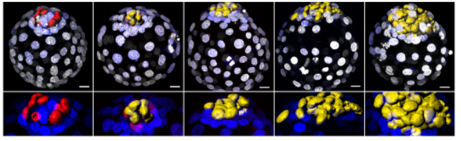
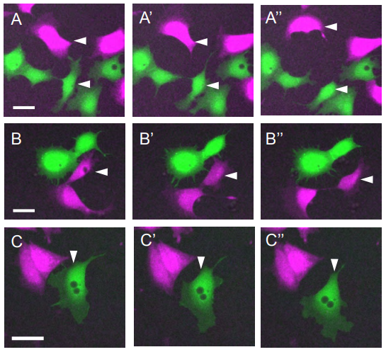
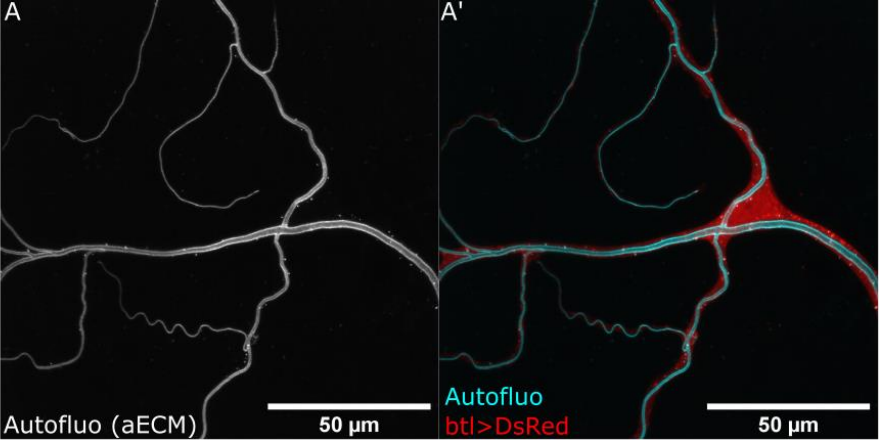
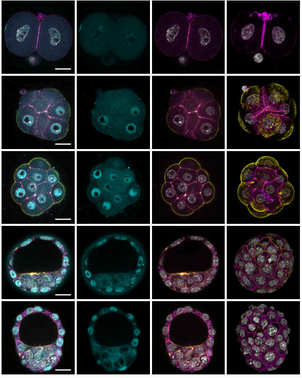
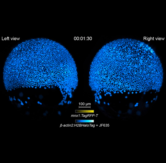
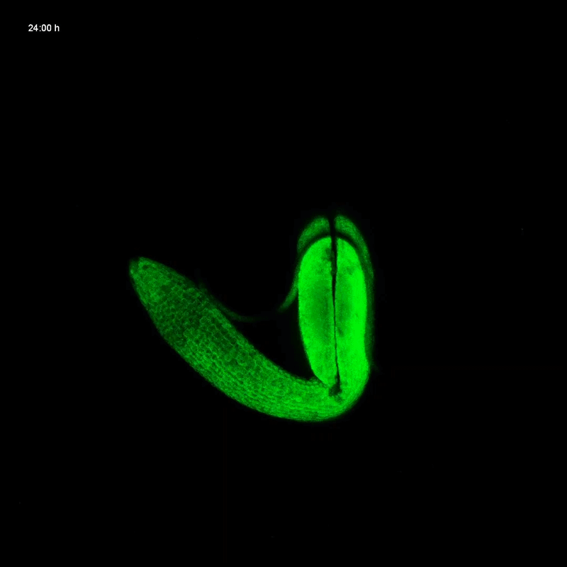
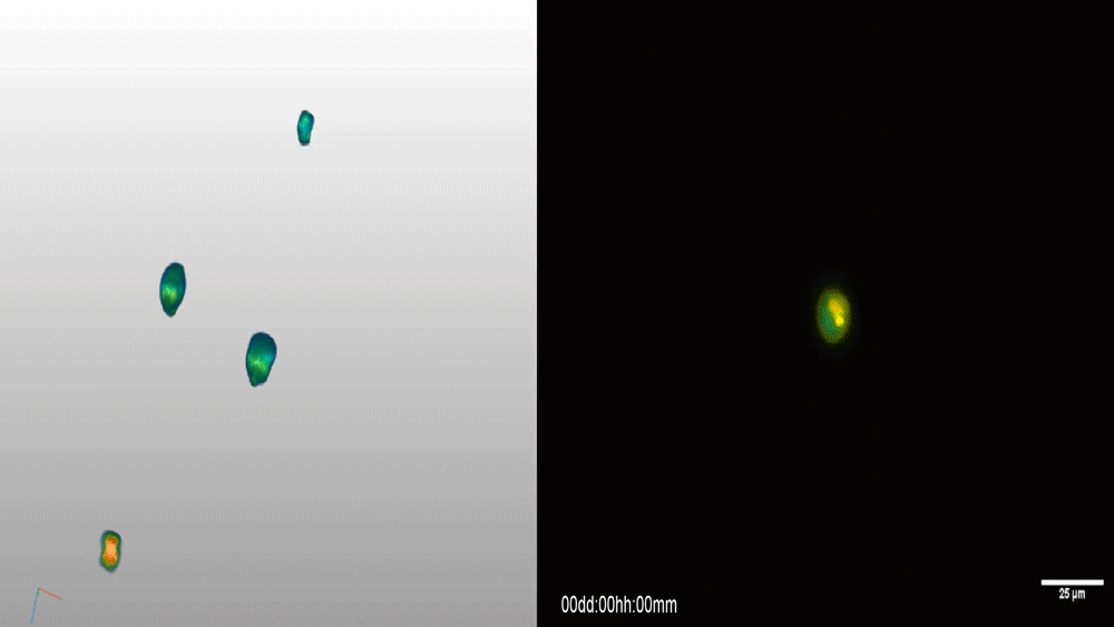
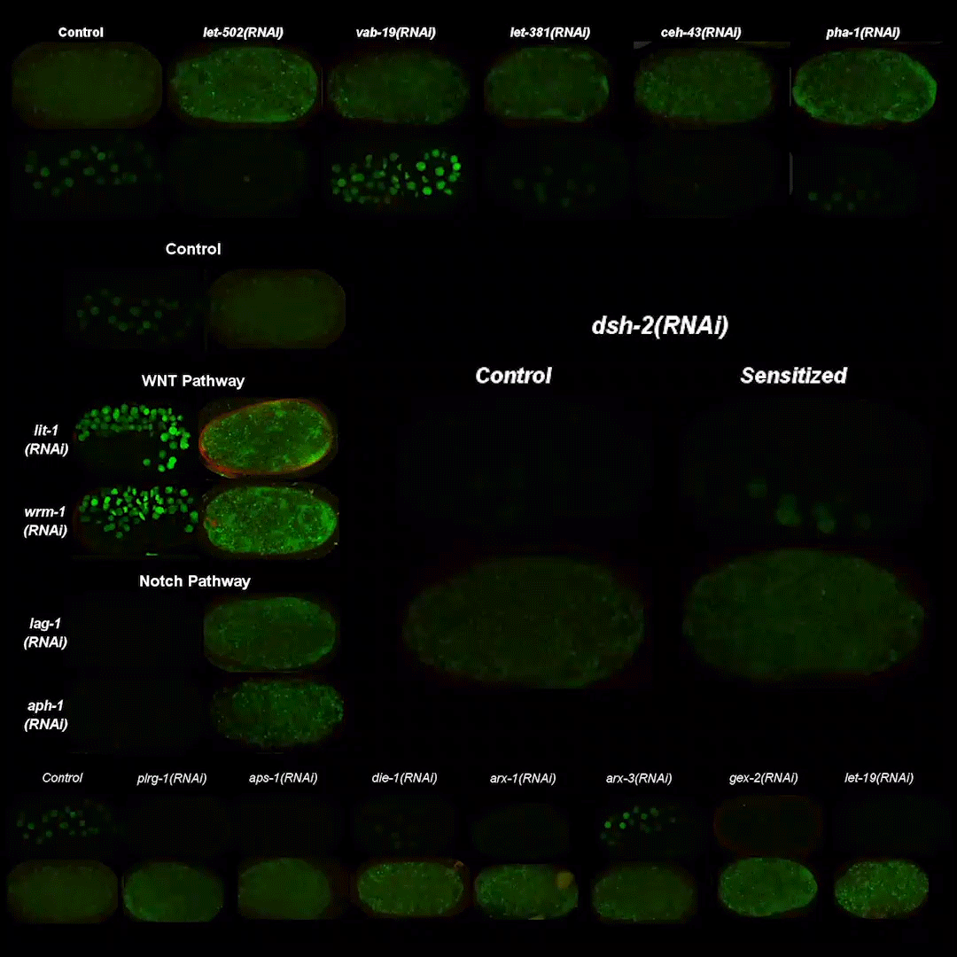
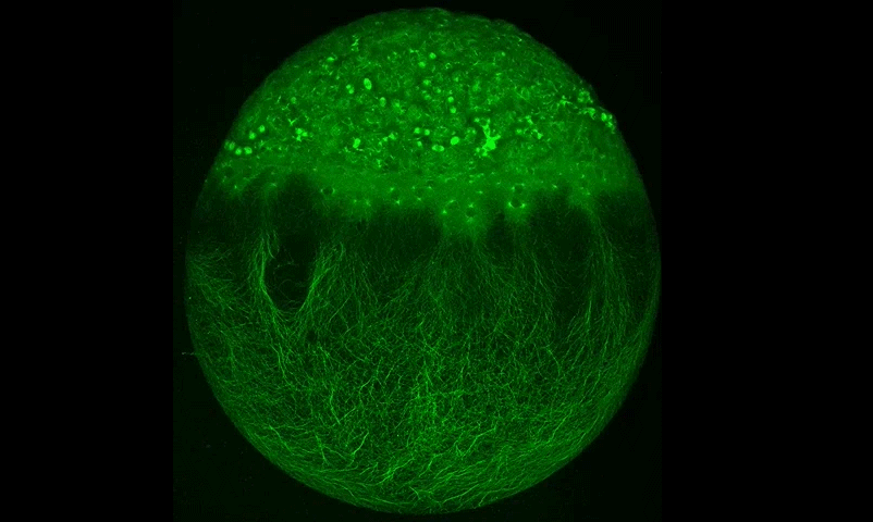
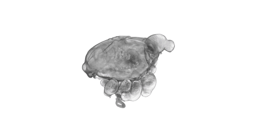
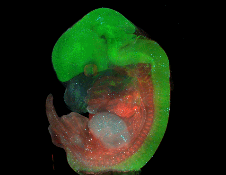
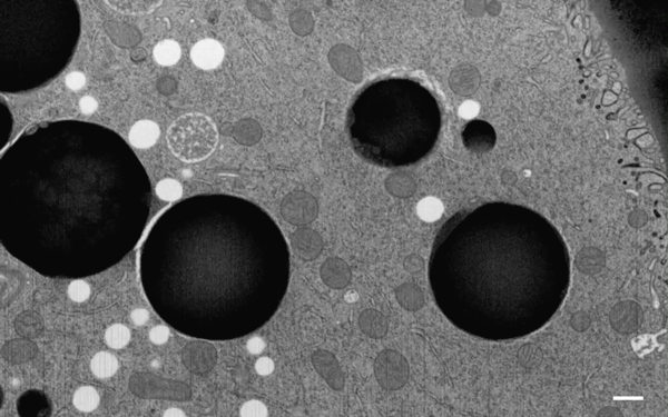
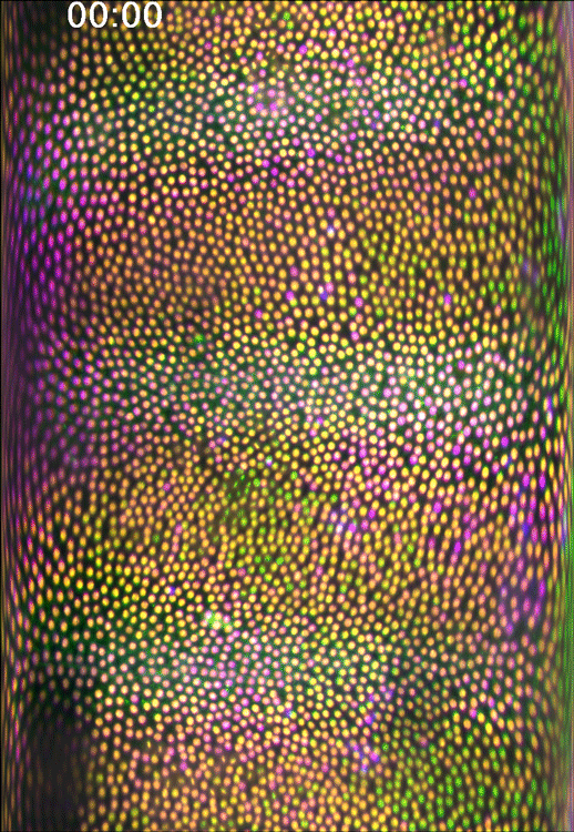
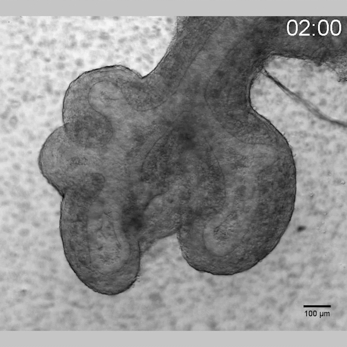
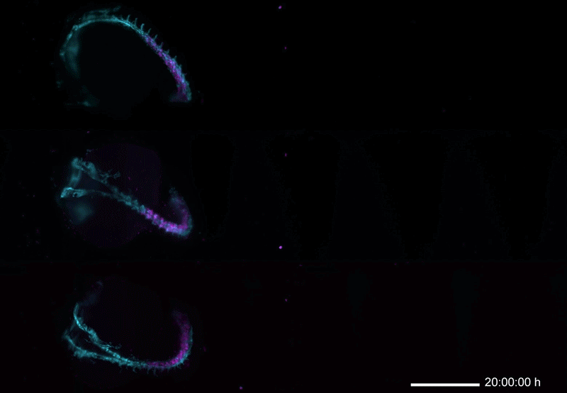
In keeping with a time-honoured tradition, we recently flooded Twitter with 12 beautiful developmental biology GIFs. They came from papers published this year and feature all kinds of systems and visual styles – here they are for posterity! Let us know your favourite in the comments
Whole-embryo developmental imaging of zebrafish spinal cord neurogenesis
Genetics Unzipped is delighted to host a short series of podcasts recorded at the 2019 Galton Institute symposium – New Light on Old Britons – which took place at the Royal Society in London at the end of October. Reporter Georgia Mills talks to some of the leading researchers uncovering the hidden stories of the people of the British Isles.
Who were these ancient Britons? Where did they come from and what were they like? What’s the real story behind the romantic myths about the Celts? And what can modern genetic and archaeological techniques tell us about their lives and loves?
Professor Ian Barnes and Dr Selina Brace, ancient DNA researchers at the Natural History Museum in London, discuss how their work on ancient DNA is shedding light on the British population from the Mesolithic to the Bronze Age.
Professor Sir Walter Bodmer FRS from the Weatherall Institute, Oxford, explains what we know so far about genetic structure and origins of populations of the British Isles.
The Celts are one of the most famous – and misunderstood – people who lived in ancient Britain. Professor Sir Barry Cunliffe CBE, FBA from the University of Oxford explores the myths and the reality.
 (No Ratings Yet)
 (1 votes)
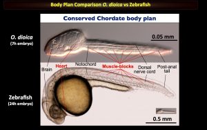


 Genetics Unzipped is delighted to host a short series of podcasts recorded at the 2019 Galton Institute symposium – New Light on Old Britons – which took place at the Royal Society in London at the end of October. Reporter Georgia Mills talks to some of the leading researchers uncovering the hidden stories of the people of the British Isles.