One scientific story has dominated the news this week: the first report of CRISPR-edited human babies being born. The story’s scientific and ethical aspects stirred up heated debate, as did its means of delivery: rather than a published paper, the story broke with reports of clinical trial documents and then a YouTube video from lead researcher He Jiankui (from the Southern University of Science and Technology in Shenzhen), all on the eve of a conference he was due to speak at (and whose organisers were seemingly unaware what we was going to speak about).
In an associated Node post, we asked developmental and reproductive biologists to give their reaction to the story (and we’d love to hear yours too), but here we’ve collated a bunch of hopefully helpful links, and some recent Development commentaries on the issues surrounding gene editing in humans.
The story breaks
On 25 November, Antonio Regaldo in MIT Technology Review reported details of the study’s clinical trial data
The He Lab YouTube channel released this video on the same day (the channel also has four associated videos about the work)
“A surgery that could save a child from a lethal genetic disease like cystic fibrosis or from a life-threatening infection like HIV doesn’t just give that little boy or girl an equal chance at a healthy life. We heal a whole family”
Statement from He’s employer, the Southern University of Science and Technology, stating that the university knew nothing about He’s work and plans to set up an independent committee to investigate (in Chinese – translates page reads quite clearly; 26/11)
Gaetan Burigo gave a helpful thread particularly regarding the science presented by He in the summit (28/11)
OK there are a lot to unravel in this He Jankui talk and panel discussion at the #GeneEditSummit on #CRISPR in human embryos leading to the birth of the twins Lulu and Nana. -> Let's analyze the science, the ethics and controversies -> thread pic.twitter.com/3qOCC6IhvJ
— Dr Gaetan Burgio @gaetan_burgio@mstdn.science (@GaetanBurgio) November 28, 2018
Peter Mills, Assistant Director of the Nuffield Council on Bioethics, gives his thoughts (28/11)
Here at Development we’ve been thinking about issues of human gene editing for some time, and have commissioned content specifically exploring scientific and ethical aspects. We recently published two Spotlight articles on the theme (published in 2017 and 2018 respectively, before the current story broke).
Stem cells are already being used in combating previously untreatable diseases. Nevertheless, stem cells are not delivering their full potential because the production of specific cell types from stem cells cannot yet be managed. Researchers have now discovered the signals that determine the fate of immature cells in the pancreas. The research shows that they are very mobile and that their destiny is strongly influenced by their immediate environment. This breakthrough will facilitate the manufacturing of pancreatic islet cells for combating type 1 diabetes.
We are rapidly approaching the era for safe mass production of specialized neuronal cell types and insulin-producing beta cells. It will then be possible to test whether transplanting such cells will enable paralysed people to walk again or people with type 1 diabetes to restart their own production of insulin. Until now, the engineering of the specialized cells from pluripotent stem cells has largely been based on empirical knowledge of what works. Results published in the prominent journal Nature by a Danish-led research project represent a major leap forward.
“We have now been able to map the signal that determines whether pancreatic progenitor cells will become endocrine, such as insulin-producing beta cells or duct cells. The cells are analogous to pinballs, whose ultimate score is based on the sum of pin encounters. They are constantly moving around within the developing pancreas, leading to frequent environmental changes. We show that the exposure to specific extracellular matrix components determines the ultimate destiny of the cells,” explains Henrik Semb, Professor and Executive Director, Novo Nordisk Foundation Center for Stem Cell Biology, DanStem, University of Copenhagen.
The matrix determines the destiny
Progenitor cells are similar to stem cells since they can both self-renew and differentiate into mature cell types. However, their self-renewal capacity is generally limited compared with that of stem cells. The dynamic behaviour of progenitors during organ formation makes them difficult to study. By seeding individual human stem cell–derived progenitors on micropatterned glass slides, the researchers could study how each progenitor, without the influence of neighbouring cells, reacts to its surroundings.
“This enabled us to discover something very surprising. Our investigation revealed that interactions with different extracellular matrix components change the mechanical force state within the progenitor. These forces result from interactions between the extracellular matrix, which is outside the cell, and the actin cytoskeleton, which is within the cell.”
Pancreatic endocrine cells include all hormone-producing cells, such as insulin-producing beta cells and glucagon-producing alpha cells, within the islet of Langerhans, whereas the duct cells are epithelial cells that line the ducts of the pancreas.
“The experiments show that exposure to the extracellular matrix laminin instructs the progenitor cells towards an endocrine fate by reducing mechanical forces within the cells. Whereas exposure to fibronectin results in a duct fate because of increased mechanical forces.”
Mechanism facilitates exploitation
To exploit their discovery, the researchers had to understand the signalling pathway. They showed that components in the extracellular matrix trigger a signal into the cell via an integrin receptor, resulting in changes in mechanical forces transmitted through the actin cytoskeleton. The yes-associated protein (YAP) then senses these forces to turn on and off specific genes.
“This cascade determines the ultimate fate of the progenitor cell. Perhaps the most astonishing achievement is that our data answer an enigma that has puzzled the field for decades. How some progenitors mature into duct cells, whereas others become endocrine cells via Notch signals.”
The researcher show that the seemingly stochastic regulation of Notch function is in fact mediated by the progenitor’s encounters with extracellular matrix interactions via the force-sensing gene regulator protein YAP. They were even able to validate the physiological relevance in vivo during pancreas development.
“We can now replace significant numbers of empirically derived substances, whose mode of action in current state-of-the-art differentiation protocols is largely unknown, with small molecule inhibitors that target specific components of the newly identified mechanosignalling pathway.”
With this new strategy, insulin-producing beta cells can now be more cost-effectively and robustly produced from human pluripotent stem cells for future treatments against diabetes.
“Our discovery breaks new ground because it explains how multipotent progenitor cells mature into different cell types during organ formation. It also gives us the tools to recreate the processes in the laboratory, to more precisely engineer cells that are lost or damaged in severe diseases, such as type 1 diabetes and neurodegenerative diseases, for future cell replacement therapies.”
”Mechanosignaling via integrins directs pancreatic progenitor fate decisions” has been published in Nature. Henrik Semb, Professor and Executive Director, Novo Nordisk Foundation Center for Stem Cell Biology, DanStem, University of Copenhagen, and head of Institute of Translational Stem Cell Research at Helmholtz Zentrum München is last author. Drs. Anant Mamidi, Assistant Professor, DanStem and Christy Prawiro DanStem share first authorship, and the work is the result of a collaboration with Professor Palle Serup’s group, DanStem.. The Novo Nordisk Foundation has awarded grants of almost DKK 700 million (€92 million) to the Center for research between 2010 and 2018.
Here at The Company of Biologists we’ve been debating the Bank of England’s decision to put a scientist on their new £50 note (the highest denomination note in England). The scientist must be deceased (only the Queen can grace notes while still alive) and ‘have shaped thought, innovation, leadership or values in the UK’.
Each of our five journals was asked to come up with their nominations for the face of the fifty. Here’s who they picked and why they picked them:
“McLaren was a towering figure in developmental and reproductive biology. She did foundational work in IVF, experimental chimeras and germ cell differentiation, contributed to regulatory policies on human embryo research, and championed pubic engagement”
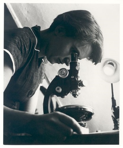
“She studied in Cambridge, and although a chemist, made a crucial, and often unrecognised, contribution to the discovery of the double helix structure of DNA”
“English fossil collector/palaeontologist. Considered an expert in her field, contributing to important changes in scientific thinking about prehistoric life, at a time when women were mostly excluded from the scientific community”
“Modern biology wouldn’t be what it is without him. Double Nobel winner known for sequencing DNA & pioneering work on the structure of proteins. Declined the offer of a knighthood, as did not wish to be addressed as Sir”
The Company of Biologists Twitter feed has a poll where you can pick your favourite out of the four:
What do you think of Development’s choice of Anne McLaren? Which other developmental biologist do you think could be honoured? Let us know in the comments
Regular meetings of scientists such as annual society conferences can create opportunities for scientists to engage the public without extensive effort, making connections between scientists and public audiences. Under the umbrella of a specific topic, events can be created to engage local communities with international researchers and foster forums for discussion of specific areas of research.
With this in mind, we created a space and a time for public engagement and a citizen’s approach to developmental biology in the recent Joint meeting of the Portuguese, Spanish and French Societies for Developmental Biology at Oporto, Portugal (http://devbiomeetingporto2018.pt/).
We invited local citizens through social media, the meeting webpage and local secondary school networks. And at the start of the meeting, which took please at the Almeida Garrett Library in Oporto, we organized an open science event for local Oporto high school students (mainly 16-17 year olds), their teachers and other members of the public.
It began with an informal conversation about what is developmental biology and why do we study it. This was done as a dialogue, with a backup of a few slides showing how embryos develop, some historical background and modern applications of the study of developmental biology. For this first part, we used some of the materials available at the BSDB as well as the Droso4schools and HHMI websites.
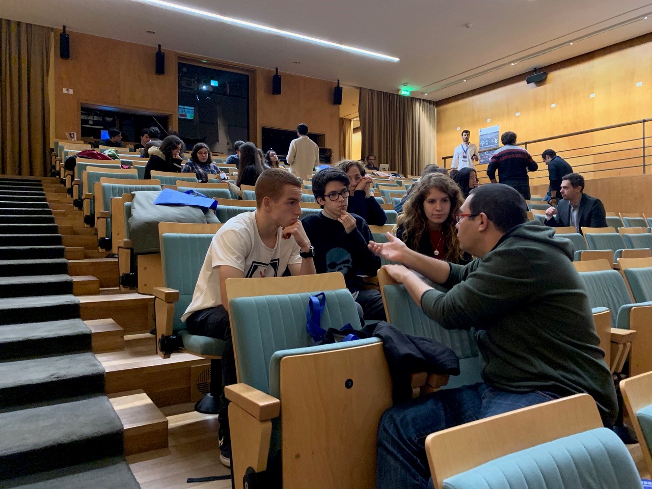
This was followed by an organized speed-dating with scientists with the help of 12 Portuguese researchers working in national institutions as well as abroad. These volunteers were asked in advance what was the main question they were trying to answer with their research, so they could start their informal conversations from this starting point. They were also asked to bring along an object related to their research as a communication “ice-breaker”. The format of the speed dating consisted of groups of three members of the public to one scientist, with seven and a half minute slots of time available. After this time, a new scientist would take the place of the previous one and the cycle would start again. We found this informal set up allowed for fluid dialogue between scientists and the invited citizens. In addition, the speed-dating format allowed for each person to have the opportunity to speak with 5 or 6 different researchers, all in about 1h. When asked for their opinion about the event, one of the teachers told us:
“As far as the activity with the scientists is concerned, the students liked it immensely. They told me that this type of interaction is much more interesting than just a conference.”
Scientific meetings can play a key role in building bridges between scientific research and public audiences. Let’s try to create more of these opportunities in many other scientific conferences.
Participants in the event:
Sofia J. Araújo, Leonor Saúde, Patrick Lemaire, João Amorim, Tomás Azevedo, Gil Carraco, Ana Gali, André Gonçalves, Sofia Moreira, Paulo Navarro-Costa, Pedro Rifes, Lígia Tavares
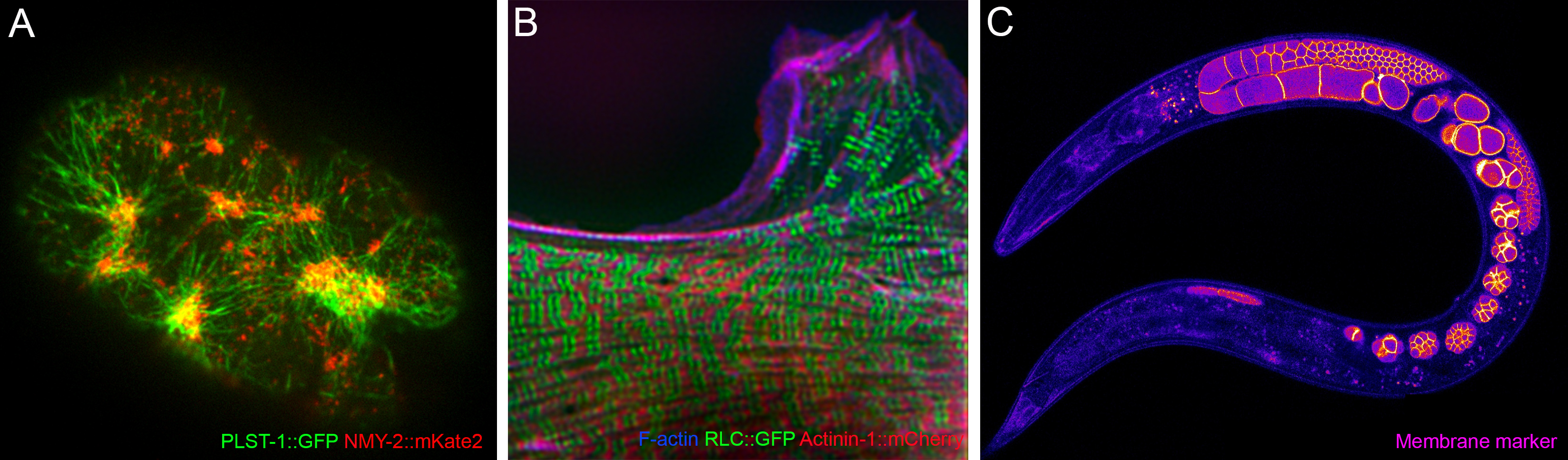
MSc/PhD and postdoc positions available in the Zaidel-Bar Cellular and Tissue Morphogenesis Lab.
We study the regulation of the cytoskeleton from single proteins to the entire organism and system levels, using multiple approaches (including bioinformatics, genetics, biochemistry and live imaging) to understand how cells and tissues change shape, move, sense, and generate forces (for more info: celladhesionlab.com).
For the last two years, our interview series ‘The people behind the papers‘ has showcased the faces of developmental biology, and we’re excited to announce that the series will now also be printed in Development.
The Company of Biologists’ journals – Development, Journal of Cell Science, Journal of Experimental Biology andDisease Models & Mechanisms – offer Travelling Fellowships of up to £2,500 to graduate students and post-doctoral researchers wishing to make collaborative visits to other laboratories. These are designed to offset the cost of travel and other expenses. There is no restriction on nationality.
They really are an amazing opportunity for ECRs to learn new things, meet new people and travel to new places.
The current round of Travelling Fellowships closes on 30 November (for travel >14 Jan 2019)
We are seeking highly motivated candidates to join us on a project beginning in 2019 on the molecular mechanisms of sensory neurogenesis. We study in particular the nociceptors, the specialized peripheral neurons that detect painful stimuli.
We aim to understand the role and mechanism of action of the Prdm12 gene. Prdm12 encodes an evolutionarily conserved epigenetic regulator of gene expression that has been found mutated in patients that suffer from a rare disease, Congenital Insensitivity to Pain, a dangerous condition that renders individuals completely unable to feel pain since their birth (Chen et al., Nat. Genet., 2015). To understand the molecular mechanisms that cause the painlessness, we are using the frog embryo and have generated Prdm12 null and conditional knock-out mouse models. We expect that the results of our work using these experimental systems as well as the identification of Prdm12 direct targets and interacting partners will help in the development of new strategies for treating pain.
The positions are funded by the Walloon government within the frame of the “Win2wal” program. They are open from 2 to 4 years and starting dates are flexible. The research will be performed in the laboratory of Developmental Genetics (Dr. Eric Bellefroid, http://gendev.ulb.ac.be/bellefroidlab/) that is part of the University of Brussels (ULB) Neuroscience Institute (UNI), the ULB Structural Biology and Biophysic laboratory (Dr. Abel Garcia-Pino, https://www.cm2ulb.be/) and the laboratory of Neuroscience (Dr. Laurence Ris, https://sharepoint1.umons.ac.be/FR/universite/facultes/fmp/services/neurosci/Pages/Equipe.aspx) at the University of Mons.
Preference will be given to applicants with a background in one of the following: mouse genetics, electrophysiology, cell and molecular biology and genome wide approaches (ChIP-seq,…).
Interested candidates should send a letter of motivation (before february 2019) describing past research experiences and full CV to:
Eric Bellefroid (ebellefr@ulb.ac.be), Laurence Ris (Laurence.RIS@umons.ac.be) or Abel Garcia Pino (agarciap@ulb.ac.be) together with the name and e-mail address of 2 references.
Selected related publications:
Thelie et al., (2015). Prdm12 specifies V1 interneurons through cross-repressive interactions with Dbx1 and Nkx6 genes in Xenopus. Development, 142(19), 3416-3428.
Nagy et al., (2015). The evolutionarily conserved transcription factor PRDM12 controls sensory neuron development and pain perception. Cell Cycle, 14(12), 1799-1808.
Written and illustrated by: Bjørt K. Kragesteen, Malte Spielmann, and Guillaume Andrey.
In early development, the forelimb and hindlimb buds of tetrapods are morphologically uniform. However, as limb development proceeds, each individual tissue attains a characteristic morphology that ultimately defines the identity of a forelimb (arm) or a hindlimb (leg). How do undifferentiated limbs bend their morphogenetic trajectories into arm and legs, since they are patterned by similar developmental processes? It is commonly accepted that the differential activities of a handful of genes instruct the formation of either arms or legs; yet the mechanism leading to their differential regulation in fore- and hindlimb buds was unknown. In our recent study, we dissected in detail such regulatory mechanisms and have revealed, once again, how important the function of the non-coding regulatory genome is, representing the overwhelming majority of vertebrate genomes.
In Stefan Mundlos’ research group at the Max Planck Institute for Molecular Genetics in Berlin, the focus is on the lessons that human limb malformations can teach us about gene regulation. Most research projects start with detecting the genomic mutation in human patients with congenital limb malformations at the Charité Berlin university hospital. These limb malformations are frequently caused by large changes in the DNA called structural variants including: deletions (removing information), inversions (displacing information), and duplications (doubling and misplacing information). One can imagine that such mutations might mess up the genetic information, and by detecting them in patient genomes, structural variants can provide a clue as to where important non-coding regulatory elements are located. From this starting point, it is possible to look for neighbouring genes that become misregulated following the mutation, ultimately changing the developmental program and the formation of the embryo.
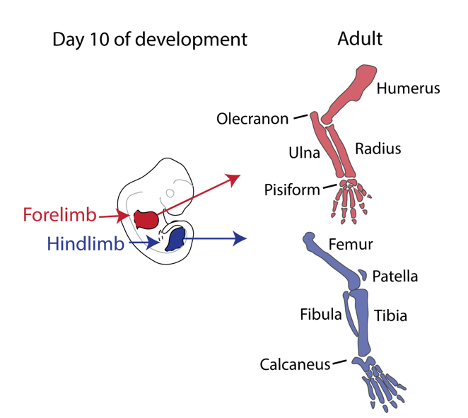
Our story started back in 2010 when Malte Spielmann, a clinical scientist interested in structural variants and congenital disease, tried to work out the genetic cause in three families with a very rare malformation syndrome affecting the arms of the patients, known as Liebenberg syndrome (named after the doctor that first described it back in 1973). When he looked at the X-rays of the patients upside down, it struck him that the arms looked very much like legs! If we think about our arms and legs, they actually share many similarities: both are formed by one long bone (upper arm/thigh: the humerus/femur), followed by two bones (lower arm/leg: ulna/fibula and radius/tibia), terminating in many small bones (hands/feet: carpals, metacarpals, and phalanges) (see Figure 1).
Figure 1. Limb development: from buds to arms and legs. Arms and legs are serial homologs; notice their similarities as well as their specialised joints that enable specific functions.
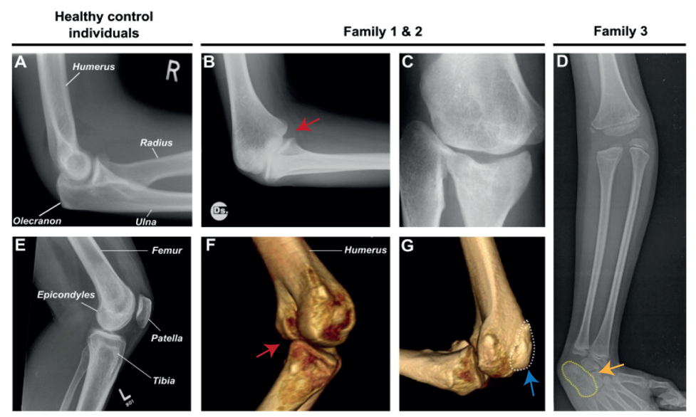
Nevertheless, they flex in opposite directions, as the arms have a specialised elbow joint, where the tip of the ulna called the olecranon (the pointy elbow) grabs around the distal end of the humerus. The legs have a specialised knee joint with a small bone, the patella (knee cap), ensuring stability and enabling weight bearing. However, the Liebenberg syndrome 3D CT scan showed the following: the fingers were very short (brachydactyly) looking very much like toes; the small bones of the wrist (pisiform, triquetral) were fused together looking like the heel bone (calcaneus), and the olecranon was reduced and no longer grabbing around the end of the humerus. Instead, the end of the humerus was broader as a patella like bone appeared fused to it (Figure 2).
Figure 2. Liebenberg syndrome: partial arm-to-leg transformation. A and E show healthy arms and legs, respectively. The rest show x-ray and CT-scan of patients with Liebenberg syndrome. Red arrow: elbow joint resembling a knee joint. Blue arrow: patella like bone fused on humerus. Yellow arrow: wrist bones fused resembling the calcaneus (heel bone).
How on earth could this happen? Malte performed DNA analysis (array-CGH) and found a large deletion (107 kb) on chromosome 5 in all affected individuals. The deletions remove the gene H2AFY. However, this gene is a housekeeping gene and in mice in which the gene had been inactivated do not show any limb phenotype. Malte then looked on each side of the gene and found two interesting things:
On the centromeric side lies a gene named PITX1, which is expressed exclusively in the hindlimb during limb development and is known to be the only transcription factor that patterns the tissue. Inactivation of Pitx1 in mice results in reduction of leg morphology, such as loss of the patella and reduction of the calcaneus and the knee joint changing into an elbow-like joint. On the contrary, misexpression of Pitx1 in mouse forelimbs partially transforms the elbow joint into a knee like joint. Thus, Pitx1 misregulation in the forelimb seemed like a good candidate to explain to Liebenberg phenotype. However, Pitx1 regulation in tetrapods was unknown. Why would a deletion 200 kb upstream of the PITX1 promoter cause Liebenberg syndrome?
Interestingly, on the telomeric side of the deletion, 300 kb upstream of PITX1, a non-coding enhancer element can be found, called Pen (pan limb enhancer) that is active in both forelimbs and hindlimbs. Enhancer elements are defined as sequence specific stretches of DNA that are bound by transcription factors that dictate the activity and ensure communication with the correct gene promoters through chromatin folding. Often several enhancer elements regulate a target promoter and the collective activity of the tissue specific enhancer reflects the promoter transcriptional output. Recent development of proximity-ligation chromatin conformation capture technology (e.g. genome wide Hi-C) has demonstrated that the genome is partitioned into topological associating domains (TADs). TADs are scaffolds of preferential interactions between cognate promoters and enhancers and thus protect them from promiscuous activity from neighbouring TADs by boundary elements. These TADs were said to be stable across cell types and evolutionary conserved. With this in mind, the following hypothesis was formulated:
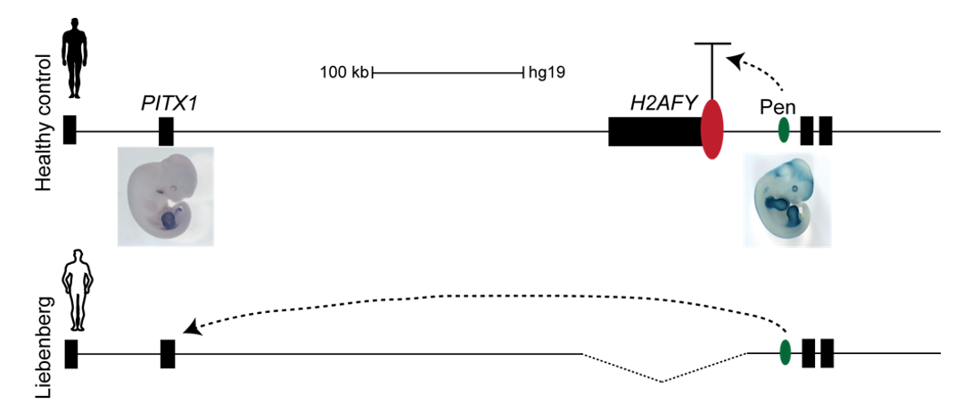
The Liebenberg deletion removes a TAD boundary element that normally separates PITX1 and Pen, resulting in PITX1 adopting a foreign enhancer, i.e. Pen, that is active in both fore- and hindlimbs. This enhancer adoption thus results in abnormal activation of PITX1 in the forelimb, where it should never be expressed, altering the patterning of the forelimb into a hindlimb, and partially transforming the arms into legs (Figure 3).
Figure 3. PITX1 locus in humans: Liebenberg deletion misplaces Pen enhancer. In control humans, PITX1 (expressed in hindlimb only) and Pen enhancer (active in fore- and hindlimbs) are 350 kb apart. In Liebenberg patients, large deletions bring Pen enhancer closer to PITX1 misexpressing the gene in forelimbs.
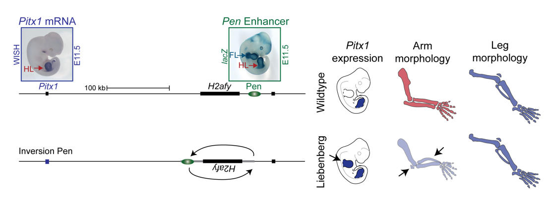
To test the hypothesis, Bjørt Kragesteen, a PhD student in the lab interested in deciphering non-coding functionality, sought to discern the molecular pathomechanism of Liebenberg syndrome together with Malte. First Bjørt created mouse mutants using CRISPR-Cas9 engineering that was newly established in the lab (back in 2013). She used two gRNAs to generate Liebenberg-like deletions and inversions at the mouse Pitx1 locus. Excitingly, analysis of Pitx1 mRNA expression in the early mutant mouse embryos showed Pitx1 misexpression in forelimbs! Skeletal analysis of adult mice showed a Liebenberg-like phenotype where the olecranon was reduced, an ectopic bone (patella-like) appeared at the humerus and the rotation of the arm was similar to the legs (Figure 4). Amazing. Case solved.
Figure 4. Pitx1 locus in mice: CRISPR engineered mice show partial arm-to-leg transformation. A 113 kb inversion misplacing Pen closer to Pitx1 resulted in its misexpression in arms and consequent partial arm-to-leg transformation with bowing of the radius and appearance of a patella-like bone (arrows).
However, many questions remained unanswered. Why is Pen, a strong fore-and hindlimb enhancer, located relatively close to Pitx1? Isn’t that too risky for a key hindlimb patterning gene?
In parallel to Bjørt’s project, Guillaume Andrey, who started his postdoc in the laboratory in early 2014, was investigating the normal regulation of Pitx1, by combining deletions and inversions of its putative regulatory landscape and enhancer assays. He observed that several engineered structural variants, which did not alter the pre-supposed boundary between Pitx1 and Pen, resulted in a mirror image pattern between fore- and hindlimb, identical to the one of the Pen activity and a Liebenberg phenotype. This observation suggested that Pitx1 could be controlled by the Pen element even when the “boundary” was intact. From that point, it became evident that in hindlimbs Pen could play a role in the regulation of Pitx1.
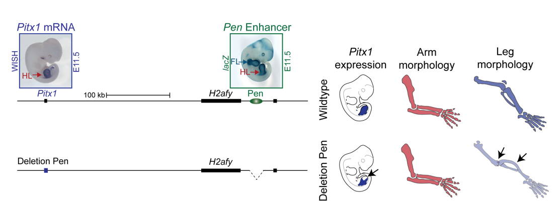
We thus next deleted the Pen enhancer to see what would happen. A visit to the animal facility some weeks later revealed that some of the adult homozygous Pen deletion mice developed club feet, dragging their hindlimbs behind. Both mouse and human patients haploinsufficient for Pitx1 develop clubfeet. Moreover, the mutants showed reduced Pitx1 expression and skeletal abnormalities whereby the patella was missing (Figure 5). This was solid evidence that Pen indeed is a Pitx1 enhancer!
Figure 5. Deletion of Pen results in reduction of Pitx1 expression and loss of hindlimb morphology. CRISPR engineered deletion removing Pen enhancer results in abnormal rotation and articulation of the knee joint and loss of the patella.
With this exciting finding we identified something not hereto described: A gene can be regulated by an unspecific long range enhancer; however, changing its location in the 2D regulatory landscape results in misregulation of the target gene and can transform tissue morphologies. We continued the search for a hindlimb specific Pitx1 enhancer, but no matter how much cloning and testing of enhancer elements we did, no hindlimb specific enhancer could be found. We thus decided to join forces, merging the projects about the normal and Liebenberg regulation of Pitx1, in order to understand how a gene that is solely expressed in the hindlimb can be controlled by unspecific enhancer elements active in both forelimbs and hindlimbs.
The research question thus became: what prevents Pitx1 expression in forelimbs in wildtype animals if its enhancer is active in both pairs of limbs?
The next obvious layer of gene regulation to scrutinise was the chromatin folding at the Pitx1 locus in limbs. We first employed circular chromatin conformation capture (4C) technology in forelimbs vs hindlimbs to detect which regions surrounding Pitx1 were in close proximity to the gene. But, using the Pitx1 promoter region as a viewpoint, no differences were observed. However, Guillaume was using a slightly alternative 4C viewpoint in the gene body, and using the same library could see very strong differences between the tissues and clear Pitx1-Pen interactions in hindlimbs, indicating that the locus structure was highly defined. This was further confirmed using capture-C variation of the method using a larger fragment as a “bait” to see the interactions with the Pitx1 promoter region. Excitingly, differences between forelimbs vs hindlimbs emerged whereby Pitx1 showed differential interactions with Pen: in hindlimbs, Pen and Pitx1 come into close proximity enabling its tissue specific activation, while in forelimbs they are kept separated! Yet, this did not give the complete picture. At that point in time (2016) a new C-method, capture Hi-C, was developed and we established it in the lab. Here RNA probes are used to pull down a region of interest, and we enriched 3 megabases surrounding the Pitx1 locus. This provides a more complete picture of the interactions over the whole locus. Contrasting forelimbs and hindlimbs interaction heat-maps showed a clear difference: in hindlimbs Pitx1 forms loops with several regions (RA1, RA3 and Pen), which were almost completely diminished in forelimbs, but with a forelimb specific loop with the repressed gene Neurog1 occurs.
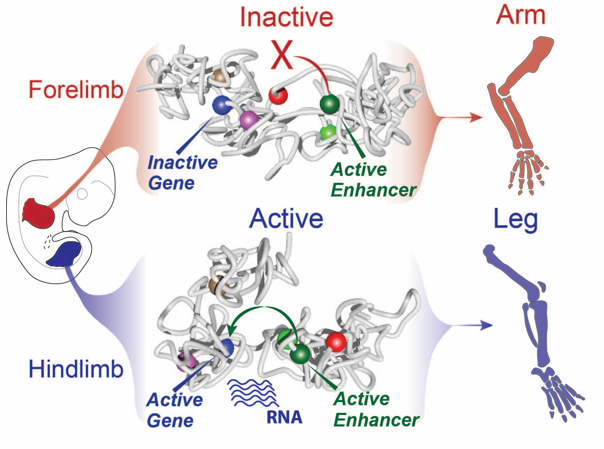
Still, this type of analysis gave us a 2D image of what was going on at the locus and we had a hard time imagining what this looks like in 3D, despite many hand drawn depictions. We thus initiated a collaboration with physicists in Italy in Mario Nicodemi’s research group. They used our capture Hi-C data and ran computational simulations using a so-called strings and binders model. They sent us the modelling results and 3D videos of forelimbs vs hindlimbs as well as an inversion mutant that shows the Liebenberg phenotype. The result was jaw-dropping: in wildtype forelimbs, the locus forms an inactive conformation whereby Pitx1 and Pen are kept a part, and where the repressed Pitx1 is embedded within its own domain next with repressed Neurog1 (neither of the genes are active in forelimbs and thus are covered with repressive epigenetic marks and hang out together). In wildtype hindlimbs, the locus folds in three domains and now Pitx1 is sitting at the surface of its domain directly facing Pen, thus enabling communication and robust transcription. Finally, in mutant forelimbs, bringing Pen closer to Pitx1 results in active folding of the whole locus, such as that in hindlimbs. Thus, not only do Pen and Pitx1 interact ectopically in mutant forelimbs, but the 3D-conformation becomes hindlimb-like, resulting in ectopic Pitx1 expression and arm-to-leg transformation.
Figure 6. Of arms and legs: dynamic chromatin folding of the Pitx1 locus ensures its correct regulation by Pen enhancer and normal morphogenesis. In forelimbs, Pitx1 and Pen are separated ensuring an inactive chromatin folding and absence of transcription ensuring normal development of arms. In hindlimbs, Pitx1 and Pen are facing each other resulting from an active folding of the locus leading to robust Pitx1 expression and normal leg development.
With all these data in hand and the genetic evidence that Pen was responsible when misplaced in the nuclear 3D space for the ectopic expression of Pitx1 in the “Liebenberg” forelimb, we could determine that the Pitx1 locus dynamic structure modulates the Pen activity in normal embryos. Specifically, in forelimb it represses the enhancer by keeping it away from Pitx1 promoter and that upon activation in hindlimb, it can participate in Pitx1 robust expression. Finally, it also showed that ectopic interaction between a gene and its own enhancer, following structural variant, in a process called gene endo-activation can cause gene misexpression and disease.
Read the full story:
Kragesteen B.K.*, Spielmann M.*, Paliou C., Heinrich V., Schöpflin R., Esposito A., Annunziatella C., Bianco S., Chiariello A.M., Jerković I., Harabula I., Guckelberger P., Pechstein M., Wittler L., Chan W.L., Franke M., Lupiáñez D.G., Kraft K., Timmermann B., Vingron M., Visel A., Nicodemi M., Mundlos S. and Andrey G.
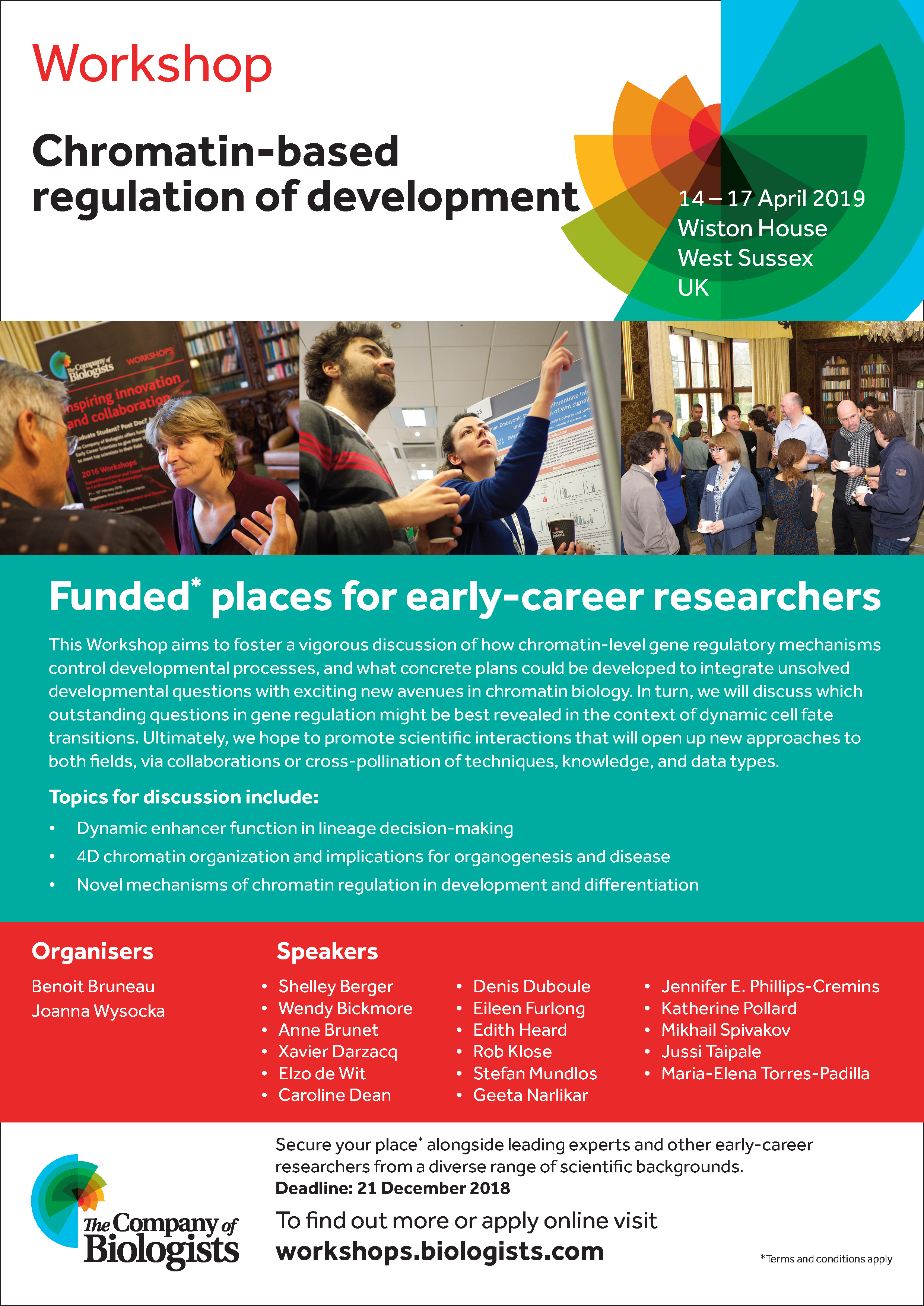
In April 2019, The Company of Biologists Workshop ‘Chromatin-based regulation of development‘ will be held in Wiston House, a 16th century Grade I listed building located at the foot of the South Downs in West Sussex.
Organised by Benoit Bruneau and Joanna Wysocka, the workshop will foster discussion of what mechanisms related to chromatin biology are informing developmental processes, and which outstanding questions in gene regulation might be best revealed in the context of dynamic cell fate transitions.
If all of this sound tantalising to you, there are around 10 funded places available to early career researchers. It’s an amazing opportunity for young researchers to participate in a relatively small (~30) group of world leading experts from across the field of gene regulation.
Deadline for applications is 21 December 2018.Find out more here:
 (2 votes)
 (No Ratings Yet)


