The community site for and by
developmental and stem cell biologists

The Kahneman Chronicles: Lessons from a Fly Lab

Posted by , on 27 September 2025


The Kahneman Chronicles #1: When a Nobel Laureate Fixed Our Lab’s Scheduling Disasters

Daniel Kahneman (1934-2024) was a legendary psychologist who revolutionized our understanding of human decision-making and became known as the “grandfather of behavioral economics.” Awarded the 2002 Nobel Prize in Economics, Kahneman’s groundbreaking research with Amos Tversky revealed systematic biases and mental shortcuts leading people to make irrational choices. 

This article series imagines what would transpire when Daniel Kahneman took a sabbatical and worked in a fly lab. 
Part of “The Kahneman Chronicles: Lessons from a Fly Lab” – A report from our imaginary interdisciplinary fellowship program


On the day Nobel Laureate Prof. Daniel Kahneman arrived for his sabbatical, our Drosophila lab buzzed with nervous excitement. Here was the legend himself—extraordinary psychologist who’d won economics’ highest prize, revolutionizing our understanding of errors in decision-making.

The ghost of Thomas Morgan urged us to do our best. We’d prepared our most impressive experiments, polished our presentations, and practiced our pitch for explaining fly development.

What we hadn’t prepared for was Kahneman spending his entire first morning silently observing us work. Often he scribbled notes in a small black notebook with the focused intensity of Jane Goodall studying chimps.

Why do we spend so much time in the lab?

“I’ll just quickly mount these embryos—twenty minutes, tops,” announced postdoc Shweta. This became a two-hour odyssey involving broken coverslips and dried glue. Followed by an existential crisis, wondering if the fluorescent blob she saw was signal or autofluorescence from a properly developing embryo.

“Quick PCR setup, maybe thirty minutes,” declared grad student Fillip, before vanishing into an afternoon-long quest. Missing primers. Buffer math. Finding the thermal cycler waited on “infinite hold” since previous Tuesday. You know the drill.

“Fascinating,” Kahneman murmured after each wildly inaccurate prediction.

By day three, a pattern was undeniable. Every time estimate in our lab was spectacularly yet consistently wrong. “Simple” tasks morphed into epic quests.

The Intervention

Kahneman approached the whiteboard where we’d sketched our weekly schedule – optimistically planning seventeen different experiments into forty work hours.

“Let’s implement realistic time budgeting,” he announced with the calm authority of someone who’d spent decades studying how humans delude themselves. Our simple thirty-minute embryo injections were now allocated one-hour blocks.

The room erupted in protests. “But we’ve done these injections hundreds of times!” “We know exactly how long they take!”

Kahneman smiled. “You’re all victims of the planning fallacy. Your System 1 is wildly optimistic about everything. Your mind accounts only for quick needle preparations while forgetting inevitable moments someone drops the cover slip itself”

“Your intuitive mind,” he explained, “only remembers the core task—actual injection. It conveniently forgets the setup, troubleshooting, inevitable equipment malfunction, and time spent staring at embryos wondering if they are worth injecting at all.”

The Planning Fallacy: The tendency to underestimate time, costs, and risks of future actions while overestimating their benefits. Even when people know similar tasks have taken longer than expected in past, they still predict future tasks will take less time.

System 1 vs. System 2 : Kahneman’s framework for two modes of thought. System 1 is fast, automatic, and intuitive (like quickly estimating “this should take twenty minutes”). System 2 is slow, deliberate, and logical (like carefully calculating each step: needle prep, embryo collection, injection setup, actual injection, cleanup, and imaging).

The Kahneman Method in Action

His solution was deceptively simple: multiply every time estimate by two, then add buffer time for “unknown unknowns.” “There are things you know will probably go wrong—known unknowns, like occasional broken needle or contaminated sample,” he explained.

“But then there are unknown unknowns—the completely unexpected problems you can’t even anticipate. The incubator that dies on a weekend, the new batch of reagent that behaves differently, or the day your hands just won’t stop shaking. You can’t plan for specific unknown unknowns, but you can acknowledge they exist.”

He made us track everything for two weeks: actual injection times, PCR setup duration…and the data was humbling. Our “standard” twenty-minute procedure had a median time of 40 minutes, with some taking over 1.5 hours when equipment misbehaved.

We tried his interventions skeptically. To our disbelief, the results were miraculous and maddening in equal measure.

For the first time in lab history, experiments actually finished when scheduled. Postdocs stopped working until midnight to complete “quick afternoon experiments.” Stress level plummeted as people stopped running late for their next commitment.

“Your emotional attachment to each experiment makes you treat it as special,” Kahneman explained. “You think ‘this time will be different’ or ‘I’m more prepared now.’ But from a statistical perspective, today’s PCR is just another data point in the distribution of ‘times PCR has taken in this lab.’
The planning fallacy tricks you into believing you can beat the historical average through wishful thinking.”

The lesson was profound: scientists are ultimately human and prone to same cognitive biases that affect everyone else. We bring these same mental shortcuts to our labs, our experimental designs, and our data interpretations. The first step toward better science maybe a more nuanced use of an important equipment—our own minds.

Have you experienced similar planning fallacies and overcome them? Do share in the comments.

What else did the Prof. Kahneman advise us on? Stay tuned for the next article in the series.

Practical Applications: The Kahneman Time Revolution

1. Track Reality First: Record actual times for routine procedures for couple of weeks.

2. Use the 1.5x Rule: Multiply routine task estimates by 1.5.

3. Use the 3x Rule: Triple your estimate for novel experiments.

4. Build Break Points: Schedule natural stopping points in long experiments, to allow buffers for unknown unknowns.

5. Try the Three-Point Method: For familiar tasks, estimate your best-case time (everything goes perfectly) and worst-case time (multiple things go wrong). Then calculate the geometric mean (root of the product) for a realistic schedule estimate.

Example: Embryo injection times (Best case 20 minutes, worst case 1 hours), geometric mean√(20 × 60) = √1200 ≈ 34 minutes.


    Sameer Thukral is a post doc in the lab of Yu-Chiun Wang at RIKEN-BDR, Kobe, Japan, where he loves discussing science in the healthy and respectful lab environment. He is a developmental biologist with a focus on mechanics of yolk-blastoderm interactions. He is also the co-founder of BDR-Launchpad, a post-doc network for supporting ECRs with the hidden curriculum of science.

    The observations made here are his own and do not reflect the opinions of the employer. This article was written by Sameer Thukral, with formatting, structuring and framing support of Claude AI.

    Thumbs up (8 votes)
    Loading...

    Categories: Discussion, Lab Life

    Navigate the archive

    Use our Advanced Search tool to search and filter posts by date, category, tags and authors.

    Remember her name?

    Posted by , on 26 September 2025

    I am an avid podcast listener, especially at the gym. Instead of fueling my reps with anger-fueled lyricism or upbeat songs that raise my bpm to 120, I noticed feeding my brain with podcasts is a better way of enduring my hour and a half workout. After all, what is a better way to feel incentivized to squat to 60 kg than to listen about how women have been overlooked in science? In the recent season of the So Cultured podcast, tears of anger fell down my face at the cruel injustices faced by Brenda Milner, Tu Youyou, and Marie Curie. Besides anger, I also felt inspired. Worried about my own academic journey, wondering if I am good enough, I found comfort in their stories.

    Conducting my own investigation, wondering whose story is not largely known, I bumped into Hilde Mangold (or Hilde Proescholdt at the time) and “The Organizer.” During her doctoral studies, she used two species of salamander with embryos that differ in pigmentation to perform 259 transplantations of the blastopore’s dorsal lip into the ventral region of a host gastrula1, 2. This experiments catapulted Spemann for the Nobel Prize. In several significant cases, she observed that a secondary axis with neural system developed in the host embryos 3. Spemann and Hilde Mangold therefore concluded that the embryonic region of the dorsal blastopore lip was able to induce embryonic development and called this specific embryonic region an “organizer.”4

    Hilde’s trajectory was impressive. At the age of 16, she attended the prestigious Gymnasium Ernestinum, at the time almost inaccessible to girls1. But like most young girls at the time, she was sent to a private institute for young ladies to learn proper housekeeping and social etiquette right after. But her curiosity and intelligence granted her a place at the University of Jena, which eventually led her to Spemann. She possessed a great deal of skill. Perhaps this dexterity to perform and master minuscule surgical operations could be attributed to the time she spent sewing in school. In fact, she had a keen eye for detail, documenting her implants with drawings in her lab notebook5, a skill that may be lost in the upcoming years with digitalization.

    Although it was Spemann’s quest started in 1903, with the production of identical twins from newt embryos using his daughter’s hair loop6. The completion of the pusruit to find the said “organizer,” couldn’t have been done without Hilde. She expanded Spemann’s techniques. Used thin glass needles, often heated, to cut certain parts from the embryos or to burn them away. She was critical in providing the empirical evidence needed, imagine your dissertation helps your professor win a Nobel. And aside from prizes, this experiments were monumental for the time, forging paths for theoretical and developmental biology, and cell to cell communication.

    Hilde’s story was short lived. Now I wonder what would’ve she further achieved is were not for dying at the young age of 26 from an accident. The best we can do is to continue living for them, and as other women at the time, to persist and lead by curiosity and resilience. Perhaps our time as graduate students are not as fruitful as Hilde’s, still, as students we understimate the work we do in the lab, and forget to advocate for our contributions. We troubleshoot for months, have failed results, or no results at all. So although we must perform the 200 experiments, we must celebrate and give some grace even if only 6 are significant. At the end, we are in fact, the sum of our parts.

    References

    1. VAN Robays, J. (2016). Hilde Mangold-Pröscholdt (1898 – 1924): The Spemann-Mangold Organizer. Facts, Views & Vision in ObGyn, 8(1), 63–68.
    2. De Robertis, E. M., Driever, W., & Mayor, R. (2024). Celebrating the centennial of the most famous experiment in embryology: Hilde Mangold, Hans Spemann and the organizer. Cells & Development, 178, 203921. https://doi.org/10.1016/j.cdev.2024.203921
    3. Kumar, V., Park, S., Lee, U., & Kim, J. (2021). The Organizer and Its Signaling in Embryonic Development. Journal of Developmental Biology, 9(4), 47. https://doi.org/10.3390/jdb9040047
    4. Spemann, H., & Mangold, H. (1924). Über Induktion von Embryonalanlagen durch Implantation artfremder Organisatoren. Archiv für Mikroskopische Anatomie und Entwicklungsmechanik, 100(3–4), 599–638. https://doi.org/10.1007/BF02108133
    5. Driever, W., Holzschuh, J., Sommer, L., Nitschke, R., Naumann, A., Elmer, J., & Giere, P. (2024). Hilde Mangold: Original microscope slides and records of the gastrula organizer experiments. Cells & Development, 178, 203909. https://doi.org/10.1016/j.cdev.2024.203909
    6. De Robertis, E. M. (2006). Spemann’s organizer and self-regulation in amphibian embryos. Nature Reviews Molecular Cell Biology, 7(4), 296–302. https://doi.org/10.1038/nrm1855
    7. Von Bubnoff, A. (2024). The Spemann-Mangold organizer discovery and society. Cells & Development, 178, 203906. https://doi.org/10.1016/j.cdev.2024.203906
    8. Brandt, C. (2022). Vitalism, Holism, and Metaphorical Dynamics of Hans Spemann’s “Organizer” in the Interwar Period. Journal of the History of Biology, 55(2), 285–320. https://doi.org/10.1007/s10739-022-09682-9

    Thumbs up (No Ratings Yet)
    Loading...

    Tags: , , ,
    Categories: Uncategorized

    Development presents… development across scales

    Posted by , on 26 September 2025


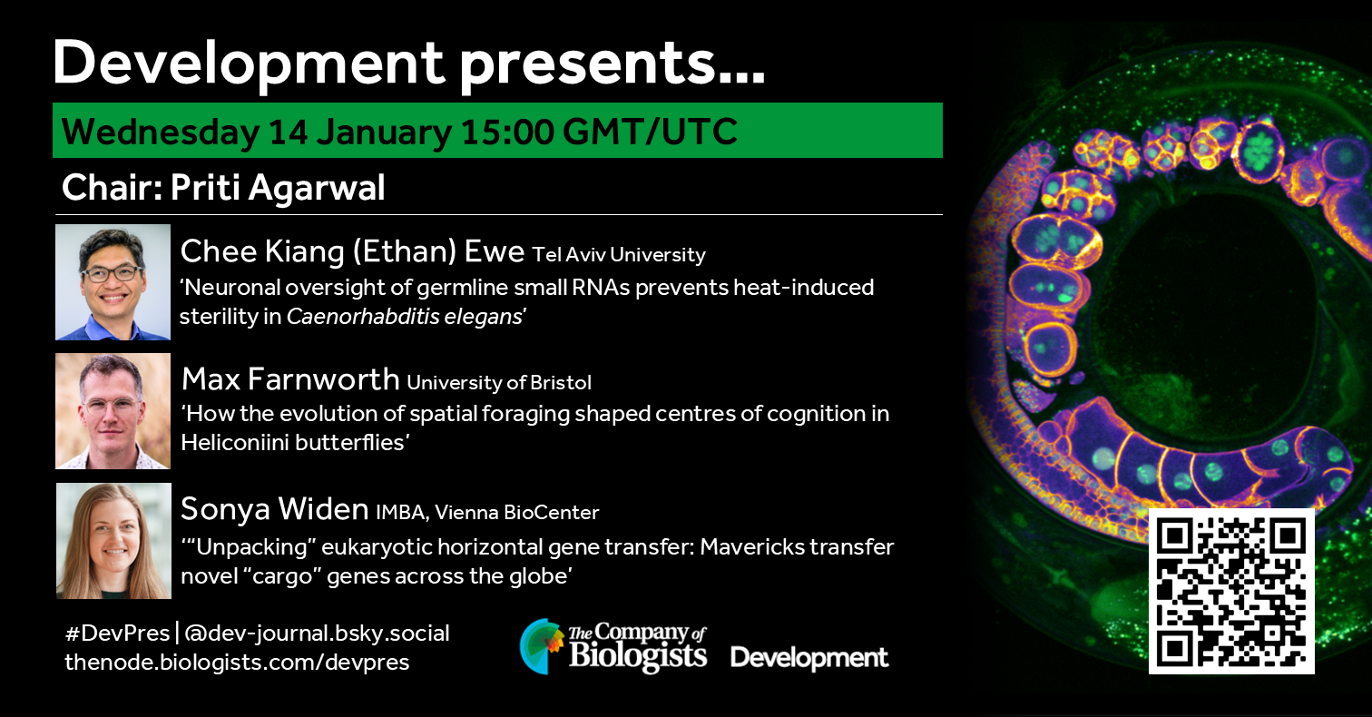
    Join us in mid-October to hear from two early-career researchers working on development across scales. Chaired by Development’s Executive Editor, Alex Eve.

    Wednesday 15 October – 09:00 BST (UTC+1)

    Osvaldo Contreras (Victor Chang Cardiac Research Institute and UNSW)
    ‘OpenEMMU: A low-cost EdU multiplexing methodology for studying DNA replication and cell cycle dynamics’

    Yinan Wan (Biozentrum University of Basel)
    ‘Whole-embryo spatial transcriptomics: from fate to form’

    At the speakers’ discretion, the webinar will be recorded to view on demand. To see the other webinars scheduled in our series, and to catch up on previous talks, please visit: thenode.biologists.com/devpres

    Thumbs up (3 votes)
    Loading...

    Tags: , , ,
    Categories: Development presents...

    The Invisible Cost: How Power Dynamics May Undermine Respect in Academic Labs

    Posted by , on 24 September 2025

    I’m a big fan of podcasts, and one of my favorites is Tim Harford’s “Cautionary Tales.” It tells true stories about disasters and what we can learn from them. One episode particularly stuck with me—the story of Tenerife.

    On March 27, 1977, two Boeing 747s collided on a foggy runway in Tenerife, killing 583 people. This wasn’t just about miscommunication, mechanical failure or bad weather. The investigation revealed something more profound: the captain was the airline’s Chief Flying Instructor, thus creating a steep “gradient” that prevented his first officer from challenging a fatal mistake.

    When the first officer realized they didn’t have takeoff clearance, he saw disaster coming but couldn’t bring himself to forcefully challenge his superior.

    This got me thinking: when power dynamics prevent people from speaking up, disaster follows. Does this also apply to academia?

    A Pattern Worth Noticing

    Tim Harford’s podcast reveals a disturbing pattern: many disasters across different fields stem from the same problem—people being unable to challenge authority when they see danger ahead. From naval catastrophes to medical errors, from financial crashes to engineering failures, a common thread is often authority gradients that silence dissenting voices.

    To be clear, most academic labs aren’t disaster zones. Most PIs, including my own, are thoughtful mentors who genuinely care about their students’ growth and scientific development. Many labs operate with healthy dynamics where ideas flow freely and disagreement is welcomed.

    But here’s a learning from other fields: even well-intentioned leaders can unknowingly create subtle power imbalances. And in science, our “disasters” aren’t plane crashes—they’re missed discoveries, delayed projects, unexplored hypotheses, and brilliant ideas that never see daylight.

    The Academic Context

    In academia, unlike most corporate environments, one person—your PI—has enormous influence over your career trajectory. As a PhD student or Post-doc, you commit years to one supervisor’s lab. They guide your research direction, allow your access to resources, and significantly influence your future opportunities.

    This isn’t inherently problematic. Expertise matters, and experienced scientists rightfully guide newcomers. The challenge is when this necessary hierarchy inadvertently creates barriers to open scientific dialogue.

    Even in the best labs, there might be subtle versions of this dynamic. A student hesitating to present data that contradicts the PI’s hypothesis. A postdoc avoiding questions that might seem to challenge established lab protocols. These aren’t dramatic confrontations—they’re quiet moments where respect for authority might overshadow respect for scientific inquiry.

    The Free Resource We Maybe Missing

    Of all the things science needs—expensive equipment, ample funding, and reagents—respect, costs nothing. Yet, it might be our most powerful tool. Every carefully planned experiment and every piece of expensive equipment depends on people thriving in an environment where they feel safe, heard, and valued.

    Science thrives on disagreement. The best discoveries often come from questioning prevailing wisdom and challenging assumptions. But when subtle power dynamics make people hesitate to speak up, we miss out on breakthrough ideas.

    The most productive labs may be doing something simple: they separate intellectual discussion from hierarchy. In these labs, everyone responds to contradictory data with curiosity, not defensiveness. Unexpected results are seen as learning opportunities, not failures.

    A Quick Self-Check for the Lab

    As an opportunity for reflection, PIs and mentees can ask themselves:How often do mentees feel comfortable disagreeing with an idea? If it’s rare, it may be worth examining why. Perhaps even create a “disagreement board” to make the act of questioning a hypothesis more salient and celebrated. What’s the atmosphere like when someone presents data that contradicts an expectation? Do people feel comfortable sharing results that go against the grain?

    These aren’t accusations; they’re simply opportunities for growth and improvement. The goal isn’t to flatten hierarchies but to ensure that authority serves discovery, not ego. Sometimes, the most junior person in the lab has a game-changing insight. But they can only share it if they feel safe to do so.

    The bottom line

    Listening to cautionary tales from other fields reminded me that power dynamics are everywhere, often subtle, and worth examining. In science, where truth-seeking is our highest goal, creating space for respectful disagreement isn’t just good mentorship—it’s essential for discovery.

    Sameer Thukral is a post doc in the lab of Yu-Chiun Wang at RIKEN-BDR, Kobe, Japan, where he loves discussing science in a healthy and respectful environment. He is developmental biologist with a focus on mechanics of yolk-blastoderm interactions. He is also the co-founder of BDR-Launchpad, a post-doc network for supporting ECRs with the hidden curriculum of science.

    The observations made here are his own and do not reflect the opinions of the employer. This article was written by Sameer Thukral, with formatting, structuring and framing support of Claude AI.

    Thumbs up (2 votes)
    Loading...

    Categories: Discussion, Lab Life

    Featured resource: Facebase

    Posted by , on 24 September 2025

    Our ‘Featured resource’ series aims to shine a light on the resources that support our research – the unsung heroes of the science world. In this post, we learn about the data and functionalities available at Facebase, and hear about new initiatives they are developing.

    What is FaceBase?

    FaceBase is a public data resource and repository dedicated to advancing basic and clinical research spanning the translational spectrum of dental, oral, and craniofacial (DOC) biology, as well as related systemic health and disease models throughout the data lifecycle. FaceBase realizes this mission by recruiting, transforming, and publicly sharing research and clinical data. 

    This freely available and public resource currently hosts over 1,100 datasets, approximately 3,000 experiments, over 210,000 images, and more than 8,000 genomics files. FaceBase exemplifies FAIR (Findability, Accessibility, Interoperability and Reusability) and TRUST (Transparency, Responsibility, User focused, Sustainability, and Technology) principles of scientific data sharing, ensuring that its clean, well-structured datasets are not only easy to find and reuse, but are also inherently AI-ready for integration into modern computational workflows.

    FaceBase hosts data from both human subjects and animal models, encompassing a wide array of experimental approaches, including multiple omics and imaging data types. This platform welcomes contributions of data from the community after going through a careful review process and quality assurance.

    • 1,170 datasets, 2,984 experiments, 210,000+ images, 8,000+ genomics files
    • Human subjects and animal model data (Current animal models include mouse, zebrafish, chimp and chick)
    • Controlled-access and public data
    • Genomic and phenotypic data from multiple species
    • Most known types of genomics and imaging data
    • Resources and strategies to enhance data reproducibility
    • State-of-the-art data science methods to support cutting edge research
    • Standards and educational resources for improving data management and sharing practices across the community

    FaceBase demonstrated itself as a credible resource for the DOC research community through its CoreTrustSeal accreditation after a two-year approval process, as well as becoming one of a select number of NIH approved Controlled Access Data Repositories (CADRs) handling genomics and other sensitive data.

    What inspired the development of FaceBase?

    In 2009, National Institute of Dental and Craniofacial Research, National Institutes of Health (NIDCR, NIH) launched FaceBase in response to the need for more comprehensive analysis of craniofacial development. With the immense amount of craniofacial data being generated, there is a danger of relevant datasets being buried in the avalanche of genomic and other data.

    The first five years (known as FaceBase 1) started with a spoke-and-hub of 10 spoke projects and resulted in almost 600 datasets and over 100 publications. The next phase of FaceBase (FaceBase 2) began in August 2014 with 10 spoke projects and a new hub that developed an updated data model allowing for more data integration and faceted searches with a new server interface. The third phase (FaceBase 3) dismantled the spoke-and-hub model in favor of a community-based model that opened submissions to any contributor. We also promoted the idea of self-curation which allowed us to scale up considerably: since opening up to community contributions, we have more than doubled the number of contributors and our dataset growth has kept pace with prior years.

    How can scientists use FaceBase in their research?

    For researchers and clinicians seeking to generate a hypothesis for a new grant, validate their own data by comparing with controls, or examine phenotypes in mutant models, the FaceBase Data Browser provides an intuitive interface. Data are represented as filtered records, with sidebar attributes that function similarly to filters on an online shopping site.

    Find Data

    You may begin your search with the BROWSE ALL DATASETS button on the homepage or you can use the DATA tab in the top navigation menu bar (available on all pages) to start with a particular model.

    When you start searching the data browser you will see:

    • Search results based on filters
    • Faceted navigation sidebar on the left
    • Search bar above the results

    By default, the data is sorted to display the most recently released data first. On the left side is the faceted navigation based on characteristics of the data and experiments. Scroll down to see all the categories of filters available to narrow down your search.

    Export Data

    All open access data can be downloaded directly from the browser without requiring login. If you want to download a large amount of data, you can use our BDBag protocol-derived tool, which allows for reliable transfer of a “bag” of digital content – in this context, a group of files that you want to export in bulk. It is available as a GUI client and a command-line client.

    For more information, see https://docs.facebase.org/docs/Exporting-Data-from-FaceBase/.

    Fill out Data Management & Sharing (DMS) Plans

    We also offer resources to help you include FaceBase in your Data Management Sharing (DMS) plan, including template text that you can copy and paste into your plan. You can find guidance on how to fill out the various fields here: https://www.facebase.org/contributing/dms/.

    Who are the people behind the resource?

    FaceBase is run by University of Southern California’s Center for Craniofacial Molecular Biology (CCMB) and Information Sciences Institute (ISI) in Los Angeles.

    Our current leadership and staff include:

    • Principal Investigators – Yang Chai (CCMB) and Carl Kesselman (ISI)

    • Co-Investigators – Robert Schuler (ISI, technical lead) and Parish P. Sedghizadeh (Herman Ostrow School of Dentistry of USC)

    • Scientific Curators – Jifan Feng, Tingwei Guo, and Thach Vu Ho (CCMB)

    • Data Management Lead – Alejandro Bugacov (ISI)

    • Collaborations and Communications Coordinator – Cris Williams (ISI)

    • Project Manager – VyVy Nguyen (CCMB)

    How can researchers help and contribute to the resource?

    The most effective ways to support FaceBase are two pronged: 1) contribute data to improve the breadth and depth of our offerings and 2) cite any data you deposit or reuse by using the citation tools embedded in the platform.

    Contribute data

    FaceBase welcomes biomedical basic and clinical research across the translational spectrum related to the DOC domains as well as those from related systems. We are also an approved repository for the HEAL Initiative, an NIH-wide effort to speed scientific solutions to stem the national opioid public health crisis.

    Our current funding phase expands our focus to accept research and data on relevant anatomical and biological health and disease models beyond DOC domains, for example the ear and eye or biomarkers that overlap with those found in DOC regions.

    Interested researchers and clinicians simply fill out a short form (https://www.facebase.org/contributing/submitting/form.html) to submit their data for review.

    After a review process from the FaceBase team and NIH program staff, approved projects will receive a one-hour one-to-one tutorial to learn how to curate their data using the online metadata forms and how to upload data. You can find more information about the process here: https://docs.facebase.org/docs/Data-Submission-Key-Concepts/.

    Note that our focus is on high quality data that conforms to FAIR initiatives that bolster or expand existing data. Find more detailed descriptions of the types of data we are especially interested in here: https://www.facebase.org/contributing/data-priorities/. If you have any questions about whether your data is a good fit, please contact us at help@facebase.org.

    Cite FaceBase data

    FaceBase has been leading the charge on effective and transparent citation of data for many years. Every data record has its own unique, permanent identifier. In addition, every Dataset and Project page has a registered Digital Object Identifier (DOI) and a “Share and cite” button that provides citation text that you can simply copy and paste into your publication.

    For more information and examples of citations, please go to: https://www.facebase.org/citing/

    What are the next steps for FaceBase?

    New collaborations and multi-tenant federation

    EarBase: As part of our new focus to include research and data from relevant anatomical and biological health and disease models, FaceBase is collaborating with the National Institute on Deafness and Other Communication Disorders (NIDCD) to migrate 3D images of the temporal bone that were previously held in a private enclave.

    CranioRate: Another new development is our collaboration with CranioRate, a user interface that is being launched in late 2025 to help surgeons and clinicians manage metopic craniosynostosis cases, a birth defect that affects the structure of the skull. In particular, FaceBase is supporting their open access human craniosynostosis image bank and working towards standardized vocabularies and ontologies to ensure the data’s FAIR-ness.

    Integrating clinical elements from Electronic Health Records (EHR)

    We are collaborating with clinician-scientists on a pilot project to integrate clinical data from patients with temporomandibular disorders (TMD) into FaceBase. Important directives of this pilot include ensuring clear patient consent for repository use (that specifically permit the use of identifiable health information for research without requiring re-authorization) and exploring the potential of AI/ML methods to analyze clinical notes and improve diagnostic accuracy.

    Advanced computation and AI-ready analytics

    By definition, aligning the data in the FaceBase repository with FAIR principles means that our data, which is clean, well-formatted, with structured metadata and provenance, is ready for a data scientist to pull into analytics platforms. In the future, we plan to continue to enhance the AI-readiness of our data, provide curated collections of “reference datasets” for training purposes, and enable interoperability with LLMs and lab notebooks and develop an AI-assisted curation bot for data contributors.

    Interoperability with external data resources

    We are also developing a pipeline to transform raw FaceBase data into a processed format that can be ingested by external resources, for example a cloud-based analytics platform.

    Where can we find FaceBase?

    You can find us at www.facebase.org and you can always contact us at help@facebase.org.

    Other ways to connect:

    Thumbs up (No Ratings Yet)
    Loading...

    Tags: , , ,
    Categories: Research, Resources

    In the apple eye of evolution: Camera-type eye regeneration

    Posted by , on 23 September 2025

    Behind the paper stories from “A genetically tractable non-vertebrate system to study complete camera-type eye regeneration

    We are all stepping into a story where evolution, development, and regeneration converge in the eye of a snail.

    Portraits of Dr. Alice Accorsi and Dr. Alejandro Sánchez Alvarado, shown alongside the apple snail, Pomacea canaliculata. Image source : Alice Accorsi and Joaquin Benitez, College of Biological-Sciences, UC Davis and Stowers Institute for Medical Research.

    Throughout their lives, organisms encounter injuries and stresses that threaten the integrity of their bodies and have evolved remarkable ways to restore lost or damaged tissues. This ability to replace body parts, which can range from reorganizing existing structures to generating entirely new ones—is known as regeneration.

    Among many forms of regeneration, the ability to rebuild eyes is especially striking. Eyes are among the most intricate organs, requiring precise anatomical organization and highly ordered neural wiring to restore function. Across the animal kingdom, eyes vary widely, reflecting adaptation to different ecological demands. While regeneration of simpler structures, such as planarian pigmented eye cups, and partial regeneration of camera-type eyes in vertebrates has been described, the idea that complete adult camera-type eyes could regenerate has long seemed improbable. These highly specialized organs, capable of high-resolution vision, present unique challenges that extend beyond conventional models.

    In a recent groundbreaking Nature Communications study, Alice Accorsi, Alejandro Sánchez Alvarado, and colleagues demonstrate that the apple snail, Pomacea canaliculata can completely regenerate its camera-type eyes. By coupling this discovery with CRISPR–Cas9 genome editing, they establish a new genetically tractable model to probe regeneration of complex sensory organs. Here are behind the scene stories from the corresponding authors – Dr. Alice Accorsi and Dr. Alejandro Sánchez Alvarado.

    Dr. Alice Accorsi
    Accorsi lab
    Bluesky: @accorsi-alice.bsky.social
    Image source : Joaquin Benitez, College of Biological-Sciences, UC Davis.

    Throughout my career I have worked with several invertebrate species, such as snails, leeches and planarians. These apple snails are originally from South America, particularly Brazil and Argentina, but have now spread to parts of Asia, Europe, and North America, where they pose a serious threat to local ecosystems. The same traits that make them invasive, such as resilience, rapid growth and prolific reproduction, also make them easy to care for. And it turns out this also makes them excellent laboratory models. My PhD mentor, Dr. Enzo Ottaviani, once purchased some apple snails from a pet shop and had them in his office. It was during one of our meetings that we wondered if we could use them as another invertebrate in my research! During my graduate studies, I was interested in studying their immune system to understand what makes them so resilient and to explore ways to affect their survival without using environmentally harmful compounds. I was also intrigued by the possibility that their immune and nervous systems might communicate with each other, as we see in vertebrates. My research uncovered evidence of this crosstalk, offering a new evolutionary perspective on neuroimmune interactions.

    Pictures featuring Dr. Accorsi visualizing the apple snail. Image source : Joaquin Benitez, College of Biological-Sciences, UC Davis.

    Moving abroad for my postdoctoral studies was a major life change. I left my family behind and immersed myself in a new culture and scientific environment. I moved from a small lab with limited resources where I was the most senior member to the Stowers Institute for Medical Research, a place with nearly unlimited possibilities and a large, diverse team of scientists, including many senior researchers.
    Despite the challenges, I never regretted the move. I learned more than I ever imagined and had the chance to connect with scientists across the country and the world. The Technology Centers at Stowers supported my work and introduced me to techniques I had only read about before. I am deeply grateful for the preparation I received through the Italian educational system, which gave me the foundation to take this leap.

    Taking on a project about complete eye regeneration in snails was both exciting and challenging. Since this phenomenon had not been studied before and this was a relatively novel model system, we had to start from scratch. We began by characterizing the morphology of apple snail eyes using microscopy and histological techniques to understand their structure and cellular composition. Then, we performed genomic and transcriptomic analyses to identify the genes involved in eye development and regeneration. Finally, we developed techniques to manipulate their genome to test gene function.
    This multi-approach research allowed us to build a comprehensive picture of apple snail eye anatomy, gene expression and regeneration, laying the groundwork for deeper investigations into the molecular mechanisms behind this process.

    Images showing A) embryonic snail eye with fluorescent photoreceptor cells in magenta, B) and C) showing intact and regenerating adult eye respectively. Picture credits : Alice Accorsi, College of Biological-Sciences, UC Davis.

    Seeing the regenerated eye for the first time was exciting, but in that moment, I was not even close to fully grasp the importance of that one piece of data. It was later on, reading literature and looking through old papers and I started appreciating how this unconventional system could reveal something truly profound about regeneration. That realization was the real turning point that deepened my commitment to this research.

    One of the most remarkable aspects of apple snail eye regeneration is how fast, precise, and reproducible it is. After complete eye removal, early signs of regrowth appear in less than two weeks, and a fully reconstructed eye, with all its components, is restored in under a month.
    What surprised me most was the efficiency and consistency of this process. The speed at which regeneration unfolds, and the minimal variability between individuals, suggest a tightly regulated mechanism. Just as striking was the discovery that many of the genes active during regeneration are also involved in vertebrate eye development. This points to a shared genetic toolkit and opens exciting possibilities for comparative studies that could inform regenerative medicine.

    Absolutely. One of our main goals moving forward is to study the behavior and visual capabilities of apple snails. We are planning to collaborate with labs that specialize in behavioral neuroscience and vision to explore what snails can see in their environment and how well regenerated eyes can function.

    The most emotional moment of this project was when I obtained pax6 homozygous mutants. I looked in the microscope without daring to hope for anything special. But after getting the embryos in focus, I saw that some of them did not develop eyes. That was the moment I knew CRISPR/Cas9 was working and the function of the gene pax6 was conserved in apple snails. It was incredibly validating and empowering. That was the moment when I truly felt I could start thinking about “the rest of my scientific career” as the leader of a lab using apple snails to study eye regeneration.

    A quirky moment that shaped not just this study but of my entire career happened during graduate school. I was so excited about regeneration after attending the MBL Embryology Course in Woods Hole that I immediately wanted to test if the apple snails I was working on were able to survive injuries and regenerate. I got dissection scissors and… well, luckily for me and for them, they regenerated!

    Pictures featuring Dr Accorsi alongside graduate student Annika Patel. Check out the Accorsi lab web page to know more about the lab and the exciting ongoing research. Image source : Joaquin Benitez, College of Biological-Sciences, UC Davis

    Dr. Alejandro Sánchez Alvarado
    Sánchez Alvarado Lab
    Bluesky : @planaria1.bsky.social
    X: @Planaria1
    Image source: Stowers Institute for Medical Research

    Curiosity has always driven my research. After years delving into planarian regeneration, I wanted to take the lessons learned and test their validity in other systems. I knew from the work of Charles Bonnet (Observations sur la Physique, sur l’ Histoire Naturelle et sur les Arts, vol. 10, Paris, 1777, in Tracts on the Natural History of Animals and Vegetables, 2nd, ed., vol. II, Edinburgh, 1803, plate 8, p. 360) that some snails could regenerate their heads after decapitation. Given that such a head included complex sensory organs such as camera type eyes, I was intrigued to see how much regeneration was possible in snails and thought of it as a great opportunity to test how far fundamental principles of regeneration extend beyond our favorite models. When Alice and I initiated the project, we aimed to develop the apple snail into a powerful system, one where we could explore not only eye regeneration but new rules for organ complexity and repair.

    In planarians, eye regeneration is fairly direct, that is, the structure is simple, and the set of participating cells is relatively constrained. Apple snail eyes, in contrast, are much more anatomically elaborate: they possess a lens, cornea, and a retina. Despite these differences, we observed the employment of a surprisingly conserved genetic toolkit, yet the deployment is tailored to the organism’s needs and eye architecture. While planarians offer lessons in simplicity and robustness, snails challenge us to understand regeneration in complex, multi-tissue architectures.

    We approached snail eye regeneration with a mix of excitement and humility. Knowing the added complexity, our first step was to characterize the anatomy and developmental processes in exquisite detail, as we’d done in planarians. When Alice showed me the early phenotypes (eyes regrowing with partial or complete restoration of layers) it was exhilarating. There was a sense of witnessing something extraordinary, something no one had seriously documented in this way before. We asked ourselves: if this is the wild type (eye regeneration) imagine what phenotypes will we get once we can begin to genetically perturb this process? We celebrated in true lab fashion: with data, good coffee, and a shared sense of purpose.

    Snail images. Image source : Stowers Institute of Medical research and Alice Accorsi, College of Biological-Sciences, UC Davis.

    I believe science advances most meaningfully when we select models tailored to address questions, not just because they’re easy or fashionable. Apple snails forced us to reconsider mechanisms dogmatically ascribed to “higher” animals. For example, we unexpectedly found developmental modules acting outside canonical developmental windows, hinting at a flexibility in the animal’s response to injury or loss. Integrating these observations required both developmental and regenerative frameworks to be more plastic and open to revision. In essence, exploring unconventional systems not only expands our sense of what is possible in biology, but also reminds us, quite humbly, that we have yet to discover the full scope of what biology is already capable of achieving.

    Pax6 is a beautiful example of deep homology: one gene at the crux of eye development in organisms as disparate as worms, snails, and humans. Our work allows us to chart the variations on a theme: the “melody” played by pax6, for example, shifts based on the “instrument.” This comparative approach helps trace evolutionary logic in how complex traits are built, lost, or re-invented, and fosters a more unified evolutionary understanding.

    Absolutely. Seeing the initial re-emergence of eye tissue in snails, especially with familiar candidates like pax6 lighting up, evoked the earliest days in our planaria research. There’s a special thrill in spotting a familiar genetic face performing in a new “play.” These moments reinforce just how interconnected biology’s solutions really are. Perhaps more importantly, it presses us to recognize that, among countless possible outcomes, biology did not have to unfold in precisely this way, yet it did. The question, then, is why? What fundamental principles have shaped these solutions over evolutionary time, and might there be yet-undiscovered rules underlying these phenomena that the study of regeneration could help us uncover?

    One of my greatest hopes is for the field to embrace genuine comparative biology across multiple scales and levels of resolution—a comprehensive roadmap that interweaves regeneration in planarians, snails, vertebrates, and beyond. By charting where regenerative capacity is retained or lost, and probing the underlying reasons, we may finally decode the molecular signatures and constraints that shape these outcomes. This is an ambitious, long-term vision that traces its roots back to my earliest work (BioEssays, 22:578–590, 2000).

    Read the paper to learn about a new protocol that enables collection of P. canaliculata zygotes and their ex ovo culture in perivitelline fluid extract — making it possible, for the first time, to observe embryonic development in real time. Images shown contain Alice showing the clutch collection process (the pink granular spheres forming a distinct speckled structure). Image source : UC Davis and Stowers Institute for Medical Research.

    Regeneration recapitulates development, sometimes literally, often figuratively. There are clear overlaps in gene regulatory networks and cell behaviors, but crucial divergences arise: injury response, aged tissue, functional integration of new tissues with old, and organismal context all shape outcomes. Examining both processes in parallel ensures our interpretations remain grounded and discerning, fostering an appreciation for both their commonalities and their distinctions.

    Mentoring is, without question, the most rewarding aspect of this work. Science is inherently a human pursuit, and watching students and postdocs mature into independent thinkers is the ultimate measure of success. My approach centers on fostering autonomy, intellectual rigor, and genuine kindness. My greatest hope is that everyone who passes through my lab carries forward a deep sense of curiosity, confidence, and thoughtful skepticism wherever their careers take them. To me, choosing to mentor means embracing the responsibility to help cultivate scientists who will one day surpass us and, in doing so, move the field forward in ways we have yet to imagine.

    I frequently remind my lab that failed experiments are the tuition we pay for discovery. I encourage tenacity by fostering a culture in which failures are shared, analyzed, and celebrated as learning opportunities. Curiosity is self-sustaining if it’s nurtured, and joy in small wins (finding a new phenotype, seeing cells behave unexpectedly) is kept front and center. It is important to emphasize that both true innovation and robust, lasting knowledge are built bit by bit, through careful testing, iterative refinement and the willingness to work patiently in the face of complexity, particularly when the prevailing winds conspire against such efforts. Our job as scientists is to contribute and continue to build a legacy of discovery that is as relevant tomorrow as it is today.

    To witness a fragment of an animal regenerate into a complex, living structure is to brush up against the truly profound. These moments evoke a sense of philosophical awe, as life reasserts itself with ancient, elegantly orchestrated mechanisms. Nature’s answers to damage and loss inspire both humility and an unshakable urge to understand how such feats are possible. In this light, every act of regeneration becomes a fresh retelling of an ancient narrative, one that has unfolded, again and again, across the history of life on Earth.

    It’s the interplay of questions, the unexpected twists, and the pure delight in discovering something genuinely new. Regeneration is a frontier: every answer spawns new mysteries, and the joy of discovery, whether majestic or subtle, never fades.

    Thumbs up (9 votes)
    Loading...

    Tags: , , , , , , , , , , , , , , , , , , , , , , , , ,
    Categories: Careers, Discussion, Interview, Research

    It’s about who you know, not what you know. Uh Oh.

    Posted by , on 21 September 2025

    When I attended my first networking event, I felt like I was sticking out like a sore thumb, but the thumb gets sorer and sorer and eventually the whole hand goes numb. I had no idea what I was doing, and how everyone else’s conversations seemed to flow and lead into a myriad of new topics so effortlessly. It was something I wanted to ‘master’ but I then realised that every connection made was not a result of mastering a skill, it was just a simple conversation.

    Before attending university, I never understood why or how valuable connections and simple conversations can be. Perfecting my knowledge and skills was always my priority, so when I knew connections are just as important, the concept of networking was alien to me. However, it is something you and I do every day. Whether its catching up with a friend or making small talk whilst waiting in line, every interaction will always simplify down to a conversation. Despite the specialist language and commonly used abbreviations, this still applies to networking. Simply asking questions about their experiences- past and present, learning as much as you can about them, will help. Relating to their experiences, finding similarities between your research and theirs, any aspect you can ‘connect’- draw that parallel.

    I am entering my third year of studies, and connections, more so simple conversations with professors and postgraduates, have helped me access opportunities I would have otherwise never heard of.

    There is also the ‘impostor syndrome’ you feel when standing in a room full of individuals who know so much and may have achieved so much more. Thoughts of ‘not being good enough’, not knowing what something means, worries of asking a ‘stupid question’. Conversation blurs into one, the slight mention of a topic you’re unfamiliar with triggers the chain reaction of panic, all because of one fleeting moment of doubt.

    Remind yourself that you have worked hard to get to exactly where you are right now, today and every day before that. That hard work is a direct testament to what you will achieve in the future.

    I feel that ‘impostor syndrome’ at this very moment. Anybody reading this is most likely 10 times more qualified and experienced than I am, an expert in their field, whereas I am ‘just an undergrad’. However, putting yourself out there can make a big difference. Taking a step outside your comfort zone can be the one step that leads to success. Whether it’s something as simple as striking up a conversation with a colleague or peer, sending a cold email to an employer, or presenting to a room full of people, even the smallest change can have a significant impact. To be completely honest, writing this article is completely new to me- who knows where it’ll get me?

    Uncertainty of the future is a pressing issue, whatever stage of academia or industry you are at, but speaking to others, asking questions, and making connections, can not only help you right there and then, but also in the future too. In ways you and I could not yet imagine, or hypothesise, bearing in mind the scientists we are.

    Thumbs up (5 votes)
    Loading...

    Tags: ,
    Categories: Discussion, Outreach, Uncategorized

    Hungary offers a dire warning about what is happening in the U.S.

    Posted by , on 18 September 2025

    I work at Northwestern University where all of our NIH funding has been frozen since March. I am increasingly concerned that too few of my fellow scientists are paying enough attention to the Trump administration’s assault on science and universities. Like many of the other assaults on things we have long taken for granted, like free speech, these things are being done to silence critical thought and dissent. It is essential that we all raise our voices and push back on what is happening while there is still time. I share below a post that recently appeared in STAT News.

    U.S. science and universities are becoming political hostages in the Orbanization of knowledge

    When an authoritarian sets out to dismantle a democracy, they rarely begin with tanks in the streets. They start with the institutions that shape how a nation understands itself — its universities, its research labs, its spaces for free inquiry. In Hungary, Viktor Orbán perfected this slow-motion coup against knowledge. Now, the same playbook is being followed here by the Trump administration and Project 2025.

    In Hungary, Orbán spent more than a decade reshaping universities and research institutes into extensions of his political machine. His seizure of the Hungarian Academy of Sciences’ research network in 2019 and the subsequent “foundationization” of public universities replaced the independent governance of these critical institutions with political appointees who often had lifetime terms. Some disciplines, such as gender studies, were banned outright. Research priorities were forcibly shifted toward politically approved agendas. If scholars dissented, they found their funding eliminated or their departments shuttered.

    Currently the Trump administration, following the Heritage Foundation’s playbook, Project 2025, is echoing this script. Federal research funding to universities including Columbia, Harvard, Northwestern, and most recently Duke and UCLA was frozen in an effort to force ideological compliance. Anything deemed “DEI,” including women’s health, HIV, and health disparities research, has been targeted for elimination. The NIH, NSF, CDC, and NOAA face cuts so deep they would gut entire branches of fundamental science. As in Hungary, the attack is not just on “what” we research but “who” gets to decide.

    These assaults on science and higher education are deliberate. The administration is moving to end independent research that threatens its ideological narratives and agendas. University campuses, with their diverse voices and global networks, are incubators of critical thought, and often dissent. By tightening control over funding, governance, and curricula, illiberal leaders can transform these institutions from watchdogs into echo chambers and megaphones for state ideology.

    In Hungary, this has meant turning once world-class institutions into rubber stampers of official policy. The Central European University was forced to relocate most of its programs to Vienna. The resulting chill on academic life was the point: Scholars got the message about which topics were “safe.”

    In the U.S., Trump’s education and science agenda, as envisioned by Project 2025, aims for a similar end state. Public universities are beginning to see shared governance gutted, and private universities are being pressured to follow suit. Federal science agencies are being reshaped to serve “America First” priorities, sidelining research on climate change, reproductive health, and social inequality, among others. Most recently, in early August, Trump issued an executive order that funding agencies should ignore expert peer review and defer to political appointees on what to fund. The intent, as in Hungary, is to redefine knowledge as the servant of ideological power.

    The results of Orbán’s policies have been disastrous for Hungary. Brain drain has accelerated — 1 in 4 early-career researchers is considering leaving. Over 37% of Hungary’s population decline in the last decade is due to emigration, much of it highly educated talent. E.U. sanctions have cut off access to Erasmus+ and Horizon Europe funding, severing international research partnerships. Innovation has slowed, and the nation’s competitiveness is slipping.

    The same consequences are emerging in the U.S. Funding freezes and politically driven grant criteria are already disrupting research pipelines, driving young scientists abroad, and undermining America’s ability to lead in fields from biotech to artificial intelligence. Slashing NIH indirect cost rates from approximately 50% to 15% would devastate lab infrastructure. Harvard, Northwestern, and other institutions, like NOAA and the CDC, have been forced to freeze hiring and lay off staff in critical research sectors.

    When governments politicize science, the damage ripples outward — shrinking innovation capacity, degrading higher education quality, and eroding a country’s economic future. In both Hungary and the U.S., the sectors under attack are the same ones that generate the technologies, medicines, and trained minds that sustain national prosperity and well-being.

    Hungarian academics did not go quietly. Open letters, protests, and legal challenges kept public attention on Orbán’s actions, but the architecture of his “reforms” made them hard to undo. Currently in the U.S., faculty coalitions and university alliances are warning that the political capture of education and science are a direct threat to the republic.

    Yet resistance has a shelf life. Once governance boards are stacked with loyalists and funding levers are rewired, reversal is difficult even after a change in government. Orbán understood this. Project 2025’s authors understand it, too.

    The Orbánization of research and higher education is not just about one country or one leader. It’s a model of governance that masks its control in a veil of supposed legality and replaces open scientific inquiry with politically curated “truth.” It corrodes the ability of science to serve the public and erodes the democratic foundations that make that science possible. If this process is allowed to run its course — whether in Budapest or Boston — we will wake up to find the very institutions meant to speak truth to power can speak only what power allows.

    The fight for science and universities is, at its core, a fight for democracy itself. Lose one, and you will lose the other.

    Carole LaBonne, Ph.D., is past president of the Society for Developmental Biology and the Erastus Otis Haven professor of molecular biosciences at Northwestern University.

    Thumbs up (5 votes)
    Loading...

    Categories: Uncategorized

    August in preprints

    Posted by , on 16 September 2025

    Welcome to our monthly trawl for developmental and stem cell biology (and related) preprints.

    The preprints this month are hosted on bioRxiv – use these links below to get to the section you want:

    Developmental biology

    Cell Biology

    Modelling

    Tools & Resources

    Research practice and education

    Spotted a preprint in this list that you love? If you’re keen to gain some science writing experience and be part of a friendly, diverse and international community, consider joining preLights and writing a preprint highlight article.

    Developmental biology

    | Patterning & signalling

    Skin capillary endothelial cells form a network of spatiotemporally conserved Ca2+ activity
    Anush Swaminathan, David G. Gonzalez, Catherine Matte-Martone, Fei Xu, Deandra Simpson, David Monedero-Alonso, Jessica L. Moore, Julia J. Mack, Chen Yuan Kam, Valentina Greco

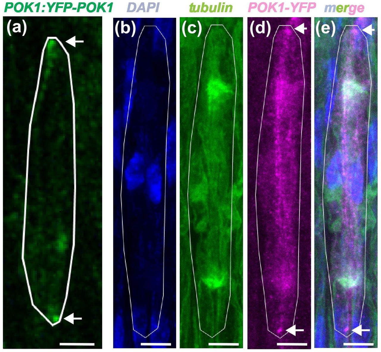
    From Swaminathan et al. This image is made available under a CC-BY 4.0 International license.

    The pro-inflammatory cytokines IFN-α and TNF-α inhibit organoid-derived extravillous trophoblast invasion
    A. Jantine van Voorden, Fangxu Lin, Souad Boussata, Remco Keijser, Liana Barenbrug, Bente Horselenberg, Ans M. M. van Pelt, Wendy Dankers, Susana M. Chuva de Sousa Lopes, Gijs B. Afink

    Differential spreading behaviors of Nodal signalling molecules in the extracellular space cooperatively shape left-right asymmetry
    Takafumi Ikeda, Toru Kawanishi, Yusuke Mii, Taijiro Yabe, Jose Pelliccia, Rebecca D Burdine, Hiroyuki Takeda

    CD44 facilitates adhesive interactions in airineme-mediated intercellular signaling
    Raquel L Bowman, Jiyea Kim, Michael J Parsons, Dae Seok Eom

    Temporal requirements of MAPK effectors reflect signalling microenvironment heterogeneity during Mesp1+ cardiac progenitor emergence and migration
    Nitya Nandkishore, Sinem Eylem Inal, Adeline Ghata, Fabienne Lescroart

    Dynamic polarization of heparan sulfate proteoglycans is involved in planar cell polarity and localization of endogenous Wnt11 during vertebrate neural tube formation
    Minako Suzuki, Ritsuko Takada, Tomoe Kobayashi, Makoto Matsuyama, Shinji Takada, Yusuke Mii

    Emergence of cell polarity by reciprocal interactions between Wnt and core PCP components
    Yusuke Mii, Minako Suzuki, Hiroshi Koyama, Kei Nakayama, Ritsuko Takada, Tomoe Kobayashi, Motosuke Tsutsumi, Tomomi Nemoto, Makoto Matsuyama, Toshihiko Fujimori, Shinji Takada

    Loss of Twist impairs tentacle development and induces epithelial neoplasia in the sea anemone Nematostella vectensis
    Patricio Ferrer Murguia, Julia Hagauer, Emmanuel Haillot, Aissam Ikmi, Alison G Cole, Ulrich Technau

    From Ferrer Murguia et al. This image is made available under a CC-BY 4.0 International license.

    3D epithelial cell topology tunes signalling range to promote precise patterning
    Giulia Paci, Francisco Berkemeier, Buzz Baum, Karen M. Page, Yanlan Mao

    Dual pathway regulation of castration response and ferroptosis in the prostate epithelium
    Weiping Li, Jing-Bo Zhou, Zejian Wang, Fereshteh Zandkarimi, Teresa A. Milner, Shouhong Xuan, John R. Christin, Caroline J. Laplaca, Andrew S. Greenberg, John P. Chute, Hanina Hibshoosh, Brent R. Stockwell, Michael M. Shen

    From Paci et al. This image is made available under a CC-BY-NC 4.0 International license.

    Wntless interacts with Notch signaling to balance the generation of neurons and gliocytes in vertebrate dorsal diencephalon
    Shu-Heng Lin, He-Yen Pan, Bo-Tsung Wu, Joe Sakamoto, Chun-Hsiu Wu, Atsuko Shimada, Yasuhiro Kamei, Hiroyuki Takeda, Yung-Shu Kuan

    Genetic mosaics reveal mechanisms of resistance to VEGF signaling loss during angiogenesis
    Irene Garcia-Gonzalez, Susana F. Rocha, Filipa V. Oliveira, Mariya Lytvyn, Maria S. Sanchez-Muñoz, Aroa Garcia-Cabero, Taija Mäkinen, Rui Benedito

    A dual role for GLI3 signaling in neural crest development
    Simon J. Y. Han, Vinit Adani, Edward Farrow, Bhavalben Parmar, Ching-Fang Chang, Kim Cochran, Paige J. K. Ramkissoon, Ezekiel Esteban, Kelsey H. Elliott, Kevin A. Peterson, Brian Gebelein, Martín García-Castro, Samantha A. Brugmann

    MAPK/ERK signaling blocks ectopic H3K9me3 heterochromatin formation to confer mesoderm and endoderm developmental competence
    Satoshi Matsui, Marissa Granitto, Samuel Sampson, Gerardo Mirizio, Ryo Maeda, Makoto Tachibana, Christopher Ahn, Hee-Woong Lim, Makiko Iwafuchi

    Caenorhabditis elegans Uses Canonical and Non-canonical Hippo signaling
    Linh Huynh, Razan A. Fakieh, C’Brionne Hendrix, Reid Powell, David J. Reiner

    Spatiotemporal WNT and BMP gradients orchestrate regional enteroendocrine cell diversity along the Drosophila midgut
    Jiaying Lv, Xingting Guo, Rongwen Xi

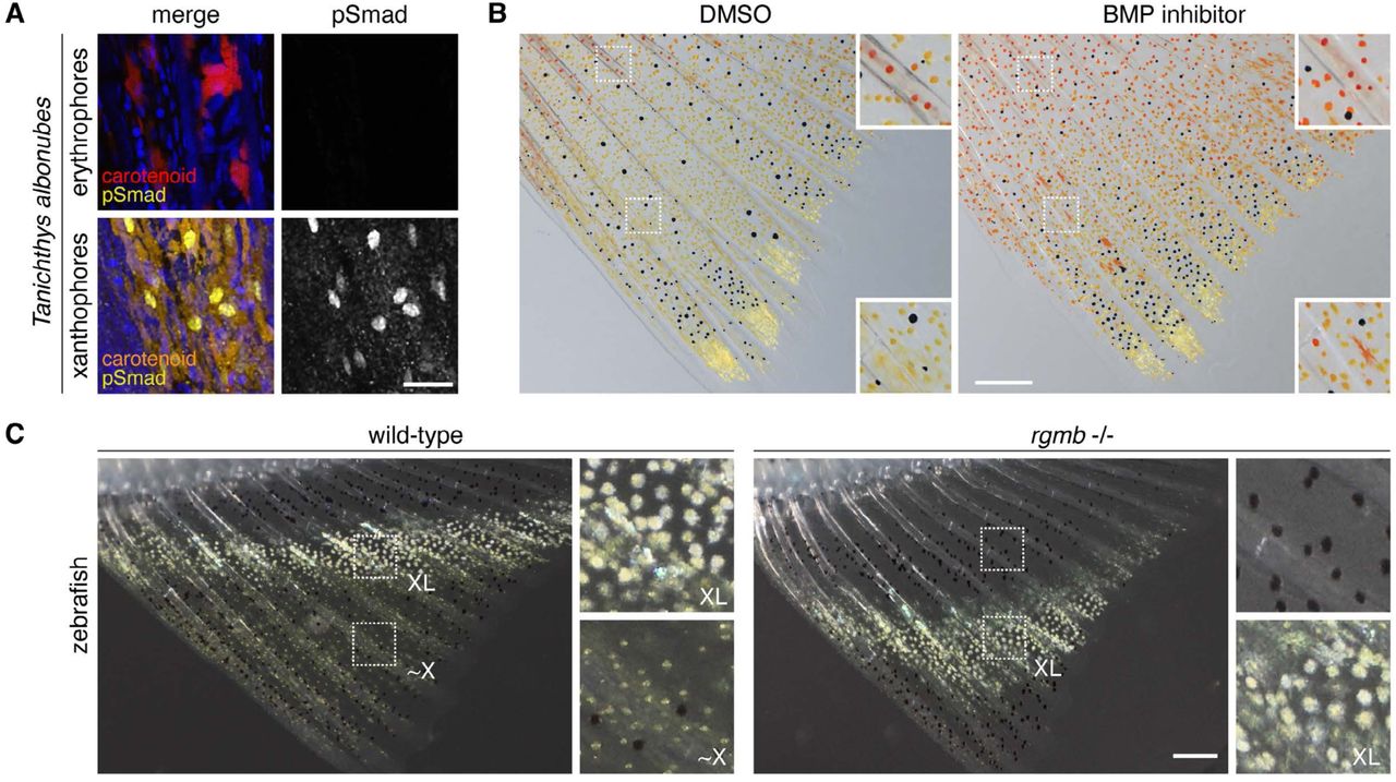
    Cell-cell communication as underlying principle governing color pattern formation in fishes
    Marleen Klann, Saori Miura, Shu-Hua Lee, Stefano Davide Vianello, Robert Ross, Masakatsu Watanabe, Emma Gairin, Yipeng Liang, Harrison W. Hutto, Braedan M. McCluskey, Marcela Herrera, Lila Solnica-Krezel, Laurence Besseau, Simone Pigolotti, David M. Parichy, Masato Kinoshita, Vincent Laudet

    Spen and Nito prevent dedifferentiation of progenitors by translationally repressing E(Spl)mγ
    Xiaosu Li, Wenwen Lu, Marisa Connell, Xiaobing Deng, Sijun Zhu

    Novel Roles of Sonic Hedgehog Signaling in Retinal Patterning and Neurogenesis During Mammalian Eye Development
    Miranda R. Krueger, Simranjeet K. Cheema, Sergi Simo, Edward M. Levine, Nadean L. Brown, Anna La Torre

    Angptl5 restricts primitive hematopoiesis by modulating retinoic acid signaling in zebrafish
    Jing Mo, Ding-Hao Zhuo, Ying Huang, Tao Cheng, Yang Dong, Yan-Yi Xing, Yun-Fei Li, Zi-Xin Jin, Xiang Liu, Guo-Qin Zhao, Hai-Rong Pu, Yu-Meng Liu, Zhi-Xu He, Li-Ping Shu, Peng-Fei Xu

    Microglia and Chek2 contribute to sex-specific organization of the adult zebrafish brain
    Paloma Bravo, Florence L. Marlow

    | Morphogenesis & mechanics

    Endothelial-zippering proceeds by sensing heartbeat-driven force through Cadherin-6 during heart-vessel connection in zebrafish
    Moe Fukumoto, Haruko Watanabe-Takano, Hajime Fukui, Ayano Chiba, Keisuke Sako, Hiroyuki Nakajima, Naoki Mochizuki

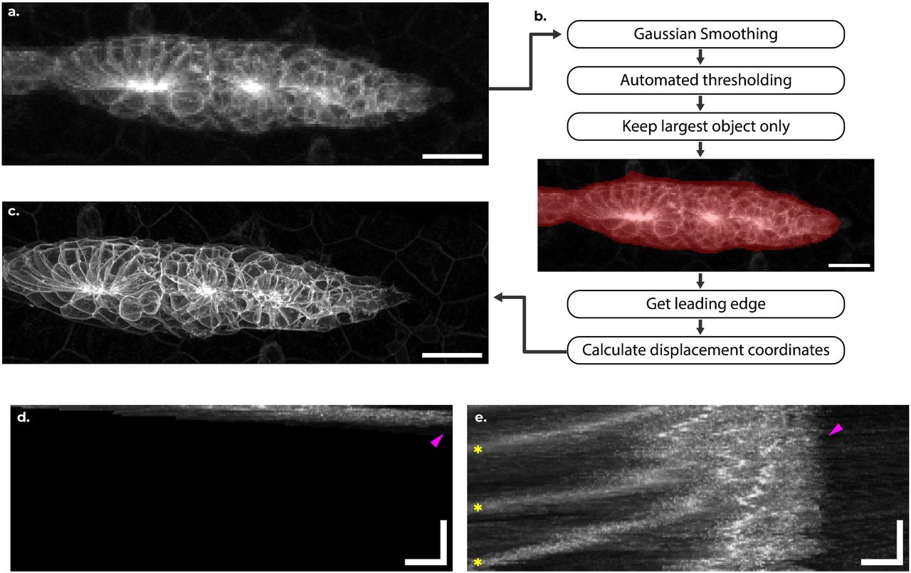
    Mechanical conditions preventing live cell extrusion during primary neurulation in amniotes
    Santiago A. Bosch Roascio, Julio A. Hernández, Flavio R. Zolessi

    From Bosch Roascio et al. This image is made available under a CC-BY-NC 4.0 International license.

    Dachsous-Fat Signaling Shapes the Drosophila Wing through Mechanical Forces
    Bipin Kumar Tripathi, Zhenru Zhou, Kenneth D. Irvine

    Epiblast Lumenogenesis is not a mammalian-specific trait
    Antonia Weberling, Natalia A. Shylo, Hannah Wilson, Melainia McClain, Richard Kupronis, Alex Muensch, Suzannah A. Williams, Florian Hollfelder, Paul A. Trainor

    From Weberling et al. This image is made available under a CC-BY-NC 4.0 International license.

    Quantitative computerized analysis demonstrates strongly compartmentalized tissue deformation patterns underlying mammalian heart tube formation
    Morena Raiola, Miquel Sendra, Jorge Nicolás Dominguez, Miguel Torres

    From Raiola et al. This image is made available under a CC-BY 4.0 International license.

    Gastruloids employ an alternative morphogenetic route to generate a posterior body axis on adherent substrates
    Guillermo Serrano Nájera, Apolline Delahaye, Benjamin J. Steventon

    Depletion of a neural cell adhesion molecule disrupts both epithelial and germ layer identity in sea anemone embryos
    Anna Postnikova, Julissa Tello, Maristhela Alvarez, Grace Lee, Ilhan Ali, Christine M Field, Timothy J Mitchison, Katerina Ragkousi

    STAT3 regulates basal cell identity and morphogenesis during early esophageal development
    Secunda W. Kariuki, Yosuke Mitani, Dominique D. Bailey, Gizem Efe, Ved V. Tripathi, Halil Tekin, Kensuke Suzuki, Jianwen Que, Joel Gabre, Ricardo Cruz-Acuña

    Microtubule architecture connects AMOT stability to YAP/TAZ mechanotransduction and Hippo signaling
    Giada Vanni, Anna Citron, Ambela Suli, Paolo Contessotto, Robin Caire, Alessandro Gandin, Giovanna Mantovan, Francesca Zanconato, Giovanna Brusatin, Michele Di Palma, Elisa Peirano, Lisa Sofia Pozzer, Carlo Jr. Albanese, Roberto A. Steiner, Michelangelo Cordenonsi, Tito Panciera, Stefano Piccolo

    From Vanni et al. This image is made available under a CC-BY 4.0 International license.

    Directional cell motility facilitates side-branching in the mammary epithelium in a tension-sensitive manner
    Beata Kaczyńska, Qiang Lan, Marja L. Mikkola, Satu-Marja Myllymäki

    Patterns of Mitochondrial ATP Predict Tissue Folding
    Bezia Lemma, Megan Rothstein, Pengfei Zhang, Bridget Waas, Marcus Kilwein, Safiya Topiwala, Sherry X. Zhang, Anvitha Sudhakar, Katharine Goodwin, Elizabeth R. Gavis, Ricardo Mallarino, Andrej Kosmrlj, Celeste M. Nelson

    From Lemma et al. This image is made available under a CC-BY 4.0 International license.

    Bidirectional translocation of actomyosin drives epithelial invagination in ascidian siphon tube morphogenesis
    Jinghan Qiao, Pengyu Yu, Hongzhe Peng, Wenjie Shi, Bo Li, Bo Dong

    Corneal lens curvature depends on localized chitin secretion
    Neha Ghosh, Jessica E. Treisman

    The Optic cup is actively shape programmed by independently patterned apical forces
    Ana Patricia Ramos, Lior Moneta, Alicja Szalapak, Louise Dagher, Malcolm Hillebrand, Carl D. Modes, Caren Norden

    From Ramos et al. This image is made available under a CC-BY 4.0 International license.

    | Genes & genomes

    Intron retention is dynamically regulated during zebrafish larval development and disrupted by high-fat diet
    Jesús Gómez-Montalvo, Jesus Hernandez-Perez, Cecilia Zampedri, Samantha Carrillo-Rosas, Daniela Fernanda Suárez-Bernal, Paula Marroquín-Morales, Marian Farrera-Borraz, Silvia Hinojosa-Alvarez, C. Fabián Flores-Jasso, Rocio Alejandra Chavez-Santoscoy, S. Eréndira Avendaño-Vázquez, Jose Mario Gonzalez-Meljem

    X-chromosome upregulation operates on a gene-by-gene basis at RNA and protein levels
    Ryan N. Allsop, Jeffrey Boeren, Beatrice F. Tan, Sarra Merzouk, Suresh Poovanthingal, Wilfred F.J. van IJcken, Jeroen A.A. Demmers, Hegias Mira Bontenbal, Cristina Gontan, Joost Gribnau, Vincent Pasque

    Genome-wide association study shows developmental robustness control by intestinal maltase via internal environment in Drosophila
    Soshiro Kashio, Izumi Enoki, Yuto Yoshinari, Takashi Nishimura, Masayuki Miura

    HDAC3 prevents enhancer hyperactivation to enable developmental transitions
    Nikolaos Stamidis, Anne Wenzel, Konrad Kacper Uściło, Smaragda Kompocholi, Sandra Bages-Arnal, Gemma Noviello, Lea Haarup Gregersen, James Alexander Hackett, Jan Jakub Żylicz

    From Stamidis et al. This image is made available under a CC-BY-NC 4.0 International license.

    Species-specific chromatin architecture and neurogenesis mediated by a human enhancer
    Federica Mosti, Jing Liu, Katie Lam, Samantha Skavicus, Victoria A. Kapps, Ketrin Gjoni, Nicholas S. Heaton, Katherine S. Pollard, Debra L. Silver

    Perturb-seq reveals distinct responses to pluripotency regulator dosages underlying the control of self-renewal and differentiation
    Jielin Yan, Hyein S. Cho, Renhe Luo, Michael A. Beer, Wei Li, Danwei Huangfu

    Genetic and physical interactions reveal overlapping and distinct contributions to meiotic double-strand break formation in C. elegans
    Marilina Raices, Fabiola Balmir, Nicola Silva, Wei Li, McKenzie K. Grundy, Dane K. Hoffman, Elisabeth Altendorfer, Carlos Jaime Camacho, Kara A. Bernstein, Monica P. Colaiácovo, Judith L. Yanowitz

    Chromatin priming by transcription factor ELF3 awakens latent competence for human amniotic differentiation
    Masatoshi Ohgushi, Kaori Honda, Hitoshi Niwa, Mototsugu Eiraku

    Transcriptional repression of central spindle factors controls endomitosis in the C. elegans intestine
    Ramon Barrull-Mascaró, Sonia Veltkamp, Samia Boutaibi, Lotte M. van Rijnberk, Rebecca Lippmann, Matilde Galli

    Specialised super-enhancer networks in stem cells and neurons
    Izabela Harabula, Liam Speakman, Francesco Musella, Luca Forillo, Luna Zea-Redondo, Alexander Kukalev, Robert A Beagrie, Kelly J. Morris, Lucas Fernandes, Ibai Irastorza-Azcarate, Ana M. Fernandes, Silvia Carvalho, Dominik Szabó, Carmelo Ferrai, Mario Nicodemi, Lonnie Welch, Ana Pombo

    Genome-wide screen for deficiencies modifying Cyclin G-induced developmental instability in Drosophila melanogaster
    Valérie Ribeiro, Marco Da Costa, Delphine Dardalhon-Cuménal, Camille A. Dupont, Jean-Michel Gibert, Emmanuèle Mouchel-Vielh, Hélène Thomassin, Neel B. Randsholt, Vincent Debat, Frédérique Peronnet

    Chamber-Specific Transcriptomic Insight into Cardiac Development using Guinea Pig and Human Heart Tissue
    Shatha Salameh, Devon Guerrelli, Luther Swift, Anika Haski, Alisa Bruce, Manan Desai, Yves d’Udekem, Nikki Gillum Posnack

    Genome-wide chromatin recording resolves dynamic cell state changes
    Yodai Takei, Jordan A. Lay, James M. Linton, Duncan M. Chadly, Yoshiki Ochiai, Ron Hadas, Andrew A. Perez, Mario R. Blanco, Paola Laurino, Mitchell Guttman, Michael B. Elowitz

    ATAC-seq of Low-Input and Cryopreserved Primordial Germ Cells Reveals Functional Enhancers
    Akane Kawaguchi, Mao Igari, Yasuto Murayama, Hiroko Iikawa, Mika Sakamoto, Yasukazu Nakamura, Shigehiro Kuraku, Keisuke Ishihara, Daisuke Saito

    REFEE Shapes ZGA Through Post-Transcriptional Regulation of Repetitive RNAs in Mouse Embryos
    Mie Kobayashi-Ishihara, Akihiko Sakashita, Youjia Guo, Hirotsugu Ishizu, Hidetoshi Hasuwa, Ten D. Li, Toshiya Nakahara, Tomohiro Kitano, Hidetaka Kosako, Kensaku Murano, Haruhiko Siomi

    From Kobayashi-Ishihara et al. This image is made available under a CC-BY 4.0 International license.

    Nanos downregulates maternal mRNAs in germline during Drosophila early embryogenesis
    Yasuhiro Kozono, Makoto Hayashi, Miho Asaoka, Satoru Kobayashi

    | Stem cells, regeneration & disease modelling

    Reduced TBX5 dosage undermines developmental control of atrial cardiomyocyte identity in a model of human atrial disease
    Irfan S. Kathiriya, Kavitha S. Rao, Alexander P. Clark, Kevin M. Hu, Zoe L. Grant, Megan N. Matthews, Zhe Chen, Jeffrey J. Saucerman, Swetansu K. Hota, Benoit G. Bruneau

    Obesogenic diet alters decidual differentiation and cell-cell communication in the mouse uterus
    Burak Koksal, Christian J. Bellisimo, Patrycja Jazweic, Deborah M. Sloboda, Alexander G. Beristain

    Momordica charantia Prevents Reproductive and Neurological Dysfunctions in Drosophila melanogaster Models of Type 2 Diabetes
    A.M. Agi, R. Abdulazeez, N. Abdulasalam, M.B. Omobalaji, O.S. Haruna, D.M. Shehu

    Patient-derived airway organoids from BAL fluid model injury and therapy responses in neonatal bronchopulmonary dysplasia
    Shilpa Sonti, Abiud Cantu, Manuel Cantu Guttierez, Connor Leek, Phinzy Pelton, Erik A. Jensen, Krithika Lingappan

    Systemic administration of a reported extracellular vesicle inhibitor, dimethyl amiloride, induces preterm birth and fetal growth restriction in pregnant mice
    Madison L. Stone, Scout Bowman-Gibson, Mili S. Bhakta-Yadav, Thomas L. Brown

    Neural Progenitors as a Novel Pathogenic Mechanism in Microcephaly
    Rami Yair Tshuva, Jeyoon Bok, Mio Nonaka, Xufeng Xue, Bidisha Bhattacharya, Aditya Kshirsagar, Tsviya Olender, Miri Danan-Gotthold, Tamar Sapir, Jianping Fu, Orly Reiner

    Glial betaPix is essential for blood vessel development in the zebrafish brain
    ShihChing Chiu, Qinchao Zhou, Chenglu Xiao, Linlu Bai, Xiaojun Zhu, Wanqiu Ding, Jing-Wei Xiong

    Developmental outflow tract abnormalities of Jag1-deficient mice are associated with abnormal ventricular activation and desynchronized contraction
    Kristýna Neffeová, Eva Nekvindová, Veronika Olejníčková, Hana Kolesová

    Human ectodermal organoids reveal the cellular origin of DiGeorge Syndrome
    Ed Zandro M. Taroc, Surangi Perera, Tunde Berecz, Jenny Hsin, Karla Barbosa-Sabanero, Sravya Pailla, Zarin Zainul, Ceren Pajanoja, Agota Apati, Daniel Martin, Laura Kerosuo

    Stem-cell modeling of cerebellar dysfunction of Angelman syndrome
    Carina Maranga, Adriana A. Vieira, João Camões dos Santos, Teresa P. Silva, Joana Gonçalves-Ribeiro, Karim Chebli, Miguel Casanova, Maud Borensztein, Laura Steenpass, Sandra H. Vaz, Tiago G. Fernandes, Simão T. da Rocha, Evguenia P. Bekman

    Characterisation of lmx1b paralogues in zebrafish reveals divergent roles in skeletal, kidney, and muscle development
    Joanna J. Moss, Chris R. Neal, Erika Kague, Jon D. Lane, Chrissy L. Hammond

    From Ferrer Moss et al. This image is made available under a CC-BY 4.0 International license.

    Disturbed mitochondrial maturation in cardiolipin remodeling-deficient cardiomyocytes
    Nanami Senoo, Macie S. Sheridan, Yvonne Wohlfarter, Mackenzie T. Primrose, Emmanouil Tampakakis, Markus A. Keller, Steven M. Claypool

    The Placental Transcriptome Serves as a Mechanistic Link between Prenatal Phthalate Exposure and Placental Efficiency
    Mariana Parenti, Samantha Lapehn, James MacDonald, Theo Bammler, Adam Szpiro, Marnie Hazlehurst, Drew B. Day, Ciara Thoreson, Kurunthachalam Kannan, Nicole R. Bush, Kaja Z. LeWinn, Qi Zhao, Sheela Sathyanarayana, Alison G. Paquette

    Regrowing the growth zone: metamorphosis kickstarts regeneration in the annelid, Capitella teleta
    Alicia A. Boyd, Elaine C. Seaver

    Biophysical dissection of SOX18/NR2F2 transcriptional antagonism reveals mechanisms of venous differentiation and drug action in vascular malformation
    Matthew S. Graus, Sawan Kumar Jha, Jieqiong Lou, Annegret Holm, Yew Yan Wong, Tara Davidson, Paul Coleman, Ella Sugo, Winnie Luu, Tara Karnezis, Jennifer Gamble, Geoff McCaughan, Scott Nightingale, Joyce Bischoff, Kazuaki Maruyama, Elizabeth Hinde, Kristy Red-Horse, Mathias Francois

    A genetically engineered vertebrate animal model of NAA15-related heart disease uncovers myocardial growth, contractility, and mitochondrial defects
    Warlen P. Piedade, Olivia Weeks, Alex A. Akerberg, Michael M. Molnar, Jennifer Galdieri, Rongbin Zheng, Kaifu Chen, Hasmik Keshishian, Patrick Hart, Steven A. Carr, Caroline E. Burns, C. Geoffrey Burns

    Deletion of KLHDC3, an E3 ubiquitin ligase complex substrate receptor, leads to obesity in mice
    Paula Armina V. Buco, Ashfaqul Hoque, Wilson Castillo-Tandazo, Alistair M. Chalk, Monique F. Smeets, Carl R. Walkley

    Frem2 Knockout Mice Exhibit Fraser Syndrome Phenotypes and Neonatal Lethality Due to Bilateral Renal Agenesis
    Rubina G. Simikyan, Xinyuan Zhang, Olga Strelkova, Nathan Li, MengYu Zhu, Andreas Eckhard, Petr Y. Baranov, Xudong Wu, Lauren Richey, Artur A. Indzhykulian

    Engulfing to Adapt: Efferocytosis by Epithelial Cells Triggers Cell State Transitions During Tissue Remodeling
    Adda-Lee Graham-Paquin, Deepak Saini, Sophie Viala, Mara KM Whitford, Mathieu Tremblay, William A Pastor, Luke McCaffrey

    Niche-targeted therapy via YAP/TAZ activation enhances hematopoietic regeneration
    Shun Uemura, Masayuki Yamashita, Takako Yokomizo-Nakano, Ayako Aihara, Takumi Iwawaki, Shuhei Koide, Yaeko Nakajima-Takagi, Motohiko Oshima, Yoshiki Omatsu, Bahityar Rahmutulla, Atsushi Kaneda, Miki Nishio, Akira Suzuki, Takashi Nagasawa, Kenta Kagaya, Taito Nishino, Atsushi Iwama

    rapunzel5 is necessary for normal hematopoietic development in zebrafish
    S Thapa, W Dowell, E Harris, DL Stachura

    Pathogenic DVL frameshifting variants in Robinow syndrome disrupt WNT signaling and cellular dynamics
    Chaofan Zhang, Rituparna Sinha Roy, Ming Yin Lun, Juliana F. Mazzeu, Janson White, Wu-Lin Charng, Nathaniel Peters, Jonas A. Gustafson, Harshini Iyer, Zain Dardas, Hyun Kyoung Lee, V. Reid Sutton, James R. Lupski, Claudia M.B. Carvalho

    A parenchymal niche regulates pluripotent stem cell function in planarians
    Skylar E. Settles, Kathleen E. Miller, Rachel H. Roberts-Galbraith

    Longitudinal Live Imaging Derived 4D Hemodynamics and Dynamic Tissue Mechanics Across Outflow Tract Morphogenesis
    Gening Dong, Jaehyun Rhee, Shivani Kumar, Molly E. Drumm, Henrik Lauridsen, Mahdi Esmaily-Moghadam, Jonathan T. Butcher

    From Dong et al. This image is made available under a CC-BY 4.0 International license.

    Identification of Human Pluripotent Stem Cell Derived Astrocytic Progenitors that Correlate with Glioblastoma Subtypes
    Stephanie Van Gulden, Anne Kathryn Linden, John A. Kessler, Chian-Yu Peng

    Transgenic mouse models for investigating human DUX4 expression during development and its roles in FSHD pathophysiology
    Yosuke Hiramuki, Charis L. Himeda, Peter L. Jones, Takako I. Jones

    Restraint of TGFβ family signaling by SMAD7 is necessary for hematopoietic stem cell maturation in the embryo
    Laura F. Bennett, Wenbao Yu, Chia-Hui Chen, Hyun Hyung An, Joanna Tober, Kai Tan, Nancy A. Speck

    Single-cell transcriptome identified ddx43+ cell types critical for maintenance of transient slow-cycling stem cells in planaria
    Nikhil Kumar Jaligam, Mohamed Mohamed Haroon, Mainak Basu, Atriya Mazumdar, Swathi Pavithran, Vinay Kumar Dubey, Praveen Kumar Vemula, Ankit Arora, Dasaradhi Palakodeti

    Adipocyte-Derived Amino Acid Storage Proteins are Required for Germline Stem Cell Maintenance in Adult Drosophila Females
    Anna B. Zike, Madison G. Abel, Robert C. Eisman, Lesley N. Weaver

    Functional classification of GNAI1 disorder variants in C. elegans uncovers conserved and cell-specific mechanisms of dysfunction
    Rehab Salama, Eric Peet, Logan Morrione, Sarah Durant, Maxwell Seager, Madison Rennie, Suzanne Scarlata, Inna Nechipurenko

    Chromosome engineering to correct a complex rearrangement on Chromosome 8 reveals the effects of 8p syndrome on gene expression and neural differentiation
    Sophia N. Lee, Erin C. Banda, Lu Qiao, Sarah L. Thompson, Karan Singh, Ryan A. Hagenson, Teresa Davoli, Stefan F. Pinter, Jason M. Sheltzer

    Single-Cell Characterization of Anterior Segment Development: Cell Types, Pathways, and Signals Driving Formation of the Trabecular Meshwork and Schlemm’s Canal
    Revathi Balasubramanian, Nicholas Tolman, Taibo Li, Abdul Hannan, Violet Bupp-Chickering, Karina Polanco, Aakriti Bhandari, Sally Zhou, Marina Simón, John Peregrin, Christa Montgomery, Krishnakumar Kizhatil, Jiang Qian, Simon W.M. John

    | Plant development

    The Moment Symmetry Breaks: Spatiotemporal Dynamics of CYCLOIDEA Expression During Early Floral Development
    Ya Min, Bianca T. Ferreira, Yao-Wu Yuan

    Developmental bias explains the evolutionary trend towards simple leaf shapes
    James S. Malone, Nora S. Martin, Samuel H. A. von der Dunk, Liliana M. Dávalos, Ard A. Louis

    Autophagic degradation of EIN3 ensures developmental plasticity and recovery from environmental stress in Arabidopsis
    Jeppe Ansbøl, Isolde Riis, Mette Stub, Rim Chaudhury, Emil Otto Kokholm Nielsen, Jonathan Chevalier, Dominique Van Der Straeten, Zhangli Thomsen Zuo, Sjon Hartman, Eleazar Rodriguez

    Spatiotemporal Dynamics of Anionic Phospholipids Orchestrate Lateral Root Initiation and Morphogenesis in Arabidopsis thaliana
    Joseph G. Dubrovsky, Juan Li, Sami Bouziri, Eric Bormann, Celine Geiger, Jazmin Reyes-Hernandez, Alexis Maizel

    Non-catalytic and catalytic TREHALOSE-6-PHOSPHATE SYNTHASES interact with RAMOSA3 to control maize development
    Thu M Tran, Hannes Claeys, María Jazmín Abraham-Juárez, Son L Vi, Xiaosa Xu, Kevin Michalski, Tsung Han Chou, Sessen D Iohannes, Panagiotis Boumpas, Z’Dhanne P Williams, Samatha Sheppard, Cara Griffiths, Matthew J Paul, Hiro Furukawa, David Jackson

    From Tran et al. This image is made available under a CC-BY-NC 4.0 International license.

    WIND1 controls cell fate transition through histone acetylation and deacetylation during somatic embryogenesis
    Akira Iwase, Arika Takebayashi, Fu-Yu Hung, Ayako Kawamura, Yetkin Çaka Ince, Yasuhiro Kadota, Soichi Inagaki, Takamasa Suzuki, Ken Shirasu, Keiko Sugimoto

    Antagonistic interactions between CLAVATA receptors shape maize ear development
    Penelope L. Lindsay, Fang Xu, Lei Liu, Panagiotis Boumpas, Andres Reyes, Byoung Il Je, Mari Ogawa-Ohnishi, Jarrett Man, Tara Skopelitis, Yoshikatsu Matsubayashi, Madelaine Bartlett, Shou-Ling Xu, David Jackson

    The mechanical properties of Arabidopsis thaliana roots adapt dynamically during development and to stress
    Luis Alonso Baez, Astrid Bjørkøy, Francesco Saffioti, Sara Morghen, Dhika Amanda, Michaela Tichá, Maarten Besten, Anastasiia Ivanova, Joris Sprakel, Bjørn Torger Stokke, Thorsten Hamann

    Clathrin-mediated endocytosis of ERECTA family receptors is essential for proper stomatal development in Arabidopsis
    Chi Zhang, Liang Chen, Suomin Wang, Chao Wang, Jianwei Pan, Suiwen Hou

    The nucleus follows an internal cellular scale during polarized root hair cell development
    Jessica Orr, Arif Ashraf

    COCHLEATA controls spatial regulation of cytokinin and auxin during nodule development
    Karen Velandia, Muhammad Nouman Sohail, Tiana Elizabeth Scott, Alejandro Correa-Lozano, Alannah Mannix, Eloise Foo

    Arabidopsis Group I Pumilio RNA-binding factors are vital for embryo development and balancing between growth and stress resistance
    Wenjuan Wu, Di Li, Danni Lin, Wangzhi Xu, Tianli Chen, Xiaomei Chen, Wei Guo, Zhengbiao Long, Xiang Xu, Xiaoyu Tu, Jirong Huang

    Epigenetic reprogramming guides sexual dimorphism during floral development in Silene latifolia
    Tomas Janicek, Vojtech Hudzieczek, Hana Polasek-Sedlackova, Marie Kratka, Vaclav Bacovsky

    TCP3-mediated regulation of cell expansion in Arabidopsis thaliana
    Tomotsugu Koyama, Tadashi Kunieda, Hiromi Toyonaga, Mika Nobuhara, Nobutaka Mitsuda, Kouichi Soga, Junko Ishida, Motoaki Seki, Koji Takahashi, Toshinori Kinoshita, Ayumu Bessho, Taku Demura, Masaru Ohme-Takagi

    PAM18-3, a J-domain protein, maintains mitochondrial integrity and plant growth and development in Arabidopsis thaliana
    Neha, S Souparnika, Chandan Sahi

    | Environment, evolution and development

    The Establishment of Cell-Type Specific Gene Regulation in the Sea Urchin Embryo
    Jonas Maurice Brandenburg, Alexandra Trinks, Dominika Voijtasova, Anna Alessandra Monaco, William Chang, Roberto Arsie, Marian Hu, Amro Hamdoun, Chloe Jenniches, Markus Landthaler, Nils Blüthgen, David Aaron Garfield

    From Brandenburg et al. This image is made available under a CC-BY-NC 4.0 International license.

    daf-16/FOXO promotes the activity of ligand-bound DAF-12/NHR to coordinate dauer exit and post-dauer seam cell fate
    Matthew J. Wirick, Isaac T. Smith, Benjamin S. Olson, Amelia F. Alessi, Himani Galagali, Kyal Lalk, Mikayla N. Schmidt, Kevin J. Ranke, John K. Kim, Xantha Karp

    Temporal orchestration of transcriptional and epigenomic programming underlying maternal embryonic diapause in a cricket model
    Kosuke Kataoka, Yuta Shimizu, Ryuto Sanno, Yuichi Koshiishi, Ken Naito, Kei Yura, Toru Asahi, Shin G. Goto

    There and back again: the dynamic evolution of panarthropod germ cell specification mechanisms
    Jonchee A. Kao, Emily L. Rivard, Rishabh R. Kapoor, Cassandra G. Extavour

    Conserved gene expression plasticity in development is more pervasive than expression divergence between species of Caenorhabditis nematodes
    Athmaja Viswanath, Daniel D. Fusca, John A. Calarco, Asher D. Cutter

    Protein sequence evolution underlies interspecies incompatibility of a cell fate determinant
    Emily L. Rivard, John R. Srouji, Anastasia Repouliou, Cassandra G. Extavour

    Evolution and Regulation of Decoupled Dimorphic Scaling Patterns in Ants
    Erica Vong, Shannon Parisien, Helene Orfali, Rajendhran Rajakumar

    Juvenile Hormone Signalling Underlies the Switchpoint and Differentiation of Soldiers in Camponotus floridanus
    Olivia MacMillan, Julia Singer, Sophia Perrakis, Alex Craig, Davina Ntanga, David Qiu, Rajendhran Rajakumar

    A Synthetic Coolant (WS-23) in Electronic Cigarettes Disrupts Normal Development of Human Embryonic Cells
    Shabnam Etemadi, Mohamed Debich, Prue Talbot

    Cracking the case: Differential adaptations to hard biting dominate cranial shapein rat-kangaroos (Potoroidae: Bettongia) with divergent diets
    Maddison C Randall, Vera Weisbecker, Meg Martin, Kenny Travouillon, Jake Newman-Martin, D. Rex Mitchell

    Genome reorganisation and expansion shape 3D genome architecture and define a distinct regulatory landscape in coleoid cephalopods
    Thea F. Rogers, Jessica Stock, Natalie Grace Schulz, Gözde Yalçin, Simone Rencken, Anton Weissenbacher, Tereza Clarence, Darrin T. Schultz, Clifton W. Ragsdale, Caroline B. Albertin, Oleg Simakov

    Cell Biology

    Microtubule acetylation by αTAT1 is essential for touch sensation in zebrafish but dispensable for embryonic development
    Samuel G. Bertrand, Daniel T. Grimes

    D-serine suppresses one-carbon metabolism by competing with mitochondrial L-serine transport
    Masataka Suzuki, Kenichiro Adachi, Pattama Wiriyasermukul, Mariko Fukumura, Ryota Tamura, Yoshinori Hirano, Yumi Aizawa, Tetsuya Miyamoto, Sakiko Taniguchi, Masahiro Toda, Hiroshi Homma, Kohsuke Kanekura, Kenji Yasuoka, Takanori Kanai, Masahiro Sugimoto, Shushi Nagamori, Masato Yasui, Jumpei Sasabe

    Ex vivo live imaging unveils the dynamics of oocyte formation in mice
    Eishi Aizawa, Junko Hara, Takaya Abe, Tomoya S. Kitajima

    The endo-lysosomal system drives lumen formation in a human epiblast model
    Anusha Rengarajan, Sicong Wang, Chien-Wei Lin, Amber E. Carleton, Nikola Sekulovski, Linnea E. Taniguchi, Mara C. Duncan, Kenichiro Taniguchi

    From Rengarajan et al. This image is made available under a CC-BY-NC 4.0 International license.

    Mouse pre-meiotic germline cysts contain fusome-like structure dependent on Dazl that mediates cyst fragmentation and oocyte development
    Madhulika Pathak, Allan C. Spradling

    Spargel/dPGC-1 influences cell growth through the E2F1-mediated endocycle pathway
    Md Shah Jalal, Atanu Duttaroy

    α-catenin phosphorylation is actomyosin-sensitive and required for epithelial barrier functions through Afadin
    Jeanne M. Quinn, Phuong M. Le, Anthea Weng, Annette S. Flozak, S. Sai Folmsbee, Erik Arroyo-Colon, Mitsu Ikura, Noboru Ishiyama, Cara J. Gottardi

    Forward Genetics Identifies ptr-18 and Other Genes as Developmental Regulators of the Four-Cell Tail Tip in C. elegans
    Uroš Radović, Marcus Henricsson, Jan Borén, Marc Pilon

    Spatiotemporal control of cortical centrin patterning by regionalized Sfi1 family scaffolding proteins in Stentor coeruleus
    Connie Yan, Niklas Steube, Gautam Dey, Wallace F. Marshall

    Membrane-tethered cadherin substrates reveal actin architecture at adherens junctions
    Sayantika Ghosh, John James, Badeer Hassan Ummat, Darius Vasco Köster

    Regulated development of cannibalistic supergiant cells
    Ben T Larson, Daniele Giannotti, Mahara Mtawali, Samuel J. Lord, Vittorio Boscaro, Patrick J. Keeling

    From Larson et al. This image is made available under a CC-BY 4.0 International license.

    Oligomerization and positive feedback on membrane recruitment encode dynamically stable PAR-3 asymmetries in the C. elegans zygote.
    Charles F Lang, Ondrej Maxian, Alexander Anneken, Edwin Munro

    Zbtb38 transcriptionally activates XIAP to regulate apoptosis in development and cancer
    Toshiaki Shigeoka, Hiroyuki Nagaoka, Nunuk Aries Nurulita, Shogo Tada, Yasumasa Bessho, Yasumasa Ishida, Eishou Matsuda

    Spatio-temporal control of nuclear mechanotransduction during EMT
    Ronan Bouzignac, Amandine Palandri, Amal Zine el Aabidine, Thomas Mangeat, Tatiana Merle, Martine Cazales, Antonio Trullo, Christian Rouviere, Virginia Pimmett, Mounia L Lagha, Magali Suzanne

    Capturing trophectoderm-like stem cells enables step-wisely remodeling of placental development
    Xinyi Jia, Bing Peng, Hongjin Zhao, Chunhui Wang, Wei Tao, Peng Du

    Cell exit during EMT is mechanically triggered independently of E-Cadherin loss
    Meritxell Font Noguera, Léa Roquin, Cyril Andrieu, Corinne Benassayag, Bruno Monier, Magali Suzanne

    From Font Noguera et al. This image is made available under a CC-BY-NC 4.0 International license.

    Oocyte polarity is established independently of the Balbiani body
    Manami Kobayashi, Joseph Zinski, Mary C. Mullins

    SLC35G3 is a UDP-N-acetylglucosamine transporter for sperm glycoprotein formation and underpins male fertility in mice
    Daisuke Mashiko, Shingo Tonai, Haruhiko Miyata, Martin M. Matzuk, Masahito Ikawa

    Modelling

    Latent Representations of Early Brain Development: A Multivariate Normative Model of Brain Structure and Behaviour
    Mariam Zabihi, Francesca Biondo, Jonathan O’Muircheartaigh, Thomas Wolfers, Sean Deoni, Andre Marquand, Muriel M.K. Bruchhage, James H. Cole

    Reconstructing Waddington’s Landscape from Data
    Dillon J. Cislo, M. Joaquina Delás, James Briscoe, Eric D. Siggia

    From Cislo et al. This image is made available under a CC-BY-NC 4.0 International license.

    Emergent Stable Tissue Shapes from the Regulatory Feedback between Morphogens and Cell Growth
    Bivash Kaity, Daniel Lobo

    From Kaity and Lobo. This image is made available under a CC-BY-NC 4.0 International license.

    Noise-driven morphogenesis independent of transcriptional regulatory programs
    Jack Toppen, Saeed Tavazoie

    Tools & Resources

    Rapid CE-MS with Real-Time Eco-AI Resolves Proteomic Heterogeneity Among Single Human Neutrophils
    Bowen Shen, Fei Zhou, Isabelle Luz, Steven J. Prior, Wagner Fontes, Peter Nemes

    7 Tesla fMRI characterisation of the cortical-depth-dependent BOLD response in early human development
    Jucha Willers Moore, Philippa Bridgen, Elisabeth Pickles, Pierluigi Di Cio, Lucy Billimoria, Ines Tomazinho, Cidalia Da Costa, Dario Gallo, Grant Hartung, Alena Uus, Maria Deprez, Sharon L. Giles, A. David Edwards, Jo V. Hajnal, Shaihan J. Malik, Jonathan R. Polimeni, Tomoki Arichi

    Trustworthy detection of exencephaly in high-throughput micro-CT embryo screens with focal-loss transformers
    Oshane O. Thomas, Rachel Roston, Hongyu Shen, A. Murat Maga

    From Thomas et al. This image is made available under a CC-BY 4.0 International license.

    Anatomy-aware, label-informed approach improves image registration for challenging datasets
    Rachel A. Roston, Nicholas J. Tustison, A. Murat Maga

    A blastocyst-derived in vitro model of the human chorion
    Luca C. Schwarz, Matthew J. Shannon, Gina McNeill, Rina C. Sakata, Viviane S. Rosa, Katherine Cheah, Laura Keller, Phil Snell, Leila Christie, Kay Elder, Anastasia Mania, Lauren Weavers, Rachel Gibbons, Tugce Pehlivan Budak, Ippokratis Sarris, Amy Barrie, Alison Campbell, Roser Vento-Tormo, Gary D. Smith, Alexander G. Beristain, Marta N. Shahbazi

    Human retinal organoid single-cell atlas allows to reconstruct retinal development at high resolution and identify nature restricted transcriptional states in vitro
    Emil Kriukov, Everett Labrecque, Nasrin Refaian, Petr Baranov

    Non-segmented unsupervised learning of multispectral whole slide images for robust analysis of tissue repair and regeneration
    Kody Paul Mansfield, Tamara Mestvirishvili, Bibi Subhan, Valeria Mezzano-Robinson, Dianny Almanzar, Sydney Hanson, Jimin Tan, Cynthia Loomis, David Fenyo, Aristotelis Tsirigos, Piul S. Rabbani

    Smart Microscopy: Current Implementations and a Roadmap for Interoperability
    Lucien Hinderling, Hannah S. Heil, Alfredo Rates, Philipp Seidel, Manuel Gunkel, Benedict Diederich, Thomas Guilbert, Rémy Torro, Otmane Bouchareb, Claire Demeautis, Célia Martin, Scott Brooks, Evangelos Sisamakis, Grandgirard Erwan, Karl Johansson, Johannes K. Ahnlinde, Oscar André, Philip Nordenfelt, Pontus Nordenfelt, Claudia Pfander, Jürgen Reymann, Talley Lambert, Marco R. Cosenza, Jan O. Korbel, Rainer Pepperkok, Lukas C. Kapitein, Olivier Pertz, Nils Norlin, Aliaksandr Halavatyi, Rafael Camacho

    ShapeSpaceExplorer: Analysis of morphological transitions in migrating cells using similarity-based shape space mapping
    Samuel D.R. Jefferyes, Roswitha Gostner, Laura Cooper, Mohammed M. Abdelsamea, Elly Straube, Nasir Rajpoot, David B.A. Epstein, Anne Straube

    Real-time feedback control microscopy for automation of optogenetic targeting
    Lucien Hinderling, Alex E. Landolt, Benjamin Grädel, Laurent Dubied, Cédric Zahni, Moritz Kwasny, Agne Frismantiene, Talley Lambert, Maciej Dobrzyński, Olivier Pertz

    From Hinderling, et al. This image is made available under a CC-BY-NC 4.0 International license.

    Cell-APP: A generalizable method for cell annotation and cell-segmentation model training
    Anish J. Virdi, Ajit P. Joglekar

    Improving RNA-seq protocols
    Felix Pfoertner, Eva Briem, Wolfgang Enard, Daniel Richter

    Low-intensity focused ultrasound enables temporal modulation of human midbrain organoid differentiation
    Jinseong Jeong, Yehhyun Jo, Youngsun Lee, Eunyoung Jang, Yeonji Jeong, Won Do Heo, Jennifer H. Shin, Mi-Ok Lee, Hyunjoo J. Lee

    A Quantitative Polymerase Chain Reaction Protocol for Sex Identification of Zebra Finch and Chicken Using Blood Samples
    Prakrit Subba, Saidat O. Adeniran-Obey, Fanny-Linn Kraft, Susan C. Chapman, Natalie A. Shay, Shannon R. Liedl, Mark D. Wild, Scarlett A. Wolcott, Julia M. George

    Neonatal Mouse Ovary Culture: An In Vitro Model for Studying Primordial Follicle Regulation
    Edgar A Diaz Miranda, Grace Anne Dyer, Faith Wilson, Mariel English, Taylor VanDeVoorde, Lei Lei

    Bridging the gap of late-gestation nephrogenesis using a non-human primate model
    Kairavee Thakkar, Sunitha Yarlagadda, Lyan Alkhudairy, Andrew Potter, Konrad Thorner, Praneet Chaturvedi, Kyle W. McCracken, Nathan Salomonis, Raphael Kopan, Meredith P Schuh

    Microinjected dsRNA triggers a robust RNAi response in Stentor coeruleus
    Makenna Kuecks, Preeti Arra, Sarah Hoffmann-Weitsman, Makenzie Funk Craig, Mark M. Slabodnick

    Research practice & education

    “Spurring and Siloing: Identity Navigation in Scientific Writing Among Asian Student Researchers”
    Devon Goss, Meena Balgopal, Shaila Sachdev, Grace Kim, LaTonia Taliaferro-Smith, Sarah C. Fankhauser

    Integrating Career Development and Immigration Planning: A Pilot Program for International Scholars
    Caroline Fecher, Natalie Chernets, Paola Cépeda

    What drives change? Characterizing scientific self-efficacy development in undergraduate research experiences
    Qiyue Zhang, Paul R. Hernandez, Benjamin S. Listyg, Erin L. Dolan

    10 recommendations for strengthening citizen science for improved societal and ecological outcomes: A co-produced analysis of challenges and opportunities in the 21st century
    Jack Nunn, Håkon da Silva Hyldmo, Lauren McKnight, Heather McCulloch, Jennifer Lavers, Julie Old, Laura Smith, Nicola Grobler, Cheryl Tan Kay Yin, Wing Yan Chan, Candice Raeburn, Nittya S. M. Simard, Adam Kingsley Smith, Sam Van Holsbeeck, Eleanor Drinkwater, Kit Prendergast, Emma Burrows, Christopher L. Lawson

    “The ship is going down and we are powerless”: The Impact of Federal Funding Changes on Researchers Training the Next Generation of Scientists
    Emily Mastej, Tiffany Do, Arghavan Salles

    Thumbs up (1 votes)
    Loading...

    Tags:
    Categories: Research, Resources

    Catch up on Development presents… webinar on gene regulation

    Posted by , on 11 September 2025

    The 27 August 2025 webinar featured two of Development’s PI fellows working on gene regulation.

    Catch up on previous webinars and sign up to the Development presents… mailing list to learn about the upcoming webinars as they are announced.

    Anzy Miller (University of Manchester)

    Talk and Q&A

    Marlies Oomen (Helmholtz Munich, Institute of Epigenetics and Stem Cells)

    Talk and Q&A

    Thumbs up (2 votes)
    Loading...

    Tags: , , ,
    Categories: Development presents..., Video