We’re now delighted to announce the results of the image competition. Congratulations to Özge Özgüç, Allan Carrillo-Baltodano and Julia Peloggia de Castro! And thank you to everyone who submitted their images to the competition. You can still view the shortlisted images on the Node. Look out for posts in FocalPlane’s ‘Featured image’ series to discover more about some of the microscopists that shared images with us in this competition.
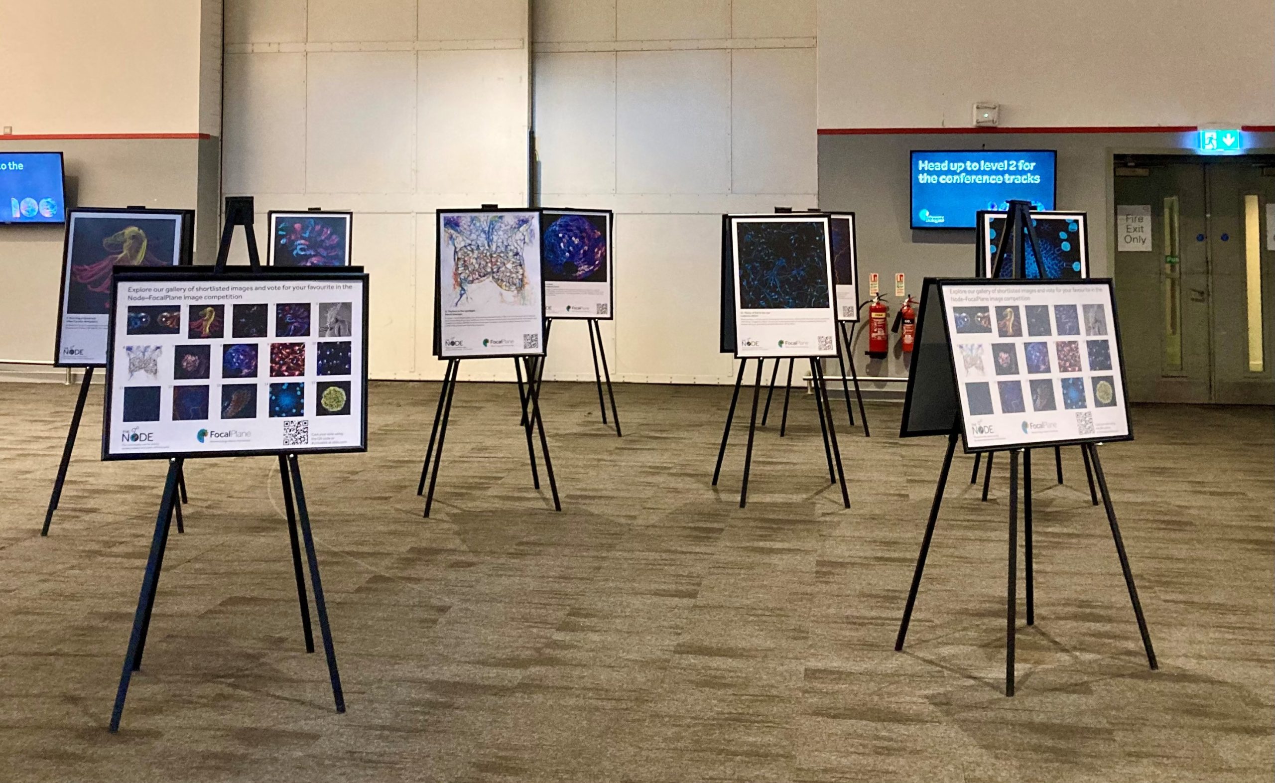
Image gallery at the Biologists @ 100 conference in Liverpool.
Winner
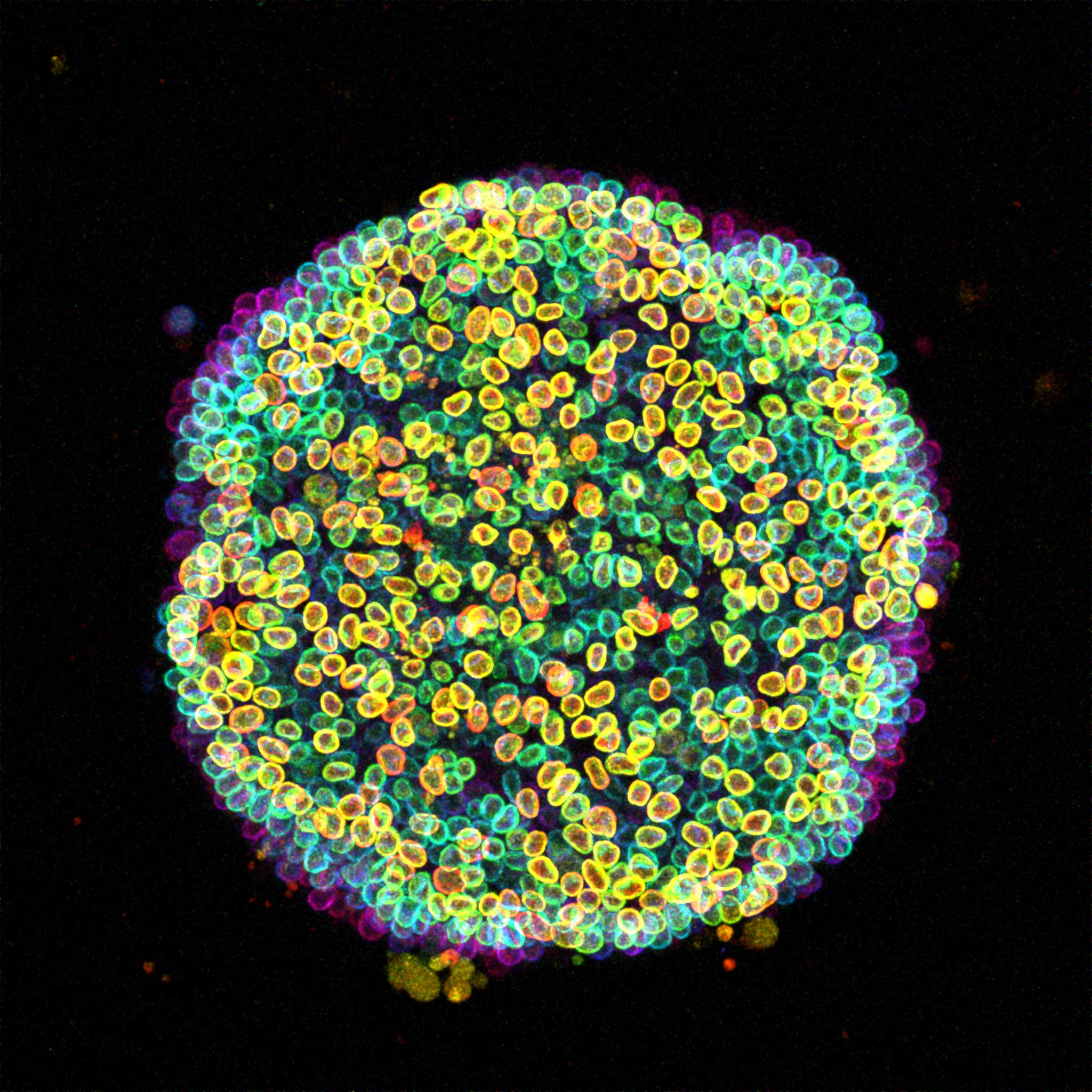
15. Cell-estial bloom Özge Özgüç A ‘Cell-estial bloom’ of human induced pluripotent stem cells (hiPSCs) flourishes on a micropatterned island. This image presents a colony of live hiPSCs, with fluorescently labelled Lamin B delineating the nuclear lamina within each cell. Acquired with a Zeiss LSM 880 Airyscan microscope, this maximum intensity projection is enhanced with depth-coded coloring to reveal the captivating three-dimensional landscape.
First runner-up
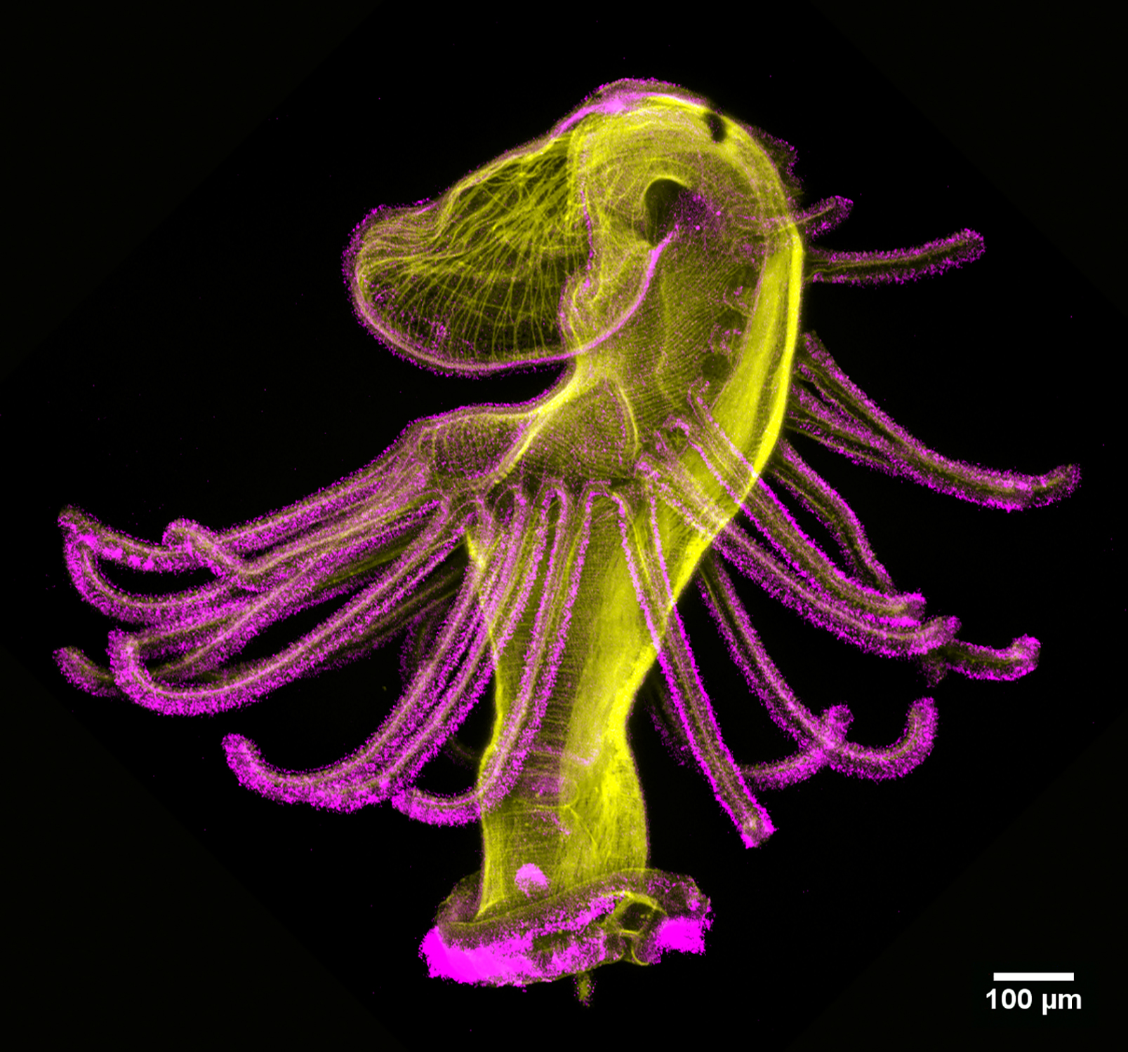
2. Dancing actinotroch Allan Carrillo-Baltodano Actinotroch larva of a phoronid worm with phalloidin shown in yellow and acetylated tubulin in magenta. Imaged with a Zeiss LSM 800 at 10 x magnification.
Second runner-up
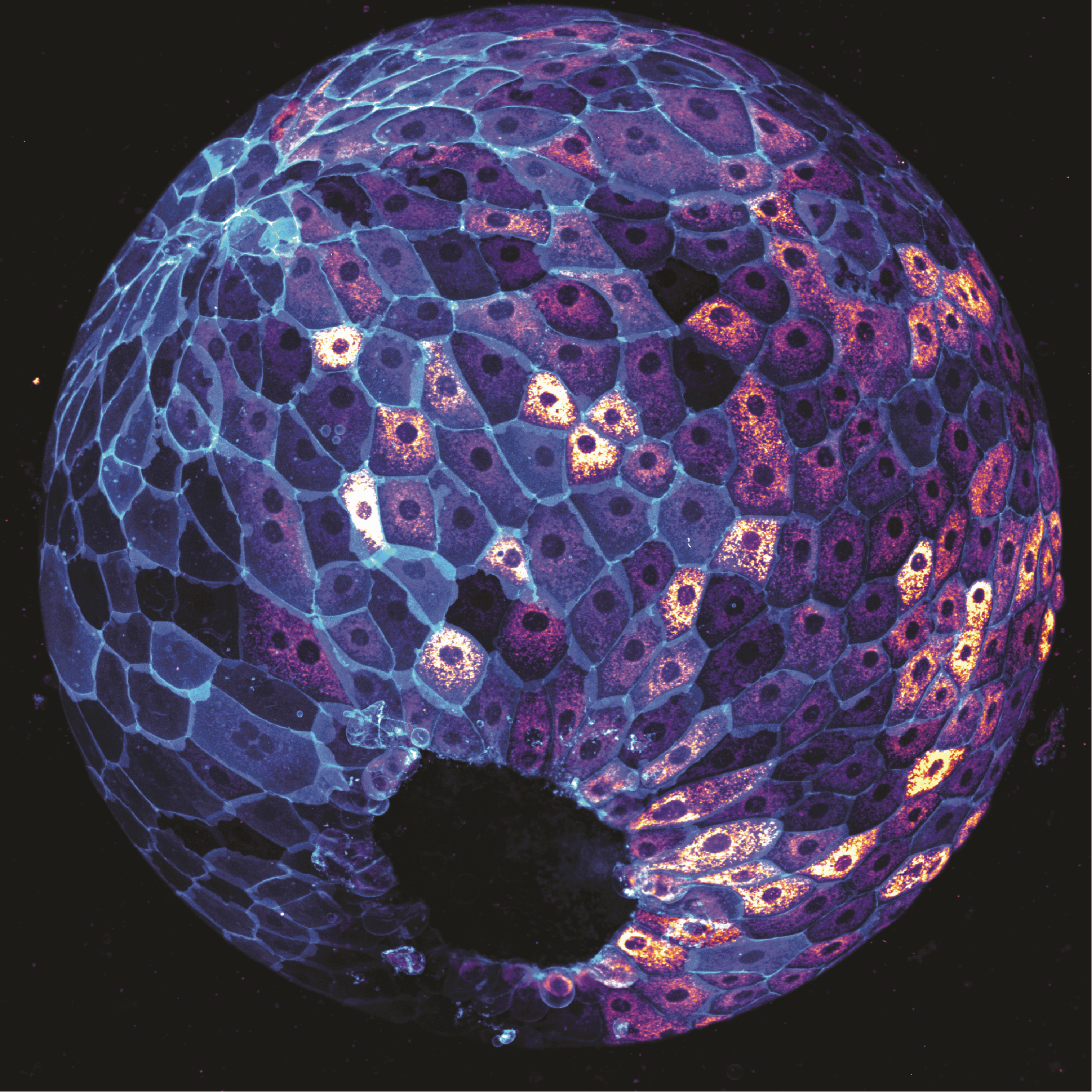
8. Who’s active? Julia Peloggia de Castro The image depicts a zebrafish embryo at 9 hours post-fertilisation on a lateral view. Cells are stained with MitoTracker, which labels active mitochondria, and cell membranes are labelled in cyan with a EGFP transgenic membrane tag. Image was taken using a 20x objective on a spinning disk confocal microscope. (No Ratings Yet) Loading...
During the coffee break on the second day of the 3rd Crick Beddington Symposium, I approached Dr Giulia Boezio, a postdoc in the Briscoe lab at the Francis Crick Institute. As a fellow chick embryologist, I was already aware of her work, but I was intrigued to learn more about the genomic barcoding technique that she has adapted for use in our favourite model organism!
Giulia presenting her poster at the 3rd Crick Beddington Symposium.
The Briscoe lab are interested in how the huge diversity of neuronal subtypes arise in the spinal cord. Different cell types originate from different positions along the dorsoventral axis, constituting 11 discrete domains of progenitor cells (Holguera and Desplan, 2018; Le Dréau and Martí, 2012; Sagner and Briscoe, 2019). But we still don’t know how these cell types arise – does one type of mature neuron come from one specific progenitor, or might two different mature neurons share a bi-fated progenitor? Or perhaps the differentiation paths converge – two different progenitors might ultimately give rise to the same final cell type. Giulia aims to shed light on these questions, which surprisingly had not been revisited in over 30 years (Leber et al., 1990; Leber and Sanes, 1991).
Giulia has adapted a genomic barcoding technique called LARRY (Lineage and RNA Recovery; Weinreb et al., 2020) – originally developed for cells in culture – for use in the chick embryo. Viral infection is used to deliver barcodes into the chick spinal cord at stage 12 (day 2 of development). Each barcode is unique, and is expressed alongside GFP, allowing the infected cells to be tracked over time. The embryos are incubated to develop for several days, before the mature cells are collected at one of three time points (stage 28, 31 and 35). They are then FACS sorted to obtain the GFP-expressing cells, and profiled using single cell RNAseq. After identifying which cluster corresponds to which cell type, Giulia can then see which barcodes are present in each cluster. Since every cell with the same barcode is derived from a single progenitor, this allows a retrospective lineage history to be assembled. I wondered how they can guarantee that each barcode is completely unique, but Giulia explained, “we have almost half a million barcodes in our library. This wouldn’t normally be enough, if we were infecting many, many cells. But the bug that actually turned out to be a (good) feature is that the infection rate is not very high.” This means that the chance of infecting two independent cells with the same barcode is extremely slim. She also checked across five separate embryos, and found no barcode overlap – so it is safe to assume that within one embryo, each barcode is unique.
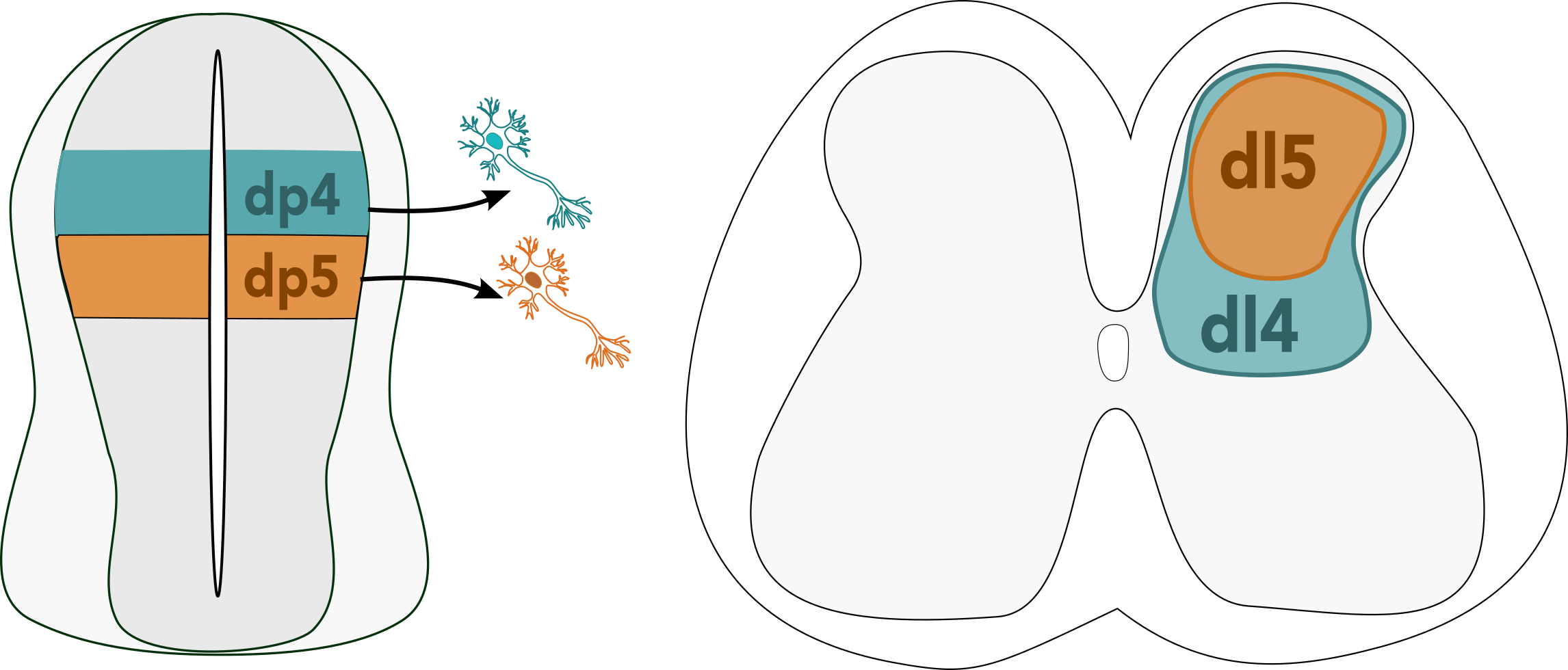
From the single cell RNAseq data, she found that spinal cord cells group into five lineage compartments – meaning that progenitors will mostly contribute to cells in one of these five lineage compartments, and rarely contribute to more than one. Some cell types were more abundant than others – this is particularly true for the dorsal dI4 and dI5 neurons, which make up more than half of all the neurons in the entire spinal cord. These sensory neurons are involved in touch and pain sensation (Lai et al., 2016), and despite their abundance, are not very well studied. Giulia observed that their ratios changed over time; at stage 28, she saw many more dI4 than dI5 neurons, but by stage 31 this had equalised to roughly 50:50 – this makes perfect sense, since one is an excitatory neuron, and the other is an inhibitory neuron in the same circuit.
A diagram showing the location of dorsal progenitor cells (dp4 and dp5) in the developing neural tube, and the final position of their mature descendants (dI4 and dI5) in the spinal cord.
But how does this imbalance arise, and how does it correct itself later on? “Initially, when I saw this, I thought that the easiest explanation would be that the blue (dI4) cells are born earlier, and then eventually the orange (dI5) cells would catch up somehow. But we had to dig a bit deeper into the clonal data, and this explanation didn’t seem to fit”, Giulia said, after observing an initial peak of dI4 neurons, followed by a peak in both dI4 and dI5 neurons later in development. It turns out that at earlier stages, most dI4 neurons derive from clones that only produce dI4 neurons, while at later stages, the vast majority of clones produce an even ratio of dI4 and dI5 neurons. This suggests that there are two different progenitor types – one that only produces dI4 neurons, and one that produces both dI4 and dI5 in equal quantities. Giulia is currently working on fitting this data to different mathematical models to find a differentiation trajectory that might explain this result.
Does this system apply to other species? To find out, Giulia used ex vivo cultures of human embryonic trunk slices – a technique (which truly sounds like science fiction to me!) in which transverse sections of the spinal cord are grown using an air-liquid interface, exposed to cell culture media from the bottom, and the air from above. Work with human embryos is notoriously difficult, as you cannot control the availability, nor what stage will be available to work on. Giulia managed to obtain some 6 week old (CS17) human embryos – older than she would have liked, but she decided to go ahead with the experiment anyway. She infected the CS17 trunk slices with LARRY barcodes and cultured them for 7 days, collecting and profiling the cells in the same way as for the chick experiment. She found that dI4 and dI5 dominated over other neurons in terms of abundance, as she saw in the chick. There were very few shared clones between ventral neural cell types; this fits with the fact that neurogenesis occurs earlier in this compartment. Some clones were shared in the dorsal neural tube, but only between other neurons of the same type. The only exception was the dI4 and dI5 neurons, which still shared the vast majority of barcodes, meaning that at the time of infection, the decision to become dI4 or dI5 had not yet been made. This appears to be the latest lineage decision of any neuron in the spinal cord. Why might this be important? Giulia speculates that their proliferation as progenitors might help to ensure that the final ratio of dI4:dI5 is balanced, since there are diseases that can arise from an imbalance between these two neurons.
Giulia’s hypothesis is that there are two different groups of progenitors in the dorsal spinal cord, one that gives rise to dI4 neurons and one that maintains progenitors with bi-fated potential. The temporal sequence is still unclear; Giulia speculates that the different groups of progenitors may mature into neurons at different rates, or begin maturing at different times, eventually resulting in an approximate 1:1 ratio. At the moment, there is no data to show what happens between the dI4-dominant stage 28 and the restoration of balance at stage 31, so it is not possible to prove when or how this change happens. Giulia is currently working on fitting a model to her data in order to test this hypothesis as well as complementing her sequencing data with live imaging to fill the temporal gaps.
Stay tuned for more poster interviews coming soon!
References: Holguera, I., Desplan, C., 2018. Neuronal specification in space and time. Science 362, 176–180. https://doi.org/10.1126/science.aas9435 Lai, H.C., Seal, R.P., Johnson, J.E., 2016. Making sense out of spinal cord somatosensory development. Development 143, 3434–3448. https://doi.org/10.1242/dev.139592 Le Dréau, G., Martí, E., 2012. Dorsal-ventral patterning of the neural tube: a tale of three signals. Dev Neurobiol 72, 1471–1481. https://doi.org/10.1002/dneu.22015 Leber, S., Breedlove, S., Sanes, J., 1990. Lineage, arrangement, and death of clonally related motoneurons in chick spinal cord. J Neurosci 10, 2451–2462. https://doi.org/10.1523/JNEUROSCI.10-07-02451.1990 Leber, S.M., Sanes, J.R., 1991. Lineage analysis with a recombinant retrovirus: application to chick spinal motor neurons. Adv Neurol 56, 27–36. Sagner, A., Briscoe, J., 2019. Establishing neuronal diversity in the spinal cord: a time and a place. Development 146, dev182154. https://doi.org/10.1242/dev.182154 Weinreb, C., Rodriguez-Fraticelli, A., Camargo, F.D., Klein, A.M., 2020. Lineage tracing on transcriptional landscapes links state to fate during differentiation. Science 367, eaaw3381. https://doi.org/10.1126/science.aaw3381
Pleasantine Mill is awarded the 2025 British Society for Developmental Biology (BSDB) Wolpert Medal, which recognises an individual who has made extraordinary contributions to the teaching and communication of developmental biology.
Pleasantine is a group leader at the MRC Human Genetics Unit at the Institute for Genetics and Cancer at the University of Edinburgh, UK. We caught up with her to talk about her research, her work with the cilia and rare disease communities, and why it’s important, now more than ever, for scientists to engage in community work.
Congratulations on winning the 2025 BSDB Wolpert medal! What does this mean to you?
It’s a really great honour. Lewis Wolpert was such an amazing figure in terms of his contributions to science and his ability to communicate science effectively to different audiences. Receiving a medal named in his honour is huge, particularly at this point in time, when science is under threat, when there is a heightened urgency of getting all parties to engage with the importance of research, basic science and discovery. This threat expedites a need to bring together communities and stakeholders to champion science. To be recognised for it means so much to me personally.
A few years ago, you were awarded the Woman in Cell Biology Early Career Award from the British Society for Cell Biology – you’ve managed to receive awards from both British societies!
It’s a lovely reflection of where we sit in terms of our research focus. What my team do is very cell biology, because we’re interested in centrioles and cilia, but leverage genetics and genomics to understand the molecular mechanisms at play in development and how they go wrong in disease. So really what we do is cell biology on an organismal scale, looking at how different cell types, developmental stages or disease states are dependent on cilia. So it’s very nice to be recognised by both societies!
Can you briefly talk about your career so far and what your research interests are?
I did my undergrad in Immunology and Microbiology at McGill University in Montreal because it seemed an exciting time to be doing immunology, but in practice, I found the coursework was a lot less engaging. I started to wander a bit and stumbled onto Genetic Engineering. I went to the University of Toronto Medical and Molecular Genetics Department for my PhD, which was a rotation-based system, and fell into by my first and lasting love with developmental biology. I joined the lab of Chi-chung Hui, a mouse developmental geneticist studying Hedgehog signalling and the role of the GLI transcription factors. Here, we had all these cool genetic tools and techniques, with which we could generate and study all these amazing ‘shock and awe’ phenotypes! I was hooked with genetics. When I left CC’s lab after my PhD, I wanted to try something really new and undertook a forward genetic screen in mice, so as not to make any assumptions about the system I was studying, in this case neural crest development. That was when I stumbled into the wonderful world of cilia. Almost all the genes we pulled out were involved in cilia structure or function, which were ironically also all involved in Hedgehog signalling. From there, the jump to human disease was easy as cilia play major roles in rare disease genetics – it was an easy segue for my independent research focus. Like our genetic screens, the unusual phenotypes in rare disease patients are sometimes the ones that are the most informative in terms of understanding how a gene works in complex processes. That’s the space that we’ve stayed in since then.
You’re currently the guest editor of Journal of Cell Science’s Special Issue – Cilia and Flagella. What are you most excited about in this special issue and in the field?
Thrilled to be guest editing it with Lotte Pedersen. We’re still accepting papers until 31 March 2025 and I’m excited about what is taking shape for this Special Issue! For example, we have commissioned both established experts and up-and-coming talent on everything from cilia disassembly, to deep dives into different ciliary compartments and we are even wading into the hot debate around condensates and their role in motile ciliogenesis. We’ve even got a perspective piece from our rare disease patient community about how to form effective partnerships and patient-centric research too.
For the field in general, I think there’s a lot of interesting work that’s going on in terms of how we understand the differences between different cilia types. There are exciting technology and tools to capture organelle-level changes in content during disease, developmental time, or even with the cell cycle, then tying this to how these change cellular signalling outputs and contribute to phenotypes. Real biology across scales! There is still a tendency to think of cilia from a cell biology aspect, as being in a dish and pointing up in the media. But in fact, as we know from being developmental biologists, cilia are often involved in much more complex interactions in the 3D space, often interacting with cellular structures or even other cilia. Live imaging, light-sheet and volumetric EM techniques- we now are really pushing our ability to look at these inter-organelle or contacts in situ, discovering new cilia synapses. Testing what they mean functionally will be our field’s next big frontier!
You are part of the team leading the UK Cilia Network. How did you get involved initially, and how’s your experience been?
Almost ten years old, the UK Cilia Network aimed to join up the existing regional cilia-focused groups across the country. It planned to hold national yearly meetings that would rotate around the country, to highlight expertise and to facilitate collaboration and exchanges. I got asked to be on the leadership team, because they were looking for more junior people to help shape the vision going forward. And then COVID hit, and the UK Cilia Network turned into something very different partly as a result of the e-symposia series. It is great – the network has now more of an online presence, with the website as an international landing page to help individuals raise their profile and build their networks. It’s got such a strong brand now. We’re discussing whether we have outgrown the UK and whether there’s an opportunity to help create a truly international ‘society’ for cilia, which the UK Cilia Network would be part. So that’s an exciting next chapter for me. We’ve already had some early meetings with leaders across Europe, North America and Asia. On an international scale, it would be a way to raise the profile of individuals, whether you have a national network or not in your country. We would look to facilitate collaborations, highlight opportunities, as well as minimise conflicts between meetings internationally and the competition for limited resources. Importantly too, it would be looking to connect ciliopathy patients and their organizations internationally and nationally too.
Another thing we’ve done recently is to formally extend our network to the centrosome community. The centrosome (and centrioles) are also very closely associated with cilia, but not in the name, and as a result some people feel excluded. There are also a group of rare diseases, the centrosomopathies, for which the patient groups and clinical teams could benefit from closer ties to researchers, as the ciliopathy community has benefited from. So, we are in the process of rebranding as the UK Cilia and Centrosome Network.
You started organizing the UK Cilia Network e-symposia during the early 2020 lockdowns and since then it has been running strong. What motivated you to start the symposia?
In 2020, I was organising the in-person UK Cilia Network Spring meeting right after the UK Microtubule Meeting in Edinburgh that early April. We’d already done tonnes of organising when everything shut down. Having just come through my own promotion and tenure process within the University, I immediately recognised how important it was for people to be invited to speak at key meetings. There were going to be damaging lapses in people’s CVs as a result of lockdowns, unless we created a forum where they could be invited to and speak. We could keep people connected and focused on the future by sharing great science. The real push for the e-symposia was making sure that we could showcase these amazing junior people even in a shutdown and create something that people could count on when everything seemed uncertain. Like real life meetings, the e-symposia also allowed people to find collaborations and build new networks. Indeed, there have been papers from collaborations that formed from the first few symposia during COVID have now been published. Amazing! We have now nearly 1500 people registered for the symposia from all over the world. We’re still hitting about 250 people with another 150 watching the recording. Even though the pandemic’s gone, the community stayed.
Apart from engaging with the research community, have you been involved in policy and public outreach activities?
On the policy side, I’ve done some advising and written whitepapers on genome editing technologies, rare diseases and expediting genetic diagnoses. We’ve worked closely with philanthropic funders for their rare disease and patient partnerships space to decide priorities and evaluate subsequent funding calls. I sit on several scientific advisory boards for various charities. Patient engagement and involvement within the rare disease space internationally is something I have been very involved with. We’ve run workshops, outreach activities and more.
When I initially started as a postdoc years ago, a bunch of us piloted an Edinburgh Science Fringe Festival that ran in parallel with the Edinburgh International Science Festival, aimed at engaging hard-to-reach groups from different backgrounds with cool science and innovating technology. We ran a series of ‘disruptive’ events on the science of beatboxing, big wave surfing and sci-art collaborations. It was a lot of fun, but a lot of work!
Any memorable/proudest moments when participating in these research-adjacent activities?
There are a lot of highs – I would struggle to find that just one! Last October, we helped PCD Research organise and run our Rare Disease Industry Accelerator Day: Getting Cilia Moving at the Crick. We attracted over 120 attendees from industry, investors, clinicians, policy makers and patient groups – from basic research through to rare disease clinical trials for the conditions I study for our research in the lab. It was a hugely successful event to join up some of the dots within this rare disease space for the benefit of patients.
Have you ever received any pushback from say, reviewers or funding bodies, about the time you spend on community work instead of ‘actual research’?
No, not yet. But your best response to any criticism like this is by evidence of the papers you publish and the funding you get. You demonstrate it by continuing to do excellent research and engage multiple stakeholders. It’s easy to get caught into these disputes with people who feel that what you’re doing is not important and question the value it brings, but you can lead by example. All the community work is incredibly important, more so than ever. We all need to do it!
How do you approach teaching and mentoring?
At the moment, I am fortunate not to have to do lot of teaching. I do a bit of graduate level teaching, usually on topics that I feel passionate about, such as ciliopathies and genome engineering. I do lots of supervision for my team, with a lab of two-three PhD students and multiple postdocs. It’s not just about science and research progress, but it’s about thinking about their career development and their next career steps, acting a lot like ‘talent management’, which needs adapting as everyone is different.
Outside my own team, I do a lot of mentoring too. I think you can be a more effective mentor if you’re not also in the role of supervising. Mentoring for me has been about helping people transition through various points, from postdoc to PI, or new PI onto tenure. Practically it means helping with writing applications, mock interviews and practice pitches, but also in terms of acting as a sponsor, putting them forward for talk invites and other ways of promoting their careers. I’m very happy to do that because I was helped very much by other people along my own career.
Any advice to early career researchers who are keen to be involved in more in these research-adjacent work?
Scientific societies and charities are always really keen to have people get involved. Whether it is being the representative of a society, writing articles, organizing events, or helping with lay summaries for websites- it’ll be time well spent. You’ll build your CV, help you with your next steps wherever you go, and hopefully make you feel more connected to the community too, outside your own project. For postdocs, most universities have these ten Professional Development Concordat days per year pro rata, which guarantee time for your own career development. This could be working with an open access publisher, a society, or a patient group too. I think in most cases as long as it doesn’t cut too much into your science, your supervisor would generally encourage it. I think everybody benefits down the way when you allow your students or postdocs to be able to take these things on.
How do you balance the time and effort required for your research and community work?
Because time is elastic, isn’t it? [laughs] When you prioritise things that you really enjoy doing, they don’t seem like much work. You can kind of create time for it. Science has a lot of ups and downs, so those community-based connections can actually help create a buffer when the lows hit.
How do you navigate the academic career alongside having a family?
Having a family is one of those things that gives people more resilience, because you have something outside of work to ground yourself with at the end of the day. Whatever that may be, it is important but doesn’t have to be family. I do think it is generally more complicated for women starting out, because at the times we’re expected to be the most productive in terms of career, we’re also the most reproductive. But being in places that support you really help and leadership that recognise that these are just short-term gaps in research outputs for key rising talent. There is also more acceptance and policies in place, such as childcare awards and ‘roving’ maternity technical cover to minimize disruption. But for every step forward, there is a shadow when we look across the pond to the US, there’s pushback on feminism, women in science and what this would mean for a generation of working moms. I think we really can’t take any of this for granted. It’s important here in the UK that we talk about it, showcase up-and-coming scientists with kids and give them the opportunities to excel.
Any final thoughts about doing community work as part of being a scientist?
I try to lead by example that community work is absolutely imperative to what we do – it makes our research more effective and hopefully sustainable in the long run. I encourage my team to be involved in whatever they’re passionate about, and that will be different for everyone. I have a senior scientist who’s extremely driven to improving research culture. She’s one of the eLife ambassadors and plays key roles locally in helping our institute shape its own research culture piece. Other members are very passionate about improving in equality, diversity and inclusion.
I think science is becoming increasingly political right now, so it’s important to take stock and fight for what we want when it’s under threat. We need to be scientists who advocate for science and do it in demonstratable ways, so that everyone understands what’s going on in this space. The more we talk about science, the more people – up to government and down to average Joe on the street – value science, and we minimize this threat. It is so important, at this particular point in time, that every scientist continue to develop these key skill sets.
Do you know that behind the Node is a real person, with the job title ‘Community Manager’, managing the site, commissioning content, and providing feedback to people’s drafts?
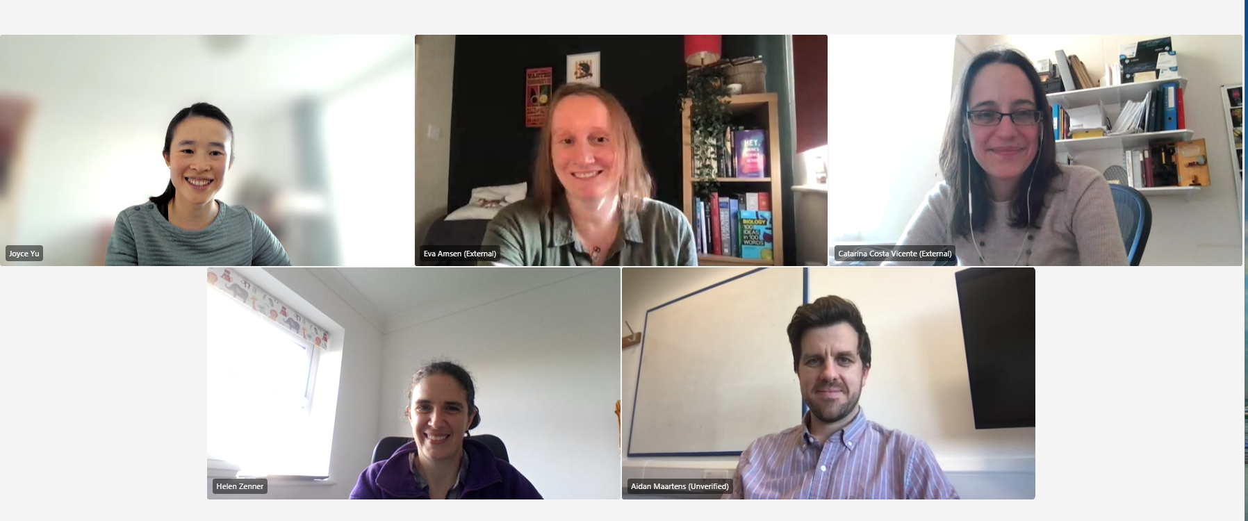
2025 is the 100th anniversary of our publisher, The Company of Biologists, and coincidentally, it’s the Node’s 15th birthday. To mark the occasion, the past and present Community Managers got together over a video call to chat about what it’s like working at the Node and how this job has influenced their subsequent careers.
Who are we?
Eva Amsen: I started in January 2010 when the Node didn’t exist yet. We launched the Node in April 2010, at the BSDB meeting. I left in 2013 to find a job in London, because I couldn’t get used to living in Cambridge. I then spent several years working on similar things as I did at the Node, such as managing blogs, talking to researchers, and writing about what they work on for different employers. But I’m now a freelance science journalist so I write a lot more now.
Cat Vicente: I was at the Node from 2013 till 2016, carrying on from Eva’s excellent work. We refurbished the website. The Company had a complete rebrand and moved into a new building. Then, because of personal reasons, I moved to Oxford. Since then, I’ve done a variety of jobs around communications and public engagement, working closely with scientists. In 2020 I decided to change tack completely. My job now is called Head of Scientific Strategy and Projects at the Sir William Dunn School of Pathology, University of Oxford. I still do a lot of writing.
Aidan Maartens: I took over from Cat in 2016 and stayed until 2021. Like Eva, I wanted to do a bit more writing. I’m working now as a scientific writer at the Wellcome Sanger Institute. It’s very varied. I edit grant proposals, papers and reviews, but I also do quite a bit of writing. I feel more engaged with the practice of science now, in a place where I interact with scientists day by day rather than remotely.
Helen Zenner: I took over from Aidan in 2021, although we didn’t overlap. I joined when everything was still remote. Then, after a year, I decided to move across to FocalPlane, the microscopy community site, still at The Company of Biologists. My research background is more in microscopy than developmental biology so I’m enjoying getting back to that.
Joyce Yu: And I’m the current Community Manager of the Node!
Top left to right: Joyce Yu, Eva Amsen, Cat Vicente; bottom left: Helen Zenner and bottom right: Aidan Maartens
What motivated us to join the Node? Was the Node our first job out of academia?
Eva: I finished my PhD about a year before I started at the Node, but I was still teaching at the University of Toronto while I searched for science communication jobs in different countries. I actually applied to the Reviews Editor role at The Company of Biologists, but when they saw my CV and my experience as a science blogger they instead invited me to apply for the Online Editor job. The Node didn’t exist yet, so they couldn’t tell me much until the interview itself, where they explained the plan to set up a community blog. I remember being asked how I would do that and I know that I had lots of ideas to make it a community rather than just a website, but I don’t remember which of these actually made it on the site!
Cat: I joined the Node straight after my PhD. I went to a couple of postdoc interviews, and it became clear quickly that while I really liked science, I didn’t like any particular aspect of science enough to dedicate several years to it. I remember the day I made the decision to apply for the Node: I was at the ASCB meeting, and I didn’t go to a single talk related to my PhD. I went to random talks I was interested in.
Aidan: I was similar. I knew during my PhD that I wasn’t going to go down the academic career path, but liked lab work and decided to do a postdoc. I really enjoyed this too, but found myself more interested in the bigger picture of science. Then the Node job came up. I always liked writing, and loved writing my thesis, which is not the case for some PhD students. And I found the communication of science to be a very important and worthwhile thing, so I applied.
Helen: I really enjoyed doing research, but I was ready to do something different. The Node really appealed to me because I remained in touch with scientists, and in some ways, it felt like you were still in academia.
What actually is a Community Manager?
Aidan: When I first saw the job advert, I didn’t know what a Community Manager was.
Eva: I actually told them to call the role ‘Community Manager’ because initially it was just called ‘Online Editor of Development’. But that’s not what the job is. If you look at other websites, the people who have similar jobs are called Community Managers.
What was it like working at the Node and Development?
Cat: For me, working at the Node made the transition out of active research relatively easy, because at The Company of Biologists you’re working with many people who used to be scientists. If I’d gone from a PhD to the job I did after the Node, it would’ve been too big a jump. It was also very good training for writing, as you constantly receive feedback from trained editors. I was a cell biologist by training, so one of the things that I was positively surprised about going into developmental biology was the sense of community. The job also involved a lot of travelling, which at that point in my life I had time to do.
Aidan: I agree that this job allowed you to have a foot in both camps. I was still talking to scientists all the time and going to conferences. While the Node was a big part of the job, a lot of my time was doing things for Development, like writing Research Highlights. I also got involved in commissioning review articles and saw the publishing side of things. The variety of tasks helps you figure out what you want to do. What I realised, quite like Eva, is that I really like writing and editing.
Cat: That’s really interesting, because I like writing, but I don’t like it enough to do it all the time. So, I really liked the interactions with different people, running projects and trying new things. It’s such a varied job that it fits different personalities!
Helen: I would be more similar to Cat in that I enjoyed talking with people, and the variety of the job. No two days are the same, and I think that different CMs at the Node can prioritise different things.
How did the role of the Community Manager evolve over the years?
Joyce: Apparently when they set the initial budget, they were hoping that our job would no longer be needed after 3 years, and that the Node could run by itself!
Eva: We thought it would become easier, but because you constantly have new PhD students and postdocs, you keep having to draw people in. The people who were regularly writing 15 years ago are now doing other things.
Joyce: How about the community aspect of the site, such as getting people to actively comment and discuss on the Node?
Eva: Back in 2010 every site like the Node had a comments section. If we didn’t have it, people would have complained, even if they never planned to leave a comment. Twitter existed, but it wasn’t as popular yet, so people did have discussions in the comments once in a while. I’m actually surprised that you still have comments, but maybe with how social media is now, it’s a good thing to have the discussion on your own site.
Aidan: By the time I joined, a lot of the discussion was on Twitter, so for me, it wasn’t a big part of the job to try to cultivate the commenting culture. From 2016 to 2018, I was going to conferences and live tweeting. That was really fun, and you met a lot of people through it, as they’d recognise the Node from Twitter. It was good for networking and for promoting what we did.
Helen: You tweeting from conferences was fantastic, Aidan, but it’s so tiring and you set quite a high bar on live tweeting!
Aidan: I think I ended up using it as a way of staying engaged, because if I had to write a tweet for each talk, I couldn’t have a nap.
Cat: I think an important thing to highlight is that the traveling was great – I loved it, but boy, was it hard work. You’d go to meetings with a list of people you wanted to talk to. You had to tweet from every talk and go to every networking opportunity. You never really turned off. I always felt like I had to explain this to people, because otherwise it looked like you were being flown around the world to have fun!
What did we take away from working at the Node?
Eva: My job now involves interviewing a lot of scientists. How to ask questions about someone’s research, how to approach a Nobel Laureate at a conference – these are things I learned to do when I was working for the Node and Development, especially when I was asked to profile the presidents of different societies. It was really scary at the start, and I would prepare for hours. Now it has become a skill that I don’t even think about anymore. And last year, I got to write about developmental biology again! I wroteBiology: 100 Ideas in 100 Words for the Science Museum. It was fun to dig up some of the old knowledge that I learned.
Cat: I think the interviews are one of the things I remember most fondly. Eva, I don’t know if you felt the same, but I think it’s quite empowering to understand the power of being a non-specialist, and asking the question that isn’t the obvious one. I also want to highlight the power of networking when you’re in a position like the Community Manager. One of the people I interviewed when I first started at the Node was Maria Leptin, who was the head of EMBO at the time. I’ve since met her in my current job, and she remembered that I’d interviewed her all those years ago. This made the introduction much easier because we had a connection already. This demonstrates that even short interactions can help you build your career.
Aidan: I developed my writing and editing skills, plus got to work with an amazing group of people in The Company of Biologists, which does so much good across life sciences. And I came to appreciate the vital importance of community for science.
Finally, what are our memorable contributions while at the Node?
Cat: I started the series called A day in the life, where you follow the day in the life of working with different organisms. Those are my favorite posts (especially the one about sharks!). The variety of research organisms is a really interesting aspect of developmental biology. Another thing I’m proud of is persuading the Company that we needed Christmas decorations. So perhaps that’s my legacy – the office has a Christmas tree now!
Eva: I have two posts I commissioned that I thought were really interesting. One is an early post from September 2010 by Kim Cooper, who’s an amazing writer and has written many posts for the Node. In her first post she introduces the jerboa as a research organism. That post was popular, and I would use it as an example to show how you can write about your research in a casual and fun way. Another memorable post I commissioned is this ‘behind the paper’ story by Tohru Yano from Japan. We noticed that an article submitted to Development was from a lab affected by the 2011 Tohoku earthquake. In the post, Tohru showed photos of the disheveled lab and microscopes on the floor. It was interesting to get the behind the scenes on how they dealt with the earthquake and how it affected their work.
Aidan: Towards the end of my time at the Node, I started ‘The people behind the papers’ interviews. These posts profile the first and last authors to find out what drives them, and to give context of the papers. That started in the Node and then went into Development – I think people really appreciated being featured. Another one is the Node Network, which is a global directory of developmental biologists for when you’re looking for a reviewer, speaker, or referee for a paper. You can filter by expertise and field, but also gender, ethnicity and other information. This idea came about around 2018, when there were lots of discussions about who gets to do science and who gets to have their voices heard.
Helen: Talking about representation, we have a lot of readers and contributors from the UK, US and Europe, but we wanted to highlight people outside of these places as well. We started the Lab meetings series, which gives each lab an opportunity to introduce themselves. We make it easy for them to participate, and they can self-nominate too.
Joyce: Those are all great examples. There’re so many amazing posts that are buried deep in the archives. When I joined, we finally improved the Node’s search functionality. You can now filter by things like author, year and category. Hopefully this makes it easier for people to revisit all the great content on the site.
A huge thank you to Eva, Cat, Aidan and Helen for building the Node community over the past 15 years!
2025 is the 100th birthday of The Company of Biologists, our not-for-profit publisher. Another birthday this year, albeit celebrating a much smaller number, is the Node’s 15th! If we take the time machine back to 2010, one of the events that happened in our little corner of the world was the launch of the Node. In June that year, Development and The Company of Biologists brought the first ever online developmental biology community site to life.
What did it take to build a community site from scratch? What even is a community site? Follow along to find out how the Node was born.
Step 1) Set the scope.
The idea of a ‘community blog’ started in late 2008, when Development surveyed members of the developmental biology community about the journal’s strengths and weaknesses. Although the survey did not explicitly ask about the journal’s website, many comments were along the lines of wanting ‘a more personal and interactive experience’ online. We called Development a community journal, and according to the survey, people wanted us to be just that: an online community for people to interact, gossip, share events and job openings, and write about research advances and all kinds of research-adjacent issues – somewhere that would be a one-stop-shop for developmental biologists.
While the community site would be hosted by Development, we didn’t want to limit the blog’s content to our journal, as we wanted to place ourselves as THE site for all developmental biologists – featuring research published in a wide range of journals and providing a forum for conference organisers, societies and other relevant organisations worldwide to reach out to the community. From the beginning, the idea was that anyone with an interest in developmental biology could post on and interact with the site.
Step 2) Set aside the budget and get the Board’s approval.
It wasn’t difficult to convince the Board to approve the proposal of setting up a community site. According to the Board meeting minutes, “it was unanimously agreed that Development should appoint an Online Editor to run their community website.” At its inception the Node was backed by a 3-year budget, which was a relatively long time for a digital ‘experiment’. The budget included the design work of the logo and the website, creation of webpages, and a lot of background coding work. Interestingly, the initial budget included ‘podcasts’, but that idea was quickly shelved.
Step 3) Pick a name for the site.
Do you know the Node could have been called ‘Out on a limb’? It is unclear how seriously this name was considered, but the name was mentioned in one of the brainstorming documents from 2009.
Eventually, ‘the Node’ came up in an email titled ‘Eureka!’ sent from Development’s then Executive Editor Jane Alfred to The Company of Biologists’ publisher Claire Moulton – two important people behind the inception of the Node – and the blog finally had a name!
“The Node felt like the perfect name for a community website that was aiming to be a hub of information and knowledge exchange for developmental biologists. It’s also a term that many development biologists will be very familiar with given that the node in developing embryos is a vitally important tissue that organises and orchestrates vertebrate development.” — Jane Alfred
Step 4) Hire a Community Manager.
Jane and Claire wanted to find someone with experience in blogging and online community building to help set up and run the site. In early 2010, Eva Amsen joined the Node as its first Community Manager.
In this conversation with the past and present Community Managers, Eva recounts how she applied to a different role within the Company but was then invited to apply to the Node position. Because Jane and Claire didn’t want to give away too much in the job advert, Eva wasn’t told what the job was really about until the interview!
Initially, we thought that once we got the momentum going, the Node would be able to run itself, without requiring the day-to-day management and commissioning of content from the Community Manager. But 15 years on, the fact that I (the current Community Manager) am writing this piece, suggests a more hands-on approach is required to keep the site running. As there’s a constant flow of new people coming into the field, it’s useful to have someone promoting the Node to new generations of developmental biologists.
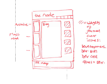
Step 5) Design the homepage.
To be the one-stop-shop for the developmental biology community, and more than just a blog, it was important to include features like jobs board, events calendar and resources (listing related societies, databases, and journals).
A freelancer was hired to set up the website and some in-house resource was available to ensure the continued smooth running of the site. Over the years, the website became more professionalised when we formed a long-term partnership with a technology consultancy – something that has proved central to our ability to grow and develop the site.
Early draft of the Node’s homepage (Reposting with permission from Jane Alfred)
Step 6) Write the policies and T&Cs.
A very important, but admittedly not as exciting piece of work was to set out the general guidelines, commenting policy, privacy policy and the Terms and Conditions. The team studied other similar science and journal blogs (there weren’t that many at that time) and came up with a set of guidelines to ensure the platform is inclusive and that discussions are carried out in an appropriate tone. One thing we worried a lot about was whether some of the topics we were likely to feature on the site might attract negative attention from, for example, anti-abortionists or creationists. While the Node is aimed at a scientific audience, we needed to have policies in place to ensure that we could handle controversial or inappropriate content should it arise. Fortunately, this hasn’t been an issue for us and in 15 years of the Node, we’ve only had to moderate its content on a handful of occasions.
Step 7) Populate the site with posts.
An empty community site would not feel very community-like. In order to make sure there was already something interesting for people to read when the Node was launched, Eva Amsen, the first Community Manager, was tasked with populating the site with content in the months leading up to the launch. That’s why even though the Node was officially launched in June, you can find posts dating back to April 2010. Curious to find out what the early posts were about? You can go down the memory lane using our (new-ish) improved search function by filtering according to the year and month.
Step 8) Organise a launch party!
After months of hard work, what better way to launch the Node than to have a party? The launch party was held at the Gurdon Institute, Cambridge, where people from the institute and developmental biologists in the area could have a browse around the Node and sign up an account on the spot.
15 years on, the (online) world has changed drastically, but the Node hopes to stick around, serving the developmental and stem cell biology community for years to come. Please continue to read and contribute to the Node – the Node wouldn’t have lasted for 15 years without your support!
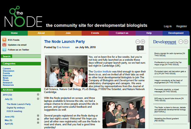
How the Node looked when it was first launched in 2010 (From the Wayback Machine)
Vicente C, Maartens A, Brown K. The Node and beyond-using social media in cell and developmental biology.Semin Cell Dev Biol. 2017;70:90-97. doi:10.1016/j.semcdb.2017.05.009 – published as part of the special issue “Science communication in the field of fundamental biomedical research”
With one week to go until Biologists @ 100, we can’t wait to see everyone there! Do you want to know more about the conference venue, the social events and where to find out more about the programme? Together with preLights and FocalPlane, we’ve recorded a video to walk you through the practicalities of the conference.
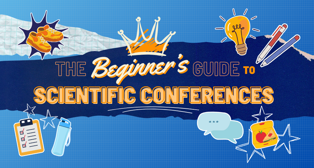
First-time conference attendee? Not sure what to prepare? Check out this ‘Beginners’ Guide to Scientific Conferences‘ from Jen Annoh, who will be attending the conference as a reporter for the Node, focusing on the cell and developmental biology scientific track of the programme.
Full transcript
Hi, I’m Helen, and I’m the Community Manager of FocalPlane.
Hello, I’m Joyce, I’m the Community Manager of the Node.
Hi. My name is Reinier, and I’m the preLights Community Manager.
We’ve made this short recording to tell you about our upcoming conference, Biologists @ 100. We’re really excited to have you join us for this special conference celebrating our 100th anniversary, and we hope that this recording helps you make the most of your experience. Let’s dive in.
So, starting with the venue. Biologists @ 100 will be at ACC Liverpool. On our conference website, you’ll be able to find all the information about travel and travelling to the conference venue. The ACC Liverpool entrance is actually not facing the river, so if you see the river, the entrance of the ACC Liverpool is on the other side. Hopefully this photo will help you to find how to get into the conference venue. There is an atrium where you enter and collect your badge.
The main auditorium is where most of the plenary talks will happen. Then the catering, exhibition and posters, will all happen in the big hall called hall two, which is in the basement. There will be lunchtime sessions held in a small theatre inside this hall. A little bit more about the exhibition hall; you will be able to find out more about the Company at our stand, and you’ll also be able to visit all the sponsor booths. Around the conference venue, you’ll find some discussion tables where you can tell us about your views on the future of publishing and help shape our journals. In the exhibition hall, there will be a sustainability area where you can find your tree in the Forest of Biologists, if you’ve ever published or reviewed for us, and you can chat to people and give suggestions about making science more sustainable. There will also be an image gallery, displaying images from the Node and FocalPlane image competition, and you’ll be able to vote for your favourite image.
As Joyce mentioned, there will be lunchtime lectures. The first one will be on Tuesday, called climate change challenges and solutions in biology. The panellists are passionate about this topic and will tell us a bit more on how to integrate sustainability in our day-to-day work. Do make sure to check it out. On Wednesday, we have a talk from Katherine Brown, Executive Editor of Development. She’ll talk about scientific publishing, and specifically, the future of scientific publishing. Then on Thursday, it will be the three of us talking about the community sites, what we do, and how you can get involved. So, please do drop by if you have any questions.
You can find us quite easily because we’ll be wearing these beautiful T-shirts. Also, we’ll have lime-green lanyards, as will our colleagues. So, you should be able to find those quite easily. Also, you’ll be able to see our conference reporters walking around the venue. They are representing the different community sites. There’s three of them, so it’s Jen, representing the Node, Jonathan for preLights and Margarida for FocalPlane.
To find out all you need to know about the conference, we’ll have a dedicated conference app, and this will have the full programme. You’ll also be able to access the abstracts for the poster sessions, find speaker information, and read about our sponsors. If there are any changes that happen during the conference, then this will be the place to find out. The conference app also has a chat function, which can be a great way to contact people and arrange to meet, so you can optimise your networking experience.
Going back to the programme, when you signed up, you’ll all have seen the wonderful diversity of speakers that we have through the sessions. The preliminary program is up on our website, and as I mentioned, the full program will be available on the app. Because this is a special conference incorporating all of the Company journals and all of the themes that link to the biology that we publish, there are a number of different strands. Importantly, we’ve made it possible for you to move between the strands. There’ll be signposts around the venue helping to direct you to the correct rooms. The sessions are running concurrently, and you will be able to move between lectures, but if you are moving in the middle of a session, we do recommend, that you sit towards the end of a row and try to minimise the disruption as you move around.
Now on to posters. All posters can be put up for the entire duration of the conference. If you have a poster, you should receive some communication about your number and when you’ll be presenting. This will tell you when you’ll need to stand by your poster during the two evening poster sessions. You’ll need to put your poster up before the morning break on Tuesday 25 March. The posters should be grouped together by topic, so if you’re interested in a specific topic, you can go and find all the related posters.
Something else that is important: food and drinks. Every day, tea and coffee will be served. There’ll be two refreshment breaks, and there will be hot food at lunch. This will all be vegetarian or vegan for sustainability reasons.
Onto other fun stuff. There will be two social events on Monday. First, there will be the welcome reception at the Museum of Liverpool, which is a 10-minute walk through the Albert Dock from the conference venue. This will be followed by the ECR dinner at Revolución de Cuba in the Albert Dock, which is a 10-minute walk from the welcome reception and a five-minute walk from the conference venue. Then on Wednesday, we’ll have the gala dinner, which is at St George’s Hall. There will be shuttle buses from the conference venue to take you to the dinner. These will start at 7pm and return shuttles will run from 10pm.
Are you attending a scientific conference for the first time? Well, we’re preparing a pre-conference 101 guide for you, together with Jen, the conference reporter for the Node. Jen has been collecting some tips from people who have been to conferences, and on the screen here, you can see some tips from our preLighters, including, ‘don’t be afraid to talk to you people’ and ‘introduce yourself and your research to others’. For the full 101 guide, check out Jen’s post on the Node. The link is in the video description below.
Okay, so that’s everything from us. Hopefully we’ve given you a little flavour of what to expect at the Biologists @ 100 conference. Please come and chat to us whilst you’re at the conference, we’ll be happy to hear from you. So, that’s everything and we’ll see you in Liverpool.
Attending your first scientific conference? In my experience, the weeks leading up to it can feel equal parts exciting and daunting. Whether you’re presenting a poster or just looking to network with fellow researchers, it is natural to feel overwhelmed if you don’t know where to start. But don’t worry! Conferences are learning experiences, not only about science but also about how to navigate these vibrant academic gatherings.
As we are preparing for the Biologists @ 100 conference hosted by The Company of Biologists, we created a guide to help you feel prepared, confident, and ready to make the most of your time at any scientific convention. We’ve covered all the basics, from what to pack and wear to note-taking and networking, and even asked the scientific community to share some insider advice! And because everyone’s experience is different, we’ve included some tips for anyone who might feel insecure starting conversations in English or spending time in crowds.
So take a deep breath, grab a notebook, and let’s dive in! You’ve got this!
How to prepare
“To be prepared is half the victory”- and that’s especially true at a busy conference. Let’s look at a few simple ways you can ensure a smooth experience before you even leave home:
Get to know the location! – Find out where the conference centre is, how far it is from your accommodation, and the best way to get there and back. Pro tip: Look up a map of the venue in advance. Conferences usually have multiple sessions running in different rooms. Knowing the layout will help you navigate smoothly – and ensure you don’t miss lunch!
List your Must-See talks – Review the schedule in advance and highlight the talks you don’t want to miss. With multiple sessions running in parallel, you may have to prioritise – ah, the agony of Session Superposition! Look out for interesting poster abstracts, and make a note of the session and poster numbers. Bonus tip: Beyond the talks, keep an eye out for networking events, workshops, and socials happening during lunch, breaks, or in the evenings.
An Easy Way to Share Your Science – Even if you’re not presenting, it helps to have a quick way to introduce your research. Create a single slide summarising your work and generate a QR code to share it effortlessly – you can do this through your browser or using an online QR code generator. If English isn’t your first language, prepare 1-3 short sentences about your research in advance. That way, you’ll feel more confident when someone asks about your project.
Double check your morning alarms! – this one speaks for itself, and yes, I’ve learned it the hard way..
What to bring
Pack smart to make the most of the conference!
The poster (if) you will be presenting – this may seem obvious, but you’d be surprised how many of us have a story of a forgotten poster and last-minute replacements. If you want to avoid bulky poster tubes when travelling, try printing on canvas! This way, you can fold your poster and pack it in your bag.
Notebook and pen for taking notes (though you’ll likely get some freebies too ;) – if you prefer to take notes on a tablet or laptop, make sure you have your charger with you, but know that power outlets might be scarce!
Comfortable but smart-ish clothes – Academic conferences tend to have a relaxed dress code, so business casual is a safe choice. Jeans paired with a shirt or blouse work just as well. The key is to wear something that makes you feel comfortable and confident rather than self-conscious. Opt for comfortable shoes – you’ll be on your feet more than you think! And don’t forget a light layer, as an experienced attendee warns: “Conference venues are often (too) well-air-conditioned.”
A small snack – like a muesli bar or an apple. You might miss breakfast, or simply get hungry during a long session. It is better to have a discreet snack than to sit and starve!
What to do once you’re there
The big day has arrived! After registering and picking up your nametag at the entrance, you’re all set to explore. Dive into the sessions, check out the posters, and start making connections! Just don’t forget to silence your phone during the talks.
Be prepared for early starts and short breaks! – Conferences usually try to pack as much science into the day as possible. Try to get there at least 15 minutes before the first talk so you can find the right room and get a good seat. If you feel you may need to step outside during the sessions, sit near the back or at the end of a row. An experienced conference attendee suggested: “Go to the freebies booth!” And they are right! It is time to collect the mug/pen/notepad combo you’ll be using in the lab for the next year.
“Go to the freebies booth! also try to come up with questions after the talks. If you’re too shy to ask in the auditorium, at least write them down on your notebook. Practice your skills in coming up with good questions.”
Ethan Ewe
Take notes! – You’ll likely hear dozens of talks a day, and trust us, you will forget more than you remember. Make a note of the speakers’ names and their contacts. Don’t try to copy everything on the slides – use shorthand and focus on key phrases. Drawing a diagram might be quicker and more effective than writing down all details of an experiment.
Write down questions you want to ask. Even if you don’t get a chance to ask during the session, you may run into the speaker again. No better way to start up a conversation than with a burning question you had about their research.
“Don’t be afraid to talk to new people! Usually, one thinks that senior PIs or other researchers are not very approachable and that’s not the case most of the time.”
Felipe Del Valle Batalla
Ask questions! – Whether at a poster session or during breaks, don’t be afraid to ask people about their research, or their conference experience. It is a sure way to start conversations, and will allow you to connect with fellow researchers.
Go to poster sessions – You may have specific targets, but also spend time roaming around. Poster sessions are a great way to find fellow researchers in your area, and have been the birthplace of many collaborations. But try not to get stuck in one spot; you can always exchange contacts. Keep circulating!
Network with your fellow academics! – It may seem awkward at first, but the main point of conferences is to connect with researchers from all around the globe. If you’re unsure how to start, a smile and a friendly question works wonders. “Hi, I’m [Your Name]. What topic brought you to the conference?” or “What did you think of the last talk?” Don’t forget to exchange contact information so you can follow up on interesting conversations.
“Try to take the first step by introducing yourself and your research and benefit from this opportunity to make new connections that might be fruitful for your career.”
Jawdat Sandakly
If you’d like a bit more advice on networking, have a look at this brilliant article by Alex Eve, the Senior Editor of Development.
Bonus tips for a stress-free conference
You don’t have to do it all – It’s okay to take breaks or even skip a session to recharge. If you feel drained by crowds and small-talk, find a quiet corridor or terrace to ground yourself.
Ask for help – organisers and fellow attendees are usually happy to point you in the right direction if you are lost or have questions about the schedule.
Don’t be too self-conscious about using perfect English. Conferences are international gatherings, and people will be more interested in what you have to say than how, exactly, you say it.
Celebrate small wins – every conversation, new idea, or spark of inspiration is a success!
Attending your first scientific conference is a big milestone, and it’s completely normal to feel a little overwhelmed. Just remember – everyone you’ll meet was a first-timer once, and most people are happy to help if you need guidance. Whether your goal is to learn more about a topic, share your research, or connect with others in your field, the experience is what you make of it. Take it one session, one conversation, and one coffee break at a time.
Above all, don’t forget to enjoy yourself! Conferences aren’t just about discoveries – they’re a chance to meet the people behind the science and share experiences. You’ve worked hard to get here, and you have everything you need to make the most of it. Put your best foot forward and have a great time!
We’ll see you at Biologists @ 100!
If you want a practical guide to the Biologists @ 100 conference, from the venue, poster sessions to the social events, check out this pre-conference recording made by the Community Managers of the Node, preLights and FocalPlane.
An Editorial from the five Editors-in-Chief of The Company of Biologists’ journals
The Editors-in-Chief have written a joint Editorial, discussing the enormous challenges currently facing researchers in the US and how members of the community can support them through this difficult time.
“To our colleagues in the USA, we stand with you during this challenging period. The international scientific community recognises your contributions and the difficult circumstances you now face. Science is a global endeavour, and setbacks to research in one nation affect us all.
In these uncertain times, we must strengthen our international scientific networks. We must build new bridges of collaboration. We must speak with a unified voice in support of evidence-based policymaking and scientific freedom. Together, we can create a more resilient scientific community, with policies that strengthen rather than diminish research capacity. Science must transcend national boundaries and short-term political considerations.”
The response of the science community to the last month is not about one statement or action. It's about all of us with our different frames. For Science's part, we are doing what we have always done and have no plans to change. My thoughts on what we can do. www.science.org/doi/10.1126/…
US President Donald Trump is taking a wrecking ball to science and to international institutions. The global research community must take a stand against these attacks.https://go.nature.com/4kd1vIu
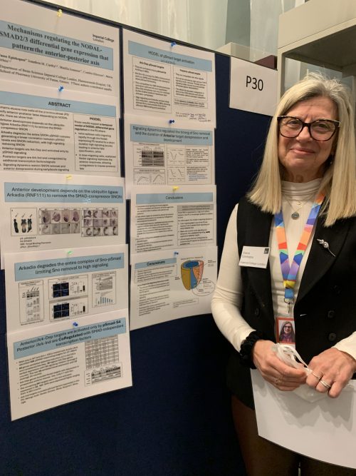
Although I originally set out to highlight the work of early career researchers at the symposium, Professor Vasso Episkopou’s overwhelmingly enthusiastic response coupled with my interest in anteroposterior patterning made her an obvious choice for an interview. At the end of the first day, although I was tired from a day packed full of interesting presentations and engaging conversations, I approached her poster – which was an unconventional, yet effective, series of laminated PowerPoint slides!
Vasso Episkopou presenting her poster.
Anteroposterior (AP) patterning in early vertebrate embryos is influenced by Nodal signalling – high levels promote anterior fates, while low levels drive posterior fates. You might therefore expect a gradient of Nodal along the primitive streak, but this is not the case; it is expressed ubiquitously in the streak. This discrepancy is the impetus of the research conducted by Vasso and her group at Imperial College London.
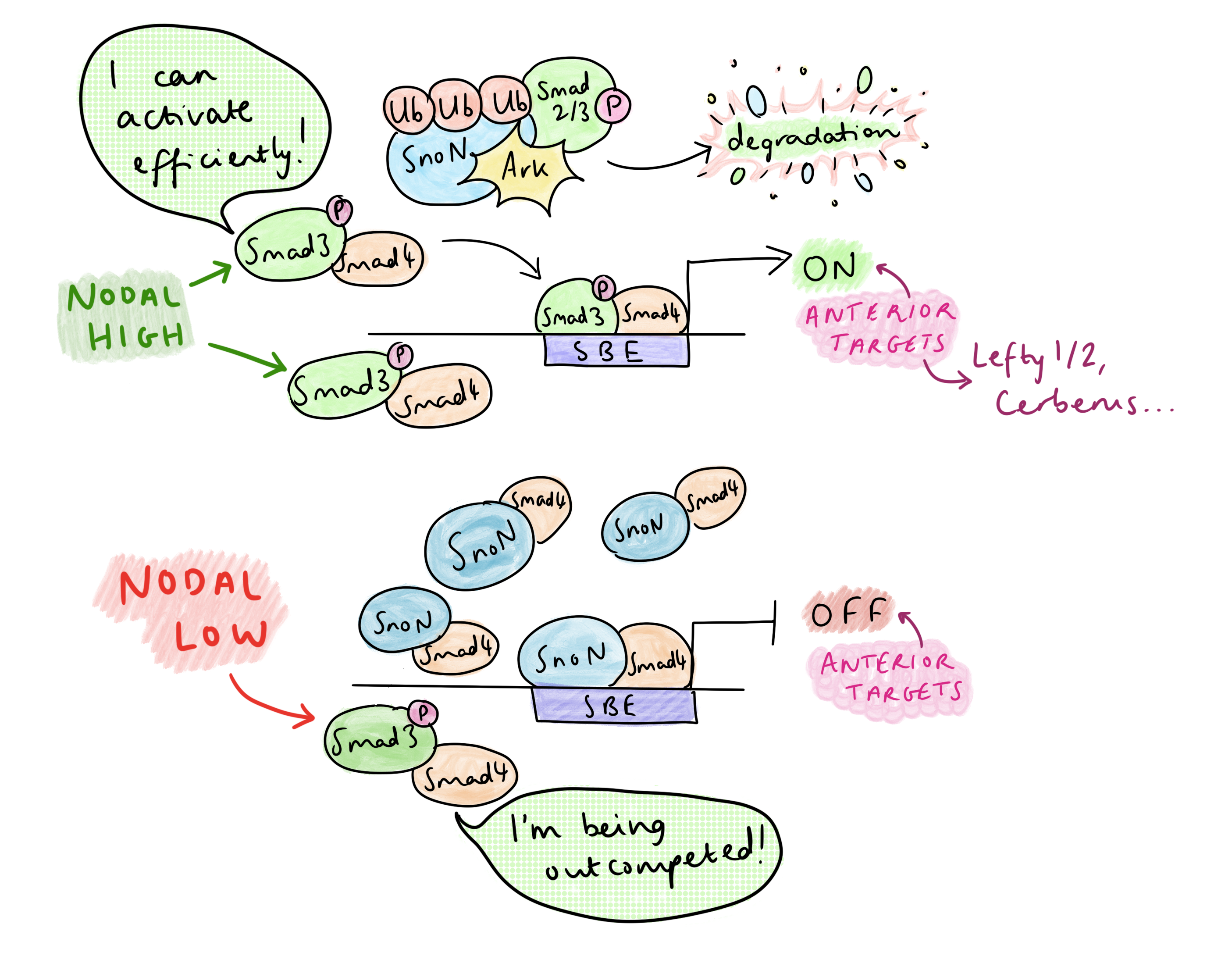
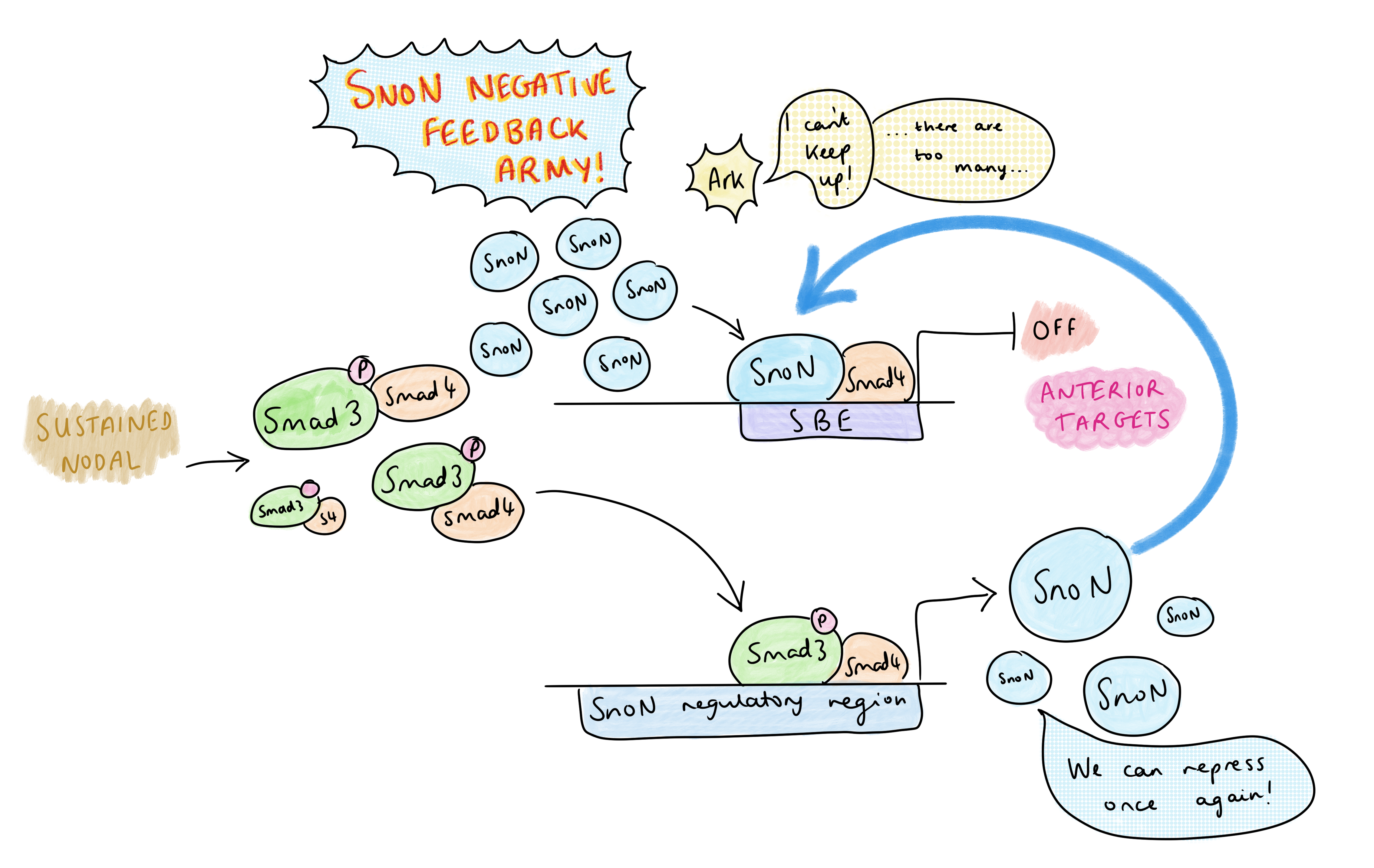
SnoN, a repressor of Nodal signalling, binds Smad4 to suppress anterior fate-promoting genes at Smad-binding elements (SBEs). Upon Nodal activation, phosphorylated Smad2/3 (pSmad) forms a complex with SnoN, which is recognized and degraded to its entirety by the ubiquitin ligase Arkadia (Rnf111). This surprising mechanism directly links pSmad levels to SnoN reduction, only forcing SnoN removal under high pSmad signaling. “The anterior target genes, they require Arkadia and high signalling, in order to degrade the repressor”, Vasso explained. The SBE is now clear, allowing pSmad-Smad4 to bind, and together with co-activators, initiate transcription of anterior genes. The necessity of Arkadia for anterior development is very clear in the headless phenotype of Arkadia-/- mice. In addition, head formation is rescued in Sno-/-;Ark-/- embryos, confirming that Arkadia is responsible for achieving the de-repression.
But Nodal signalling is abundant in the primitive streak – so how is AP patterning regulated? Vasso’s research group have identified that the dynamics of Nodal signalling control AP patterning in the primitive streak, rather than morphogen gradients. A TGF-B time course treatment of embryonic stem cells showed that, at high levels of signalling, there is a temporary decrease in SnoN at 1-2 hours due to its degradation by Arkadia, then SnoN levels increase again at 4-6 hours; this temporary reduction frees up space on the SBE for pSmad-Smad4 to activate transcription of anterior genes. The rising of SnoN levels at 4-6h is due to a negative feedback effect: one of pSmad’s early targets is SnoN itself, and the resulting increase in SnoN overwhelms Arkadia. This leads to a very transient activation of Arkadia-dependent anterior targets.
A sketch to show how, in the presence of high levels of Nodal signalling, Arkadia degrades the SnoN repressor complex, which allows the binding of pSmad at SBEs (Smad-binding elements) to activate transcription of anterior target genes.Sustained Nodal signalling causes anterior gene transcription to switch off, since pSmad activates SnoN transcription, leading to negative feedback.
On the other hand, in the presence of lower levels of Nodal, SnoN is not degraded by Arkadia, so there is competition between SnoN-Smad4 and pSmad-Smad4 for binding the SBE. Posterior Nodal targets do not require the SBE to be cleared completely – only a low level of Nodal signalling is required to relax the chromatin by pausing histone deacetylation, which permits co-regulators of posterior targets to bind (at other sites) and activate transcription. Since posterior genes are co-regulated by other transcription factors, they only need Nodal signalling for partial de-repression to become activated. This contrasts with the anterior targets, which can only be activated by pSmad-Smad4 after clearing of the SnoN from the SBE.
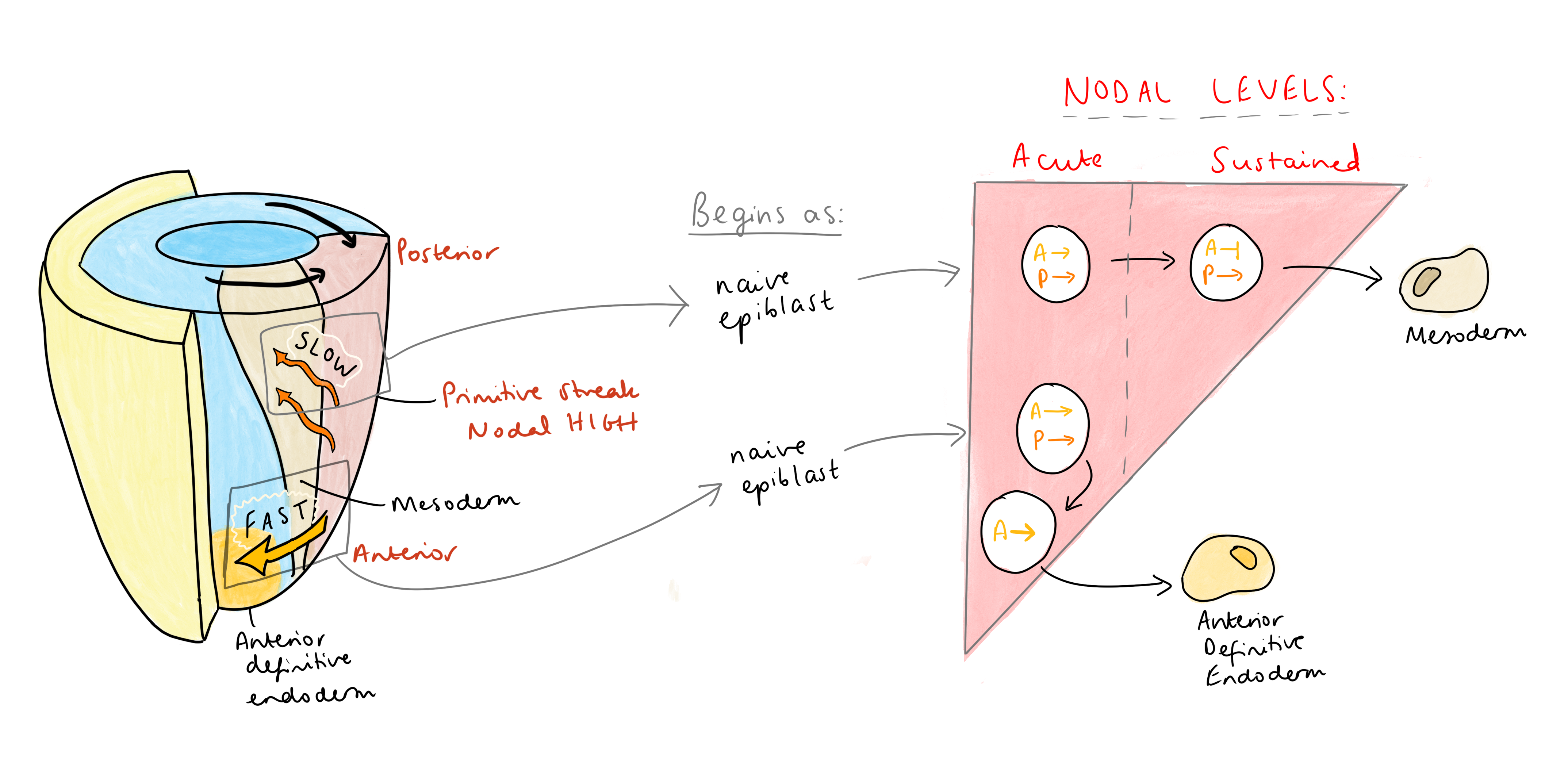
Altogether, this means that anterior identity in the primitive streak must be acquired in a very short time window, in response to acute, high levels of Nodal signalling, before any negative feedback can kick in. Vasso has put this together with what we know about cell migration in the streak – the first cells to exit, the fastest migrating cells, leave the streak via anterior migration and become the anterior endoderm. These cells also express Nodal antagonists, such as Lefty 1/2 and Cerberus, doubling down to ensure that their exposure to Nodal is very short-lived. “Cerberus is a triple attack,” Vasso explained, “a Nodal, Wnt, and BMP inhibitor. So, these cells express antagonists to shield themselves from sustained (posteriorising) signals”. These antagonists activate the transcription of immediate early genes, ensuring that anterior identity is swiftly acquired. In contrast, slower migrating cells acquire posterior identity, since remaining in the streak for longer exposes them to sustained Nodal signalling, leading to the repression of anterior genes and allowing co-regulators to impose a posterior fate.
In the early mouse embryo, slow migrating cells remain in the primitive streak for longer, so are exposed to sustained Nodal signalling, and acquire a more posterior mesoderm fate. Whereas fast migrating cells leave the primitive streak first, so are only exposed to Nodal signals for a short period. This allows them to acquire an anterior fate, such as anterior definitive endoderm.
Vasso suggests that this dynamic regulation of signalling may be a widespread mechanism in fate determination. A similar principle applies to T lymphocyte differentiation: high TGFβ levels drive Treg differentiation in an Arkadia-dependent manner, whereas low levels drive Th17 differentiation independently of Arkadia (Xu et al., 2021). This model may extend to other biological contexts yet to be explored.
You can read Vasso’s group’s preprint on Nodal dynamics here: Carthy, J.M., Ioannou, M. and Episkopou, V. (2019) ‘Arkadia via SNON enables NODAL-SMAD2/3 signaling effectors to transcribe different genes depending on their levels’. bioRxiv, p. 487371. Available at: https://doi.org/10.1101/487371.
Read about how the same system acts in T lymphocyte differentiation here: Xu, H. et al. (2021) ‘Arkadia-SKI/SnoN signaling differentially regulates TGF-β–induced iTreg and Th17 cell differentiation’, Journal of Experimental Medicine, 218(11), p. e20210777. Available at: https://doi.org/10.1084/jem.20210777.
Stay tuned for more poster interviews coming soon!
 (No Ratings Yet)
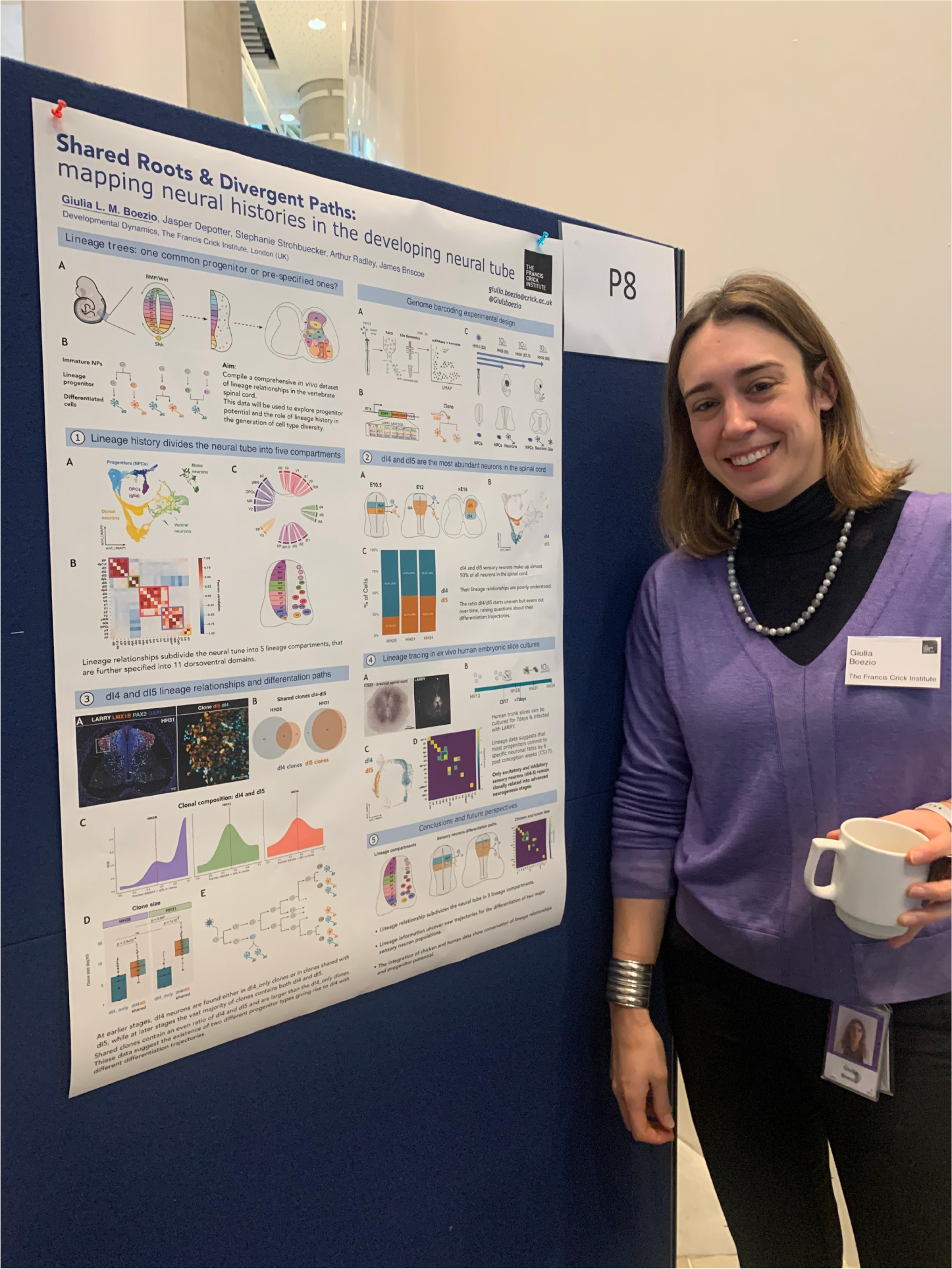
 (1 votes)