We have an opening for a postdoctoral position that will address fundamental questions in small RNA biology, genomic conflict, and speciation.
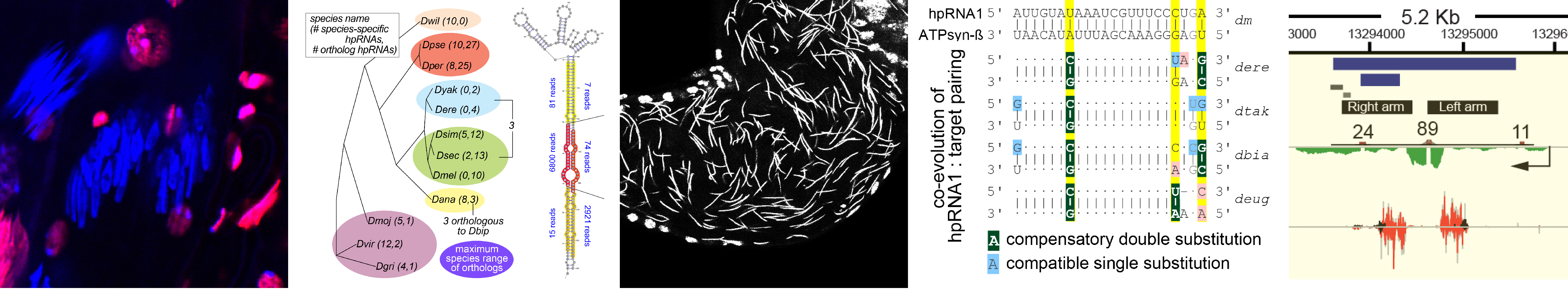
What is endogenous RNAi utilized for? We earlier described a mysterious class of endo-RNAi substrates termed hpRNAs (Okamura Nature 2008), and recently recognized that these mediate adaptive gene regulation in testis (Wen Molecular Cell 2015). These data open a window on a vital biology of RNAi, and now lead us to explore the evolution and function of RNAi systems across the Drosophilid phylogeny.
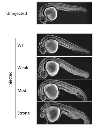
We discovered networks of rapidly evolving RNAi substrates we hypothesize resolve intragenomic conflicts, and successfully started to use CRISPR/Cas9 in non-model fly species to test some of these ideas (Lin 2018, in submission). Amazingly, while endogenous RNAi was not historically thought to have much phenotypic impact, we find that rapidly evolving genomic battles are being waged and critically depend upon the powerful weapon of RNAi silencing to propagate the species. Reciprocally, we are fascinated to understand by what novel molecular mechanisms de novo selfish meiotic factors can paradoxically drive population extinction.
We seek a motivated postdoctoral fellow with strong Drosophila molecular genetics experience and interest in integrating genome engineering, biochemistry, and bioinformatics to analyze the evolution and function of RNAi silencing systems in resolving deleterious intragenomic conflicts in testis. Although our entry point is RNAi biology, we anticipate that selfish factors yet to be discovered may mediate their effects through chromatin mechanisms.The successful candidate will integrate into a team that is actively engaged in diverse topics in gene regulation in Drosophila and mammalian models, and the Sloan-Kettering Institute provides a vibrant research community.
Funded position with housing and medical benefits are available immediately. Please provide CV, motivation letter and references to Eric Lai, laie@mskcc.org.
Relevant recent papers on hpRNAs and testis post-transcriptional regulation.
see also https://www.mskcc.org/research-areas/labs/eric-lai
Lin, C.-J., F. Hu, R. Dubruille, J. Wen, J. Vedanayagam, P. Smibert, B. Loppin and E. C. Lai (2018). The hpRNA/RNAi pathway is essential to resolve intragenomic conflict to preserve balanced sex ratio. Submitted.
Mohammed, J., A. Flynt, A. Panzarino, M. Mondal, M. DeCruz, A. Siepel and E. C. Lai (2018). Deep experimental profiling of miRNA diversity, deployment, and evolution across the Drosophila genus. Genome Research28: 52-65.
Kondo S., J. Vedanayagam, J. Mohammed, S. Eizadshenass, L. Kan, N. Pang, R. Aradhya, A. Siepel, J. Steinhauer and E. C. Lai (2017). New genes often acquire male-specific functions but rarely become essential in Drosophila. Genes and Development31: 1841–1846. (Highlighted in Genes and Dev 31: 1825-1826.)
Lin, C.-J., J. Wen, F. Bejarano, F. Hu, D. Bortolamiol-Becet, L. Kan, P. Sanfilippo, S. Kondo and E. C. Lai (2017). Characterization of a TUTase/RNase complex required for Drosophila gametogenesis. RNA23: 284-296.
Wen, J., H. Duan, F. Bejarano, K. Okamura, L. Fabian, J. A. Brill, D. Bortolamiol-Becet, R. Martin, J. G. Ruby and E. C. Lai (2015). Adaptive regulation of testis gene expression and control of male fertility by the Drosophila hairpin RNA pathway. Molecular Cell57: 165-78.
We are at the National Institute of Child Health and Human Development (NICHD) at NIH, Bethesda, Maryland USA. Our lab is interested in understanding cell lineage differentiation, gene regulation and how non-coding DNA elements and the 3D architecture of chromosomes contribute to these processes during early mouse development.
You share our enthusiasm for epigenetics, gene regulation, nuclear organization and mouse development.
You have PhD-experience in one or more of the following: mouse development, mouse genetics, epigenetics, massively-parallel sequencing techniques or computational biology.
What we offer:
Fully-funded postdoc positions.
Opportunity to start your own research program or lead ongoing projects.
2 paragraph cover letter explaining your scientific trajectory and why you would like to join us.
CV and email contacts for 3 references.
The NIH is dedicated to building a diverse community in its training and employment programs.
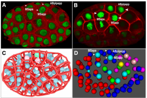
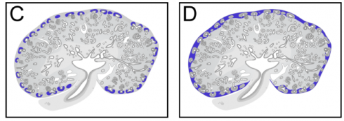
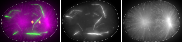
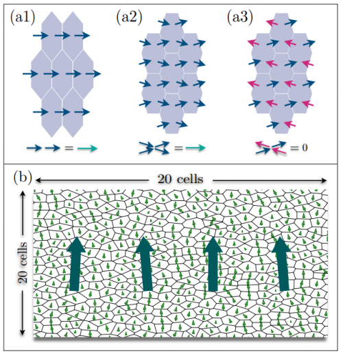
We combine imaging techniques in both fixed and living cells with sequencing- based genomic techniques that assess DNA-DNA interactions. (A) Hi-C and CTCF ChIP- seq of GM1278 cells (B) dCAS9 MCP-EGFP and PCP-CHERRY live imaging of the Igh and Akap6 loci. The mouse embryo is an unparalleled system in mammalian biology for understanding how tissue- specific gene expression is achieved. (C) Whole mount in-situ hybridization for patterning markers in mid and late gastrulating embryos. (D) Tetraploid aggregation with GFP ES cells allows generation of fully ES-cell derived embryos.
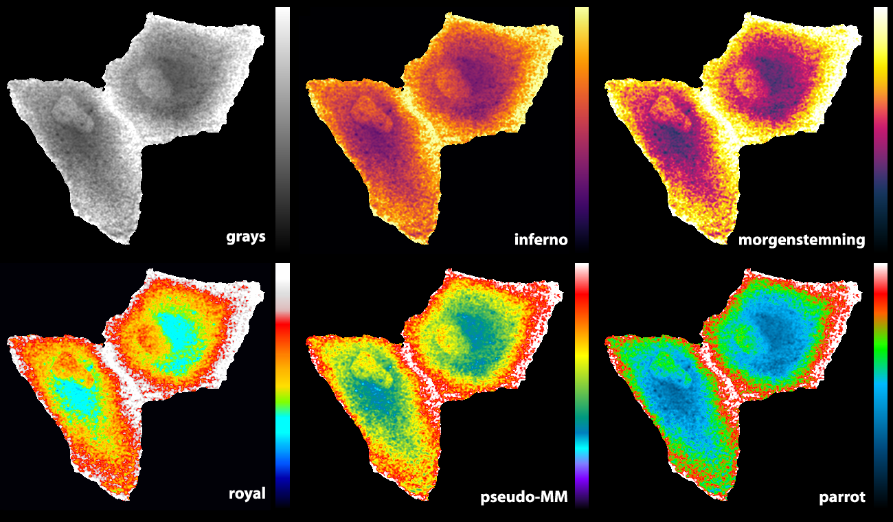
In a previous blog, I have disgraced parrots by associating them with P-values and discrediting them for their mechanic repetition. Nevertheless, I admire the vivid colours of these multifaceted birds. Here, I want to make it up by dedicating a pseudo-colour look-up table (LUT) to parrots.
The images produced by fluorescence microscopy are best displayed in grayscale for optimal contrast, using a linear relation between gray-level and pixel intensity (footnote 1). However, the number of gray levels that can be distinguished by humans is orders of magnitude smaller than the number of colours that can be discerned. Therefore, pseudo-colours (also often named false colours) can be used to achieve better contrast and convey a finer level of detail (Sheppard et al., 1968), at the cost of loosing the relation between intensities in the image and the actual pixel value. Thus, the choice between grayscale representation and pseudo-colour will depend on the information that is visualised.
A monochrome digital image can be converted into a pseudo-colour image by replacing each pixel value with a colour (Fink, 1976). The conversion is defined by a so-called look-up table (LUT). The LUT contains 256 RGB codes that define a colour for each of the pixel values between 0 and 255. The combinations are basically endless. A collection of LUTs for cartography, technical illustration and design is available here and the popular image processing software ImageJ/FIJI comes with a set of LUTs.
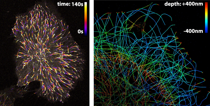
The LUT that is applied to an image largely depends on the information that one wants to visualise. Since the LUTs are critical as to the level of detail that can be discerned, careful selection is important. Here, I highlight different LUTs in the context of fluorescence images. Besides fluorescence intensity, several other parameters can be encoded by colour. Examples are (i) temporal encoding (figure 1 left), (ii) depth encoding (figure 1 right, and see several other examples here), and (iii) encoding of a spectroscopic parameter, e.g. fluorescence lifetime (Joosen et al., 2014).
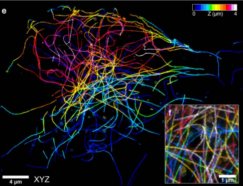
Figure 1: Pseudo-colours are used (in the left panel) to encode time to show the dynamics of microtubule plus ends or (in the right panel) to encode depth in a 3D STORM image of microtubules. Image credits: Timelapse movie of EB3-mNeonGreen by Anna Chertkova, 3D STORM image by Christophe Leterrier.
Here, I will give examples of pseudo-coloured images that were obtained by FRET ratio-imaging. In FRET ratio-imaging the response of a biosensor is monitored by emission ratio-imaging. Both the spatial differences and the changes over time can be of interest. The spatial differences are usually presented with pseudo coloured maps of the FRET ratio. Movies of these FRET maps are generated to depict the ratio changes over time.
The ‘Fire’ LUT that comes with ImageJ works well for temporal colour coding (figure 1, left panel). A variant of the Fire LUT was generated and named ‘MorgenStemning’. This LUT is colourblind friendly and has a linear increase in luminance (Geissbuehler and Lasser, 2013). Similarly, several “perceptually uniform” colour palettes were generated that are also colourblind friendly. These LUTs, ‘Inferno’, ‘Viridis’, ‘Magma’ & ‘Plasma’, are described here. The Inferno LUT is a good choice, since it starts with black at zero intensity. This LUT does, however, not use white for maximal intensity, thereby not providing maximal contrast.
Some of the LUTs that we previously used are the ‘royal’ LUT of ImageJ (Unen et al., 2015) and a pseudocolor LUT that is part of MetaMorph software (Reinhard, 2016). Since I was not entirely satisfied with the existing LUTs, I designed a new LUT (footnote 2). The basic features are that it starts with black and ends with white to achieve maximal contrast between the minimal and maximal pixel values. The false colours that correspond with lower half of the pixel values does not have a red component to get a cyan colour which blends into bright green. For higher pixels values the false colours turn from green via orange and red to white. As the colours in the LUTs reminded me of parrots, I call this LUT ‘parrot’. Figure 2 shows how the parrot LUT compares to other LUTs. The parrot LUT is colour-blind-friendly (footnote 3), but it is not perceptually uniform and is suboptimal in this respect.
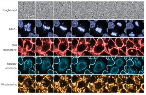
Figure 2: An overview of different LUTs applied to the same grayscale image that displays Rho GTPase activity in endothelial cells (Reinhard, 2017). The image and LUTs are available here.
Final words
The choice of a LUT is often based on a qualitative assessment of how it visualises the information. By trying a number of LUTS, the LUT that best conveys the information can be selected. Therefore, it useful to have a broad panel of LUTs to choose from and the parrot LUT is just another option. I encourage designing and sharing of LUTs (either as supplemental data to go with a paper or on data sharing platforms – footnote 4) to increase the number of options and the chances of finding the right LUT.
Acknowledgments: Thanks to Jakobus van Unen for tracing back the origin of the ‘pseudo-MM’ LUT to metamorph and to Marten Postma for making the LUT editor available.
Footnotes
Footnote 1: Achieving a true linear relationship between pixel value and displayed grayscale value may be impossible and irrelevant.
Footnote 2: The parrot LUT was created with a MatLab based LUT editor that was generated by Marten Postma
Footnote 3: I have a colour vision deficiency that prevents me from distinguishing blue from purple and bright green from yellow. This is the main reason to remove most of the red component and only have a limited band with a green/yellow component. Since I designed it, it is intrinsically colour-blind friendly. However, as there are several types of colour vision deficiency it may not work for everyone.
Footnote 4: The LUTs and related material described in this blog are available at Zenodo (Doi: 10.5281/zenodo.1211690).
Development and homeostasis depend crucially on the maintenance of cell identity, and in gamete-producing tissues the somatic/germline distinction is paramount. A recent paper in Developmentexplores how cell identity is secured in the Drosophila ovary by studying the function of the conserved tumour suppressor L(3)mbt. To find out more about the story, we caught up with first author Rémi-Xavier Coux and his supervisor Ruth Lehmannof the Skirball Institute at New York University School of Medicine.
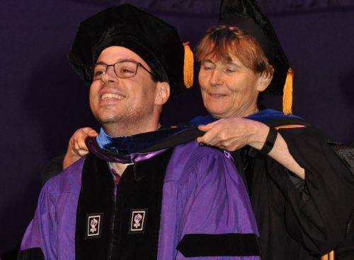
Remi and Ruth at graduation
Ruth, can you give us your scientific biography and the questions your lab is trying to answer?
RL I grew up in Germany and received my PhD in the lab of Christiane Nuesslein-Volhard. I was in her lab at the very beginning when the genes required for establishing embryonic polarity and pattern were identified. My project involved the genetic analysis of gap genes and a group of maternal effect genes, we termed the posterior group of genes, as mutation in these genes affect the development of the embryonic abdomen and my cytoplasmic transplantation experiments suggested a gradient of patterning activity emanating from the posterior pole. I became intrigued by this posterior pole plasm as it is also the site of germ cell formation in Drosophila. So, I made the more unusual and often discouraged decision to stay with my graduate project, yet decided to delve into molecular analysis of these genes. By the time I arrived for my first independent position at the Whitehead Institute at MIT in 1988, I had molecular entry points for the identification of the nanos, oskar and pumilio genes in my suitcase.
Analysis of these genes was really exciting as it showed that the mRNAs encoding Oskar and Nanos were localized to the posterior pole and that the translation of these RNAs was specifically regulated such that only the localized RNA (and now we know that is only a very small fraction of the total RNA) is translated while unlocalized RNA is translationally repressed. From the beginning, I have been amazingly lucky to have incredibly talented graduate students and postdocs in my lab, not only did they make all these discoveries but they also taught me a lot.
In 1996, I was recruited to the newly founded Skirball Institute at NYU School of Medicine. Here my lab focused completely on germ cell biology studying the germ line life cycle. We are particularly interested in three areas:
1. How the germ line-soma dichotomy is initially established in the early embryo. Critical for the initial distinction between soma and germ line are the properties of membraneless germ granules that co-ordinate the posttranscriptional regulation of RNAs specifically needed for germ cell formation, specification, transcriptional silencing and germ cell migration to the somatic gonad. Crucial for progress here has been the development of ever so powerful imaging modalities (from light sheet microscopy and super resolution microscopy to electron microscopy) and the development of many different ways to mark molecules and observe them in vivo.
2. How interactions between cells from different origins coordinate growth and differentiation of the gonad so that primordial germ cells mature into germ line stem cells and deposit eggs during adult life. Initially, we relied on forward genetic screens to address this big and fascinating problem of organ development but now we are increasingly take advantage of whole genome genetic analysis by RNAi and Crispr/Cas9 as well as high-throughput molecular analysis such as single cell sequencing at different developmental stages.
3. Finally, we are interested in how the unique role of germ cells as the only cells of the body with the potential to give rise to a complete new organism manifests specialized adaptions. So, we have become intrigued by the broadest sense of mutual ‘host-pathogen’ interactions as they relate to the germ line. Here, we are interested in how germ line regulatory mechanisms manage to control transposable elements activity, the interplay between genome and mitochondria during the germ line life cycle and emerging optional relationships like the ability of the intracellular bacterium Wolbachia to grow in the Drosophila host without direct harm. We reason, that such germline specific control and defence mechanisms protect the germ line, but may also provide opportunity for species to evolve via changes to the germline.
And Rémi-Xavier, how did you come to be involved with this project?
RXCl(3)mbt was identified in the laboratory of Elisabeth Gateff, who was the first to use Drosophila to identify tumor-suppressor genes (Gateff, 1978, Gateff et al., 1993). As the name says, ‘l(3) malignant brain tumor’ mutations cause brain tumors in Drosophila larvae. Many years later, Chris Yohn, a postdoc in our lab, identified several alleles of l(3)mbt in a clonal screen for maternally expressed genes that affected germ cell formation in the embryo. While Chris determined that the PGC formation defect was a secondary consequence of L(3)mbt’s role in early embryonic nuclear divisions, he also observed that l(3)mbt females were viable at the permissive temperature (brain tumors form only at high temperature) but they were completely sterile and produced no eggs. This suggested an additional role for L(3)mbt in gonadogenesis (Yohn et al., 2003). The project rested for a while, until Cayetano Gonzalez and his group at the IRB in Barcelona published an intriguing observation. The Gonzalez lab profiled l(3)mbt larval brain tumors and showed that in these tumors many germline genes were apparently derepressed (Janic et al., 2010). We were intrigued by this possible soma-to-germline transformation, so when I joined the lab as a graduate student I started two projects on l(3)mbt. First, I asked whether l(3)mbt brain tumor cells indeed behaved like bona fide germ cells and could be used to identify novel regulators of the germline fate. Second, I wanted to test whether the sterility phenotype was also due to a soma-to-germline transformation. Our paper describes the results of the later study that proved much more successful than the former.
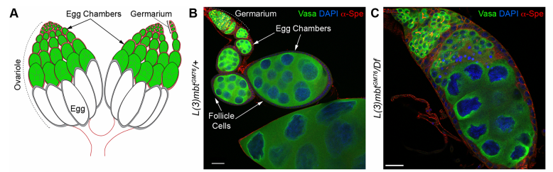
Drosophila ovaries and the l(3)mbt phenotype, from Fig. 1 in the paper
Can you give us the key results of the paper in a paragraph?
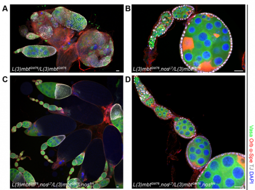
RL&RXC In addition to its role in suppressing brain tumors in the larvae, L(3)mbt functions in two tissues of the ovary to safeguard oogenesis: the somatic support cells and the germline. We found that in both tissues, L(3)mbt prevents expression of genes incompatible with normal development: in the somatic ovarian cells, L(3)mbt represses genes normally expressed in the germline while in the germ cells, it silences testis and neuronal genes. This, and the fact that l(3)mbt mutant tissues still express genes characteristic of the tissue of origin revealed a function broader than previously thought. Our study therefore suggests that L(3)mbt functions as a tissue specific transcription repressor rather than simply silencing germline genes in somatic tissues. L(3)mbt binding sites overlap with insulator elements (Richter et al., 2012) so it is also possible that L(3)mbt functions in insulator complexes.
Do you think L(3)mbt work with different partners in different tissues? Any ideas how it plays tissue-specific roles?
RL&RXC Indeed, L(3)mbt has been found to function with the dREaM/MMB and LINT complexes in S2 and Kc167 somatic embryonic cells (Georlette et al., 2007; Meier et al., 2012). However, our genetic studies suggest that L(3)mbt functions independently of dREaM in the ovary. Several components of the dREaM complex are required for endo-replication (polyploidization) of both somatic ovarian cells and nurse cells in the germline, but these processes seem not to require L(3)mbt function. It would be interesting to test if L(3)mbt functions in only one of these two chromatin complexes in other tissues besides the ovary and how the potential switch between complexes is regulated.
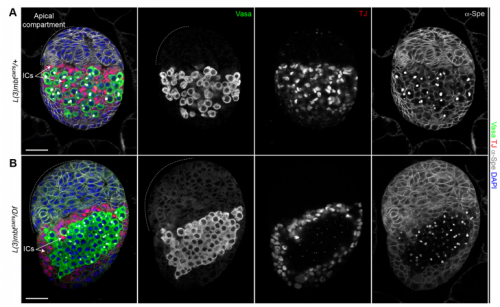
Larval ovaries from Fig. 3 in the paper
Your results suggest that rather than guarding against transdifferentiation, L(3)mbt guards against adoption of mixed identity. Where does this leave the concept/importance of transdifferentiation in development?
RL&RXC This is a conceptual question we really struggled with: the “orthodox” definition of transdifferentiation is complete acquisition of another cell identity. However, functionally testing fate switching in vivo is very challenging. Jarrault and colleagues beautifully showed that in C. elegans the Y epithelial cell transdifferentiates into a fully functional neuron (PDA) in wild-type larvae (Jarrault et al., 2008). It is one of the only examples of complete transdifferentiation to our knowledge. There are many more examples, especially with mutations in chromatin factors, where cell-specific gene signatures are misexpressed. At the end, it comes down to the assay that is used to define a ‘cell fate switch’. For example, can we call a cell that aberrantly expresses most of another cell type’s transcriptome trans-differentiated without a functional assay?
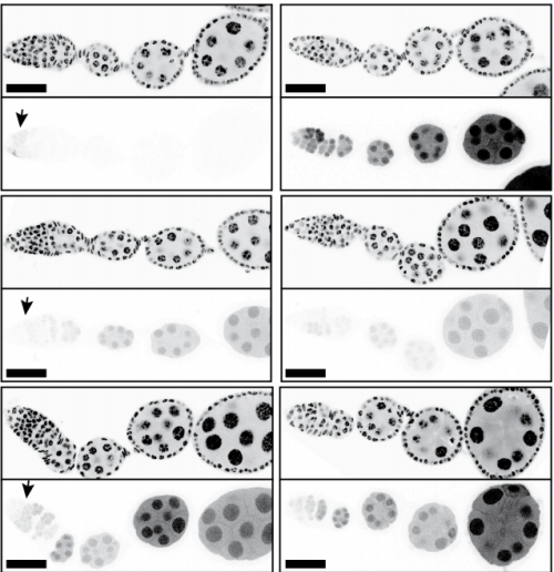
The l(3)mbt, nos double mutant ovaries, from Fig. 5 in the paper.
When doing the research, did you have any particular result or eureka moment that has stuck with you?
RXC Yes, I was very glad to observe that nanos mutations suppress the l(3)mbt mutant ovarian phenotypes. It was late at the confocal and I immediately emailed Ruth. The morning after, we checked that embryos laid by l(3)mbt, nos double mutant females had the typical patterning defects caused by nos mutations. When this control was done, we were really excited!!!
And what about the flipside: any moments of frustration or despair?
RXC I tried transplanting l(3)mbt tumorous brain cells into embryos devoid of germline to test if they could, at least partially, behave as germ cells. This experiment was quite challenging, and we could not detect the cells a few hours after transplantation, which was very frustrating! We now know that this experiment likely did not work because these neural-origin cells may not be completely transformed.
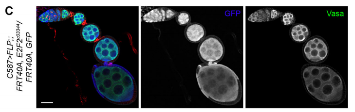
E2f2 mutant clones from Fig. 7 in the paper
What next for you Rémi-Xavier?
RXC I just started a postdoc in the Cohen-Tannoudji and Navarro-Gil labs in the Stem Cell Biology and Development Department, Pasteur Institute in Paris. I will study and characterize Transcription Factor bookmarking in the early mouse embryo.
Where will this work take the Lehmann lab?
RL Further study of L(3)mbt may provide us with clues about the transcriptional mechanisms underlying germline-soma dichotomy. We found in our study that mutating a key regulator of germline fate, the translational repressor Nanos, suppresses the somatic gonadal defects of l(3)mbt mutant ovaries almost completely. Thus, Nanos targets may be key regulators that distinguish between the germline and soma program and the l(3)mbt mutants may guide us towards their identification.
Finally, let’s move outside the lab – what do you like to do in your spare time?
RXC In my free time, I enjoy music, modern art, playing rugby and outdoor activities such as sailing and mountain activities.
One of the most talked about preprints this month was a corrigendum to that Nature Methods paper reporting widespread off-target mutations following CRISPR-Cas9 editing in mice. The paper was challenged quickly on bioRxiv, and four days after the corrigendum went up, it was retracted, as reported by Retraction Watch! Blink and you’ll miss it.
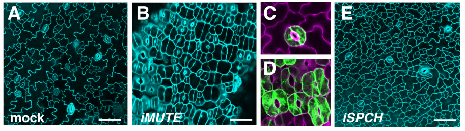
Otherwise this month we found plenty of plant patterning, lovely larval lampreys and ovulating roundworms, and the developing diaphragm finding its voice.
The preprints were hosted on bioRxiv, PeerJ, andarXiv. Use these links to get to the section you want:
Megan B O’Hare, Alamin Mohammed, Kyle J Connolly, Katelyn C Aitchison, Niki C Anthoney, Amy L Roberts, Matthew J Taylor, Bryan A Stewart, Richard I Tuxworth, Guy Tear
Analysis of novel domain-specific mutations in the zebrafish ndr2/cyclops gene generated using CRISPR-Cas9 RNPs
Ashley N Turner, Reagan S Andersen, Ivy E Bookout, Lauren N Brashear, James C Davis, David M Gahan, John P Gotham, Baraa A Hijaz, Ashish S Kaushik, Jordan B McGill, Victoria L Miller, Zachariah P Moseley, Cerissa L Nowell, Riddhi K Patel, Mia C Rodgers, Yazen A Shihab, Austin P Walker, Sarah R Glover, Samantha D Foster, Anil Kumar Challa
Deep proteomic analysis of chicken erythropoiesis
Marjorie Leduc, Emilie-Fleur Gautier, Anissa Guillemin, Cédric Broussard, Virginie Salnot, Catherine Lacombe, Olivier Gandrillon, François Guillonneau, Patrick Mayeux
A critical role for miR-142 in alveolar epithelial lineage formation
Amit Shrestha, Carraro Gianni, Nicolas Nottet, Ana Ivonne Vasquez-Armendariz, Susanne Herold, Julio Cordero, Indra Bahadur Singh, Jochen Wilhelm, Guillermo Barreto, Cho-Ming Chao, Elie El Agha, Bernard Mari, Jin-San Zhang, Saverio Bellusci
Chromatin accessibility dynamics across C. elegans development and ageing
Jurgen Janes, Yan Dong, Michael Schoof, Jacques Serizay, Alex Appert, Chiara Cerrato, Carson Woodbury, Ron Chen, Carolina Gemma, Ni Huang, Djem Kissiov, Przemyslaw Stempor, Annette Steward, Eva Zeiser, Sascha Sauer, Julie Ahringer
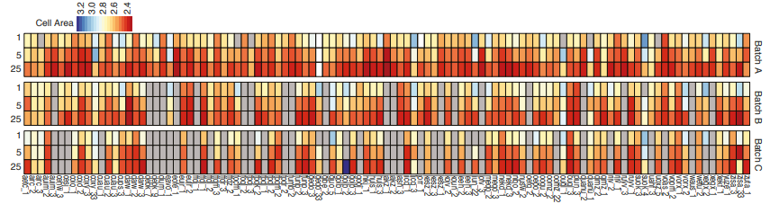
Measuring cell area in iPSC lines, from Vigilante, et al.’s preprint
Identifying the genetic basis of variation in cell behaviour in human iPS cell lines from healthy donors
Alessandra Vigilante, Anna Laddach, Nathalie Moens, Ruta Meleckyte, Andreas Leha, Arsham Ghahramani, Oliver J. Culley, Annie Kathuria, Chloe Hurling, Alice Vickers, Mukul Tewary, Peter Zandstra, HipSci Consortium, Richard Durbin, Franca Fraternali, Oliver Stegle, Ewan Birney, Nicholas M Luscombe, Davide Danovi, Fiona M Watt
Placozoa and Cnidaria are sister taxa
Christopher E. Laumer, Harald Gruber-Vodicka, Michael G. Hadfield, Vicki B. Pearse, Ana Riesgo, John C. Marioni, Gonzalo Giribet
Frederic Delsuc, Herve Philippe, Georgia Tsagkogeorga, Paul Simion, Marie-Ka Tilak, Xavier Turon, Susanna Lopez-Legentil, Jacques Piette, Patrick Lemaire, Emmanuel J. P. Douzery
Quantifying the impact of public omics data
Yasset Perez-Riverol, Andrey Zorin, Gaurhari Dass, Mihai Glonț, Juan Antonio Vizcaino, Andrew Jarnuczak, Robert Petryszak, Peipei Ping, Henning Hermjakob
Helena Cousijn, Amye Kenall, Emma Ganley, Melissa Harrison, David Kernohan, Thomas Lemberger, Fiona Murphy, Patrick Polischuk, Simone Taylor, Maryann Martone, Timothy Clark
Gerald G Singh, Vinicius Farjalla, Bing Chen, Andrew Pelling, Elvan Ceyhan, Martin Dominik, Eva Alisic, Jeremy Kerr, Noelle Selin, Ghada Bassioni, Elena Bennett, Andrew Kemp, Kai MA Chan
Single-cell approaches are revolutionizing developmental biology. We can now trace in time the behavior of each cell in a live developing organism (1). In parallel, single-cell transcriptomics and genomics gives access to the transcriptional state of each cell (2). Combination of these two approaches promises to unravel how genomic information translates into individual cell behaviours.
We are harnessing these approaches to the embryos of ascidians (Tunicates, 3,4), a group of marine invertebrate chordates. These embryos develop with such stereotyped cell lineages that each cell of an embryo of one species has an exact homolog in all embryos of different, even distantly related, species. Contrasting with this extraordinary morphological conservation, the genomes of ascidians are widely divergent.
We are looking for a PhD student, who will study how the transcriptional program of homologous cells has evolved between distantly related tunicate species. She/He will focus on two classes of genes, those building the gene regulatory networks (GRN) driving development and the effector genes controlling cell behaviours through their action of cytoskeletal architecture.
The selected student will first establish single-cell RNA-seq for a range of developmental stages in 4 ascidians and 2 thaliaceans (a different class of tunicates, 5). From this dataset, She/He will analyse the level of conservation of the transcriptional programme across species, which could lead to a classification of genes/subnetworks according to their level of expression conservation. In a second step, she/he will select a few genes based on their pattern of expression conservation and their functional annotation and study their function in cell fate specification or embryonic morphogenesis using CRISPR/Cas9 technology.
The project is mostly experimental. Bioinformatics knowledge or a desire to acquire it will be a plus. More about the host group and institute can be found on the CRBM website: http://www.crbm.cnrs.fr/.
Collaborations with the Christiaen (NYU, New-York, USA), Zinzen (MDCC, Berlin, Germany) and Aerts (KU, Leuven, belgium) labs are foreseen.
References:
1) L. Guignard*, U.-M. Fiuza*, B. Leggio, E. Faure, J. Laussu, L. Hufnagel, G. Malandain, C. Godin#, P. Lemaire# (2017) Contact-dependent cell communications drive morphological invariance during ascidian embryogenesis. bioRxiv 238741 https://www.biorxiv.org/content/early/2017/12/24/238741 2) Karaiskos N, Wahle P, Alles J, Boltengagen A, Ayoub S, Kipar C, Kocks C, Rajewsky N, Zinzen RP (2017) The Drosophila embryo at single-cell transcriptome resolution. Science. 358:194-199 3) Lemaire P. (2011) Evolutionary crossroads in developmental biology: the tunicates, Development, 138(11):2143-52. 4) Lemaire P. (2009) Unfolding a chordate developmental program, one cell at a time: Invariant cell lineages, short-range inductions and evolutionary plasticity in ascidians. Developmental Biology ;332(1):48-60. 5) Piette, J. and Lemaire, P. (2015) Thaliaceans, the neglected pelagic relatives of ascidians: a developmental and evolutionary enigma. The Quarterly Review of Biology. 90(2):117-145.
How to apply?
Interested candidates are encouraged to contact Patrick Lemaire (patrick.lemaire@crbm.cnrs.fr) directly prior any formal application to the Life Science doctoral school in Montpellier (Deadline for application May 18th 14:00, French time).
International students are encouraged to apply, provided their master English. Understanding or speaking French is not necessary.
►Criteria to apply to the call
The contest for a doctoral contract is open to all candidates regardless of their nationality and institutions of graduation.
Twenty-four 3-year doctoral positions will be funded by the doctoral school
To be eligible, candidates must hold a Master’s degree (or diploma recognized as equivalent to a Master’s degree).
►Timeline for the 2018 campaign (All times are in Paris time zone.)
18th May 2018 at 14:00. Deadline for submission of applications
4th June: short listing of candidates
28th and 29th June 2018. Interviews of shortlisted applicants (video-conference for distant applicants should be possible)
Applications (single pdf file) should include the following documents:
A cover letter and CV, including academic background and professional experience, in particular internships accomplished as part of the study program.
Copies of diplomas. It is mandatory to hold a Masters degree by the beginning of the 2018/2019 academic year).
Academic transcripts of all courses taken as part of university degrees *.
At least two letters of recommendation. Referees are expected to explain how they have known the candidate, their frank evaluation of his/her capability and motivation to undertake a Ph.D., as well as of his/her creative and analytical skills. It is recommended that referee rank the student among all students they mentored (Top 5%, 10%, 20%).
The Company of Biologists is looking to recruit a Scientific Copy Editor for the journal Development – one of the leading international journals in the field of developmental biology. This is a permanent full-time position.
The role entails copyediting articles to a high standard, compiling author corrections, overseeing the journal production process, and liaising with authors, academic editors, external production suppliers and in-house staff to ensure that articles are published in a timely and professional manner.
Candidates should have a degree (ideally a PhD) in a relevant scientific area, and previous copyediting experience is strongly preferred. Additional requirements include excellent literacy skills, high attention to detail, a diplomatic communication style, good interpersonal and IT skills, a flexible approach and the ability to work to tight deadlines.
The position represents a unique opportunity to gain experience on our highly successful life-science journals and offers an attractive salary and benefits. The position will be based in The Company of Biologists’ attractive modern offices on the outskirts of Cambridge, UK.
The Company of Biologists (biologists.com) exists to support biologists and inspire advances in biology. At the heart of what we do are our five specialist journals – Development, Journal of Cell Science, Journal of Experimental Biology, Disease Models & Mechanisms and Biology Open – two of them fully open access. All are edited by expert researchers in the field, and all articles are subjected to rigorous peer review. We take great pride in the experience of our editorial team and the quality of the work we publish. We believe that the profits from publishing the hard work of biologists should support scientific discovery and help develop future scientists. Our grants help support societies, meetings and individuals. Our workshops and meetings give the opportunity to network and collaborate.
Applicants should send a CV to recruitment@biologists.com, along with a covering letter that summarises their relevant experience, why they are enthusiastic about the role, and their current salary.
Applications should be received by 16 April and we welcome early applications or expressions of interest. Late applications may still be considered.
As many of you will be aware, The Company of Biologists initiated a search last year for a new Editor-in-Chief for Development, after Olivier Pourquié announced his intention to step down in September 2018. Following community consultation and a shortlisting and interview process, we are delighted to announce that James Briscoe will be the journal’s new Editor-in-Chief.
Many of you will already be familiar with James and his research. As a developmental neurobiologist working on the vertebrate spinal cord, he has a particular interest in using quantitative approaches to understanding how signalling pathways (particularly sonic hedgehog) regulate gene expression networks to control patterning, cell fate specification and growth of this tissue. His lab uses a range of in vivo, in vitro and in silico models to gain insight into this question at the molecular, cellular and tissue scales. For those interested in finding out more about James, we invite you to read our Spotlight interview with him elsewhere in this issue.
The position of Editor-in-Chief of Development is an important one in the developmental biology field – given Development’s position as a key community journal. The Editor-in-Chief is responsible for (among other things) appointing the editorial team, overseeing the handling of papers submitted to us, and setting new priorities for the journal. It was therefore important to us to gather input from the community as we set about appointing a successor to Olivier. We are hugely grateful to those of you – editorial board members, referees, authors and readers – who took the time to respond to our community consultation and provide feedback, not only on who they would like to see running Development, but also more broadly on how we are doing as a journal. Many of you are aware that, as a member of The Company of Biologists’ Board of Directors, James was part of the advisory group who initiated the consultation. He stepped away from any involvement in the process as soon as his name began to appear in nominations. The responses were collated by the three of us, and here we would like to share with you some of that feedback.
Your responses were, in general, consistent with our own assessment of the journal’s standing. We heard that Development is seen as a high-quality, rigorous venue for publication, with excellent academic editors, a ‘tough but fair’ review process and good production values. We were delighted that many respondents picked up on some of our more recent innovations – cross-referee commenting during peer review, openness to preprints, and our strong online presence, particularly through our community blog the Node. Given that the Node was launched at least in part in response to the consultation we conducted when looking for a new Editor-in-Chief to replace Jim Smith, it was fantastic to see how much traction the Node has now gained in the community. A recurring theme in the feedback on the journal’s strengths was that Development is the ‘journal of reference’ for the field, and that Development papers ‘stand the test of time’. If this is how we are seen in the community, we are clearly doing something right!
You also told us that Development can be seen as ‘hard to get into considering its impact factor’ and that competition from newer journals means that Development is sometimes seen as a less attractive choice, especially for early career researchers. These are of course issues of which we are all too aware. As signatories of the San Francisco Declaration on Research Assessment (DORA), we would argue that impact factor is a poor proxy for journal quality, and an even worse one for individual papers. That this measure still holds so much sway, particularly in certain geographic regions, is disappointing, and is something that The Company of Biologists – as a supporter of the newly revamped DORA project – is trying to change. Nevertheless, Development recognises that you as authors have a wide choice of journals to which you can submit, and the team will continue to work to make the journal an attractive option.
Looking to the future, we were delighted to hear that many respondents feel it is important for Development to maintain and strengthen the focus on stem cells, regeneration and human development. However, this is clearly not the only area in which developmental biology is growing: there was strong support for increased visibility in the genomics, biophysics, quantitative and systems biology, and evo-devo fields. These are all fields that Development has highlighted as future priorities for the journal, and the team looks forward to working with many of you to realise the potential of these areas.
Away from consideration of specific research areas, we heard that Development should do more to support and advocate for the field (this is actually something the Development team is actively working on – look out for news in an Editorial in the near future!) and that we should continue to support our communities through our charitable activities – and, if possible, grow these. Development and The Company of Biologists see these activities – meeting grants, travelling fellowships, workshops and so on – as a key part of our ‘raison d’être’, and you can rest assured that The Company will continue to give back to the community as much as we can.
On the specifics of the choice of new Editor-in-Chief, your feedback helped us to draw up a ‘wish-list’ for the kind of person we wanted to lead the journal. We were looking for an individual whose research is at the cutting edge of developmental biology, with interdisciplinary skills, broad interests in the field and a strong vision for the future of the journal – to build on the developments that Olivier initiated, bring new ideas to further strengthen the journal, and be an active advocate for Development. James fulfils all these criteria and more, as evidenced by the fact that his name came up over and over again in the feedback we received; in fact, he was suggested by almost half of those who provided specific names – around four times as often as any other single individual. The unanimous view was that James is an outstanding candidate to succeed Olivier, and we are delighted that he has agreed to take on this role.
Over the next few months, James will be working alongside Olivier to ensure a smooth handover when Olivier steps down in September. You’ll be hearing more from him later in the year when he officially takes over the editorship. As James gets ready to take the reins, he (and the journal more broadly) welcomes any feedback or suggestions that you might have for how you would like to see the journal develop. In the meantime, we hope you will join us in congratulating James on his appointment, and in wishing him luck as he steps up to this challenging and important position!
An interview with James Briscoe
Katherine Brown
James Briscoe is a group leader at The Francis Crick Institute in London. His lab’s research focusses on the developing vertebrate spinal cord, with a particular interest in how sonic hedgehog gradients, and the downstream signal transduction and transcriptional networks, regulate the development of this tissue. In September 2018, James will become the new Editor-in-Chief of Development. We met with James to discuss his career and research interests, the importance of interdisciplinary thinking in developmental biology, and his views on the current state and future opportunities in scientific publishing.
Let’s start at the beginning: how did you first become interested in biology?
There was never a moment of epiphany and I didn’t have a well thought out plan. At school I enjoyed science and did well at it, but I was not aware that a career in academic research could be a possibility. Around the time I was thinking about going to university, I watched the film Life Story, based on Jim Watson’s book The Double Helix, and read Richard Dawkins’ The Blind Watchmaker. I think both of these tipped me towards biology. I also remember reading Microbes and Man by John Postgate, which is less well known but a really superb book, and it’s probably at least partly a consequence of this that I ended up at Warwick University studying microbiology and virology.
During your PhD, you worked on interferon signalling in cell culture. What attracted you to that field, and what then prompted you to move into developmental neurobiology for your postdoc?
During my undergraduate degree, I became fascinated by how the study of microbiology and virology had taught us fundamental aspects of eukaryote molecular biology. This led to an interest in interferons – the secreted cytokines that are the major cellular response to viral infection – and I joined Ian Kerr’s lab at the Imperial Cancer Research Fund (ICRF; which later became Cancer Research UK and then merged to form part of The Francis Crick Institute) to study the mechanism of interferon signalling. By chance my timing was perfect, as we were identifying what became known as the Jak-STAT signalling pathway. Our neighbouring labs were also investigating signal transduction and elucidating pathways such as MAP kinase, RAS, PKA and more. I became very interested in understanding how cells perceive and respond to extracellular signals. Most of the groups at ICRF used cell lines but there were a few developmental biology labs, including those of David Ish-Horowitz, Julian Lewis and Phil Ingham. I went to their seminars and this introduced me to the beauty of embryos and to hedgehog signalling. I decided I wanted to continue studying signal transduction but to learn developmental biology so that I could investigate signalling in its natural environment rather than just in cell culture.
You joined Tom Jessell’s lab at what must have been an exciting time: sonic hedgehog (Shh) had only recently been cloned and it was known to be involved in spinal cord patterning, but little was understood about how the system worked. What were the questions you set out to answer during your time there?
Again, I was lucky with my timing. As you say, it was a really exciting time to be in the Jessell lab and at Columbia University. I wanted to understand Shh signalling and how it could function in a graded manner to control cellular responses. Very little was known about the molecular mechanism of signal transduction – I think even today we still have an embarrassingly poor understanding of this pathway – and I thought we could figure this out using the neural tube. Being in the Jessell lab, however, I soon became fascinated with and distracted by broader aspects of developmental neurobiology and how the spinal cord forms and functions. It became apparent that understanding the role of Shh in the spinal cord required studying the gene regulatory network that it controls and this has become the passion I’ve pursued ever since.
So what are the problems you’re working on now?
Broadly, they’re still the same problems that I was interested in when I was in Tom’s lab. For me, the fundamental question in developmental biology is how the right cells are produced in the right place, at the right time and in the right amounts in a developing tissue. The spinal cord turns out to be a fantastic system to address this and it’s revealing general principles that are applicable to many, if not most, developing tissues. Addressing these issues covers some of the most basic questions in biology. How is gene activity controlled? How is cell function determined? How are tissues shaped and organised from cells? Over the last couple of decades, developmental genetics has identified at least some of the key players in these processes. Now, new approaches and technology, from imaging to genomics to genome engineering, are providing unprecedented insight and resolution. I’ve been keen to move beyond purely qualitative explanations to a more dynamic and quantitative understanding of how the neural tube is formed and patterned. We’re trying to bridge scales from molecules to cells to tissues, to explain how the cells make the key decisions and how this guides the assembly of a functional, well-organised neural tube. I think this is of fundamental interest, but it also has practical implications for understanding disease and for progress in regenerative medicine and tissue engineering.
You’ve embraced mathematical modelling of developmental systems: why do you think this is important, and what are the challenges involved in trying to model something as complex as the developing spinal cord?
This was a decision I made about 10-12 years ago and, in part, it is because of the complexity of the problems we were investigating that I thought mathematical modelling was important. We were analysing increasingly complicated gene regulatory networks and we were trying to get away from simplistic reductionist descriptions to find explanations of how the system functioned as a whole. It’s often difficult to form an intuitive understanding of even relatively small systems if there are multiple interactions and feedback – it’s very easy to fool or confuse yourself. Mathematical modelling provides a rigorous way to describe and investigate a system. It tests whether your assumptions and interpretations are compatible. And different kinds of modelling can be useful at different levels of organisation, from the molecular to the tissue level, allowing us to look at the problem at different scales. I’ve been fortunate to have had great collaborators over the years that have taught me a lot of maths and physics. One thing I’ve learnt from this is that developing productive interdisciplinary collaborations takes a long time and requires a lot of trust and patience, but the investment can be very rewarding. We now think of mathematical modelling as just one of the available techniques that we use to tackle a problem, alongside more conventional molecular and cellular experiments.
More broadly, how important do you think it is for today’s young scientists to think interdisciplinarily?
Perhaps because it operates across multiple scales (both spatially and temporally), I think developmental biology has always been interdisciplinary. The molecular genetics revolution that transformed our field over the last 30-40 years is just one example of this. The importance of genetics is now taken for granted but it was once seen as pioneering and innovative. Today, it’s increasingly recognised that biologists need to be more quantitative and computationally literate and many of the applicants we see at The Francis Crick Institute have these skills. Nevertheless, it is something that needs to be strengthened through training at undergraduate and graduate levels. Having said that, one of the pleasures of academic research is that you’re continually learning and challenging yourself, so there are always opportunities to fill any gaps in your knowledge and learn something new. I often participate in advanced study courses run by organisations such as the Marine Biological Laboratory at Woods Hole and the Kavli Institute for Theoretical Physics at UC Santa Barbara. Even though I’m nominally ‘faculty’ on these courses, I usually end up learning at least as much as the students.
Developing productive interdisciplinary collaborations takes a long time and requires a lot of trust and patience, but the investment can be very rewarding
You recently moved into The Francis Crick Institute, which formed through the merger of NIMR at Mill Hill (where your lab was based), Cancer Research UK Lincoln’s Inn Fields and a number of other partners. How has the move been?
The planning took a long time, but the move itself went much more smoothly than I had anticipated. It’s been less than 18 months since we moved but it feels much longer; I guess that means we’ve settled in well. While I miss the charm and familiarity of our old institute, the new building more than makes up for it. The merger of the two institutes and our new partners mean I have new colleagues, which is stimulating and invigorating. I also love being in the centre of London – both because of the proximity to other academic institutions, and because we’re in a very vibrant part of town just minutes from the West End.
You’re active on Twitter: where do you see the value in social media for science and scientists?
I firmly believe that communication is a central part of science and academia: knowledge that isn’t passed on is wasted. Although not everything that appears on Twitter necessarily contributes to human progress, I enjoy being involved and I find it very useful. I get a lot of scientific information from Twitter. Particularly in fast-moving fields; for example, when CRISPR/Cas9 was coming on to the scene, I found Twitter was a great way to stay up to date and hear the latest developments. Also, like many scientists, I have friends and colleagues all over the world and Twitter is an easy way to stay in touch as well as to share and discuss (albeit briefly) papers and ideas.
You’ll be taking over as Development’s Editor-in-Chief in September, but you’ve been associated with our publisher, The Company of Biologists, for many years. Can you tell us a bit about your role as Director on The Company’s board and why you chose to get involved?
Yes, I’ve been involved a long time – since 2004. The Company of Biologists is a not-for-profit scientific publisher that publishes five journals: Development, Journal of Cell Science, Journal of Experimental Biology, Disease Models & Mechanisms and Biology Open. But unlike commercial publishers such as Elsevier and Springer Nature, we put the money that we make back into the scientific community. We give away about £1 million per year across our range of charitable activities: funding scientific societies such as the British Society for Developmental Biology (BSDB), sponsoring conferences, running workshops, and promoting research collaboration through travelling fellowships. We also support non-profitable, community-focussed activities such as the Node and preLights. There are 17 Directors of The Company and we’re almost all active scientists. We aren’t directly involved in the day-to-day business but we are responsible for the major strategic decisions, such as deciding to launch a new journal or start a big new project. In my role as Director, I was very involved in setting up the Workshops programme, and I’ve chaired the Grants Committee for the last few years, which decides how we spend our charitable funds. I’ve really enjoyed being on the board: it’s been a great opportunity to support the scientific community and I hope we’ve done at least a little bit of good.
Remember, by submitting your paper to Development or reviewing a paper for us, you’re supporting not only the journal but also all our other charitable activities
Scientific publishing is going through some significant changes at the moment, with the open access movement, developments in online publishing technology, the rise of preprints and so on. What do you think the future holds for small not-for-profit publishers like The Company of Biologists?
It’s definitely an exciting time in the publishing world. While all the changes create a lot of uncertainty both for people working in publishing and for scientists, I think there are lots of opportunities for small publishers such as The Company of Biologists. One of our advantages is that we’re run by scientists and we can respond in ways that help scientists and science and we’re not driven by profit margins. We’re also small enough and bold enough to experiment occasionally. Eight or nine years ago, we saw the increasing use of social media in our labs and heard calls from the community for an online discussion forum, so we launched the Node as a way to encourage informal discussion and communication about science within the developmental biology community. This has continued to grow in popularity. Just last month, we launched another experiment – preLights, the preprint highlighting and commenting service. This was in response to the growing numbers of preprints and it will be interesting to see how this develops in the coming months and years. More broadly, The Company’s goal is to support and inspire our fields and the people working in them and these aims will continue to drive our thinking and any future innovations.
Fundamentally, though, the journals and the research papers we publish remain at the heart of The Company of Biologists and it’s crucial that we all continue to read, write and referee for Development, if we are to continue to benefit from all the other initiatives and charitable support that The Company offers. Remember, by submitting your paper to Development or reviewing a paper for us, you’re supporting not only the journal but also all our other charitable activities.
Given the huge volume of published research, it’s increasingly difficult to keep up with the scientific literature. How much do you read and how do you choose what you read?
I agree it can be daunting keeping up with literature. I have a routine where I read a paper first thing in the morning when I get in to work (often I’ll choose it the night before). So I read at least one paper every day, although usually I’ll also read or review an article during the day. I try to read things outside my own research area, as this can provide ideas for my own work. I often pick up paper or preprint suggestions from people I follow on Twitter and I expect I will increasingly use preLights to get recommendations. I also think this is where community journals such as Development play a crucial role. Every issue of Development has papers handled by academic editors who are leaders in the field, so it offers a curated collection of the latest developmental biology research selected by experts. I always browse through the list of newly published papers and I often find myself picking one or two to read – it might be on neural development but as often as not it’s a Drosophila paper, an evo-devo study or even sometimes a plant paper.
And how would you like to see the journal (Development) evolve under your editorship?
I’m not taking over until late September 2018, so although I have lots of ideas I’m still refining my plans and taking suggestions. Olivier has done a great job during his time as Editor-in-Chief. He’s strengthened the journal, incorporating and encouraging new areas of research, and I certainly want to keep building on his accomplishments as developmental biology continues to change and grow over the next few years. I’m also very keen that Development continues to innovate and support our community. I’d welcome suggestions from readers, authors and referees about what we should do. What areas of science should we be encouraging in Development? What more can Development do to help our field? I’d urge anyone with thoughts or ideas to contact me.
Finally, is there anything that Development readers would be surprised to find out about you?
Some people find it surprising that I’m really a country boy at heart. Despite having lived most of my adult life in big global cities – London and New York – I was brought up in a 300-year-old thatched cottage on the Sussex Downs. I spent most of my teens working in stables and riding horses. We always kept lots of animals. As well as cats, dogs and rabbits we also had hens, ducks and goats; as a consequence, I can milk a goat.
The Department of Pediatrics at the University of Colorado School of Medicine invites applications for a faculty position in the tenure track. Appointment is expected at the Associate Professor level, but more senior individuals may apply. Applicants must have a Ph.D. and/or M.D. degree and demonstrated excellence in research.
We are particularly interested in individuals studying cardiac cell diversity, cardiovascular cell lineages and embryonic origins, gene regulatory networks and systems biology, cardiac cell regeneration and reprogramming, cardiovascular matrix biology and developmental models of cardiac biology. Applicants should have a record of creative and cutting-edge research, consistent and impactful publication and external funding. Applicants also should have a strong commitment to graduate and medical education and a successful record of trainee mentorship. Individuals who can enhance the diversity and accomplishment of our campus academic community are especially encouraged to apply.
Dr. Bruce Appel, Head of the Section of Developmental Biology, is Chair of the search committee. Questions can be sent to bruce.appel@ucdenver.edu.
 (1 votes)
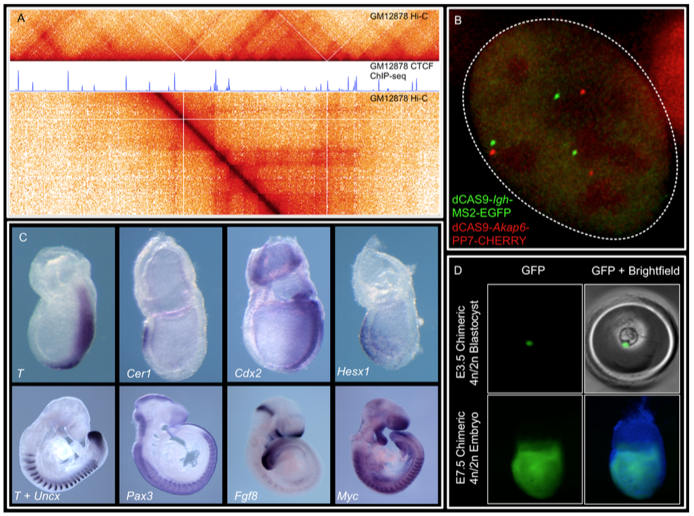
 (No Ratings Yet)