The community site for and by
developmental and stem cell biologists

Nominate BSDB committee members,

Posted by , on 16 February 2018

BSDBlogo
Sear BSDB Members,


I am writing to ask you to consider nominating individuals for membership of the BSDB committee.   Due to committee retirements, three new individuals are needed to serve as ordinary committee members, beginning in October 2018.   Their term of office will be five years and they will be required to (make every attempt to) attend two committee meetings per year.

All nominations must be proposed by a member of the BSDB, and seconded by another member, and must also be accompanied by a statement from the nominee that they are willing to serve if elected. The nominee has to already be a member of the BSDB.

Please send your nominations to me ASAP and by Friday 2 March 2018 at the latest.

In the event of a vote being required to decide between >2 candidates, the election will take place at this year’s BSDB AGM, which will be held during the BSDB Spring Meeting 2018 in Warwick (15th –  18th April, 2017). If there is an election, I will require someone, preferably the proposer or seconder, to give a short verbal statement of support of their nominee.

The BSDB committee also invites self-nominations for a new post graduate representative and a new post doctoral representative. Please, include a short text (max. 1 page) explaining your motivation and intentions to serve on the committee. Deadline will also be Friday 2nd March 2018 .

Please email these to me at: secretary@bsdb.org

Best wishes,

Kim Dale

Thumbs up (No Ratings Yet)
Loading...

Tags: ,
Categories: News, Societies

Navigate the archive

Use our Advanced Search tool to search and filter posts by date, category, tags and authors.

PhD positions in Marseille (France) – Turing Centre for Living Systems (CENTURI)

Posted by , on 15 February 2018

Closing Date: 15 March 2021

The Turing Centre for Living Systems aims at attracting students willing to work in an interdisciplinary life-science environment and with backgrounds in cell or developmental biology, immunology, neurosciences, theoretical physics, biophysics, computer science, bioinformatics, applied mathematics, engineering.

Candidates are asked to select the projects in order of interest (up to 3) and rank them in the application form.


Link to the PDF file.

Deadline for application: 15th April

Informal enqueries: info@centuri-livingsystems.org


PHD2018-01 – Using single cell transcriptomics to classify neuron types in two brain regions relevant to social behavior: the olfactory bulb and the hippocampal CA2

PHD2018-02 – Visualizing deep hippocampus neuronal functional activity in vivo using ultra-thin 2-photon endoscopes

PHD2018-03 – Understanding uORF functions through dendritic cells biology and computational approaches

PHD2018-04 – Using higher-order biological networks to explore temporal patterns in cell cycle control

PHD2018-05 – 3D optical microscopy for quantifying T lymphocyte activation

PHD2018-06 – Life and death of Salmonella-containing vacuoles

PHD2018-07 – Dynamics of a self-organized multicellular system

PHD2018-08 – Bacterial adaptation by OXPHOS dynamics: from single cell biology to gut microbiota

PHD2018-09 – Emerging near-infrared chromophores for photoacoustic bio-imaging

PHD2018-10 – Fluid-induced self-organization of ciliated-cell activity in human lungs

PHD2018-11 – Muscle building: bridging molecular order to macroscopic morphogenesis

PHD2018-12 – Deciphering the activation states of plasmacytoid dendritic cells, their dynamical relationships and their molecular regulation

PHD2018-13 – Mathematic modelling of cell migration in confined domains

PHD2018-15 – A versatile virtual reality system to understand animal navigation

Thumbs up (No Ratings Yet)
Loading...

Categories: Jobs

Nominate your Drosophila genes of interest for CRISPR fly stock production

Posted by , on 13 February 2018

The DRSC/TRiP-Functional Genomics Resources is a Drosophila community resource with three main focus areas: (1) cell-based Drosophila cell screening, (2) fly stock production, and (3) bioinformatics. Our TRiP fly stock production platform is taking nominations for production of CRISPR sgRNA fly stocks. Researchers can nominate genes for knockout (TRiP CRIPSR-KO) or activation (TRiP CRISPR-OE), or both. To search existing and in-production stocks, and to nominate genes for production of new stocks, please visit the gRNA Tracker website. In keeping with our long-time policies, all fly stocks are deposited to the Bloomington Drosophila Stock Center. We look forward to continuing to serve the needs of Drosophila developmental biologists and others through this effort!

Thumbs up (2 votes)
Loading...

Tags: ,
Categories: Research, Resources

Research Assistant/Associate: Zebrafish Optogenetics and Live-Imaging

Posted by , on 13 February 2018

Closing Date: 15 March 2021

The Buckley lab at the department of Physiology, Development and Neuroscience (PDN), University of Cambridge is recruiting a postdoctoral research associate or research assistant. The lab uses cutting edge optogenetic and live confocal imaging approaches within the whole zebrafish neural tube to manipulate the polarity of single cells (Buckley et al., 2016, PMID: 26766447). In combination with CRISPR-mediated functional knock down experiments, we are directly testing the role of cell polarity in building epithelial integrity during organ development and in breaking it during developmental processes such as EMT and diseases such as carcinoma. The department of PDN is home to world-leading research in development, neuroscience, zebrafish live imaging and optogenetics. It hosts the Cambridge Advanced Imaging Centre (CAIC), which provides cutting edge microscopy systems, bespoke development of new imaging equipment and expert support.

We are seeking an enthusiastic and proactive candidate to join the team at the beginning of this exciting research. There are two main projects with which the successful candidate could be involved, depending on their interests and expertise. The first is to use optogenetics and tissue-specific CRISPR to determine how cell polarity and cell division are linked during epithelial establishment (we previously discovered a novel mechanism of cell polarisation that occurs independently to cell division: Buckley et al., PMCID: PMC3545300). We will do this within zebrafish embryos and, in partnership with our collaborators, in mammalian stem cell culture systems. The second project is to test the role of polarity dysregulation in tissue disruption. We will do this by optogenetically manipulating polarity-linked signalling pathways (such as the PI3K pathway) in the already established zebrafish neural tube epithelium. We will use 4D imaging to assess the cellular consequences of these manipulations and will model how signalling dynamics are propagated through the tissue in real time.

The successful candidate should have or be near completion of a PhD (or equivalent) in a relevant field and have a competitive history of research achievements. We are interested both in candidates with a background in developmental cell biology and those coming from a more biophysical background. Experience in molecular biology and genetics is essential and ideally the candidate should have experience in CRISPR technology. Candidates must also have a good understanding of data analysis and bioinformatics. Experience in advanced imaging and analysis would be a great advantage, as would specific knowledge of zebrafish genetics. Knowledge and interest in cell polarity and epithelial development, biochemical signalling pathways and optogenetic techniques would be desirable.

Although this is a full-time post, part-time working i.e. 80% of full-time over 4 days may be possible.

Fixed-term: The funds for this post are available for 3 years in the first instance.

To apply online for this vacancy, please go to the University job pages: http://www.jobs.cam.ac.uk/job/16315/

Thumbs up (No Ratings Yet)
Loading...

Categories: Jobs

2-year Postdoctoral position (University of Birmingham) – Research Fellow in Cardiovascular genetics and development

Posted by , on 13 February 2018

Closing Date: 15 March 2021

Applications are invited from outstanding individuals to work under the supervision of Dr Rui Monteiro, Birmingham Fellow, on a BHF funded research project to study the role of TGFb signalling in angiogenic and haemogenic endothelial cell programming. The Monteiro Lab are interested in learning how extrinsic signalling impinges on lineage fate decisions in development and how progenitors and stem cells carry out those decisions, with a particular emphasis on the Transforming Growth Factor β (TGFβ) pathway. Previous work in the lab demonstrated that TGFb1 and TGFb3 play different roles in programming haemogenic endothelium to become of blood stem cells in vivo (Monteiro et al, 2016). The Research Fellow will study the gene regulatory network that carries out the ligand-specific functions for TGFb1 and TGFb3 ligands in haemogenic and angiogenic endothelium in vivo. They will make use of several approaches, including genome editing with CRISPR/Cas9, transgenesis, fluorescence activated cell sorting and transcriptional and epigenetic profiling using zebrafish as a model.


The successful applicant will have a first degree and a PhD in developmental biology, molecular genetics, biology or in a related discipline relevant to the project. They will also have a strong background in molecular biology and previous experience with model organism and/or analysis of transcriptomic and epigenetic data.


Informal enquiries should be directed to Dr. Rui Monteiro (R.Monteiro@bham.ac.uk)

Starting salary is normally on Grade 7 according to experience.

Closing date: 10 March 2018            Reference:  58652


To download the details of this position and submit an electronic application online please go to https://www.birmingham.ac.uk/staff/jobs/index.aspx. Please quote the appropriate Job Ref in all enquiries, alternatively information can be obtained from www.hr.bham.ac.uk

Thumbs up (No Ratings Yet)
Loading...

Categories: Jobs

2-Year postdoctoral position (Marseille, France): Computational study of cell pattern emergence in embryonic and tumorigenic tissues

Posted by , on 9 February 2018

Closing Date: 15 March 2021

PD2018-03 – Computational study of cell pattern emergence in embryonic and tumorigenic tissues

Project abstract – The ability of cells to self-organize into patterned tissues composed of multiple cell types is central to animal morphogenesis and relies on both biological and physical factors. We propose to investigate numerically and from a biophysical point of view the pattern formation observed in two different complex tissues respectively characterised by their stereotyped vs seemingly disorganised structure: the embryonic epithelium of Xenopus (with A. Pasini) and Drosophila brain tumors (with C. Maurange).

In Xenopus, we will study how multiciliated cells (MCCs) distribute in a regularly spaced pattern during intercalation into an epithelial layer, and explore how the pattern is established and maintained through a balance between mutual repulsion among MCCs and attraction between MCCs and epithelial layer cells. In Drosophila, we will study how clusters of brain cancer stem cells (CSCs) form and how they affect tumor progression. We will investigate how physical (tension, adhesion) and biochemical (growth and differentiation factors) cues contribute to segregate clusters of cells with different self-renewing potentials, regulate their size distribution and density, and thus determine tumor growth rate.

The computational tools envisaged for the project involve the numerical implementation of energy minimization algorithms such as the Cellular Potts Model (with R. Clément). We plan to model both systems with an energy function encompassing the different biological and physical interactions suspected to play a role in the processes. Such energy functions can comprise adhesion, tension, affinities or repulsions among cell types. Motility and cell proliferation can also be implemented at given rates, depending on cell types. Models with be implemented in light of the experimental results, and we expect that simulations will in turn guide the design of new biological experiments.

Expected profile – Candidates should have a robust background in physics and numerical simulations, and ideally be familiar with the Potts Model and its cellular version. As the project is strongly interdisciplinary and involves close collaboration with experimental biologists, previous experience in developmental biology or biophysics will be appreciated. A strong interest in biological questions, in particular in the principles of morphogenesis, is mandatory.

Scientific environment – The recruited post-doc will benefit from a world-class interdisciplinary environment, both within the IBDM (Marseilles Institute for Developmental Biology) and among the other institutes taking part into the CENTURI program.

Supervisors

Raphaël Clément (raphael.clement@univ-amu.fr)- IBDM, UMR 7288 – Cell and tissue physics – Team Lenne

Cédric Maurange (cedric.maurange@univ-amu.fr)- IBDM, UMR7288 – Neural stem cell plasticity – Team Maurange

Andrea Pasini (andrea.pasini@univ-amu.fr) – IBDM, UMR7288 – Biology of ciliated epithelia – Team Kodjabachian

Deadline for application: 28th February

Please apply online on the Centuri website.

Recruitment Form – PD2018-03 – Computational study of cell pattern emergence in embryonic and tumorigenic tissues

Thumbs up (No Ratings Yet)
Loading...

Categories: Jobs

Postdoctoral Position in Developmental Signaling at St. Jude Children’s Research Hospital

Posted by , on 8 February 2018

Closing Date: 15 March 2021

A postdoctoral position is available to study mechanisms of Sonic Hedgehog signal transduction in Stacey Ogden’s lab at St. Jude Children’s Research Hospital, Memphis, TN. The successful candidate will join a collaborative work group aimed at understanding how the Sonic Hedgehog pathway is regulated during development, and dissecting how its regulation is usurped in cancer. Areas of interest include biogenesis and secretion of the Hedgehog family ligands, contributions of lipid metabolism to pathway activity, regulation and signaling of the signal transducer Smoothened and investigation of the downstream effectors to which it signals. Research projects in the lab will entail use of biochemical and cell biological techniques and mouse model systems.


Applicants should have or expect a PhD degree at the time of application. The selected postdoctoral fellow will actively develop their own research project, perform laboratory experiments with minimal supervision, develop new procedures as needed and interact collaboratively with other members of the lab. The successful candidate will also actively participate in the publication and presentation of research results. Prior experience with signal transduction research, lipid metabolism or mouse model systems is preferred.


www.stjude.org/ogden


Email: Stacey.ogden@stjude.org

Phone: 901-595-6281

Application website: https://postdoc-stjude.icims.com/jobs

Job number: 38158


Thumbs up (No Ratings Yet)
Loading...

Categories: Jobs

Updates from DORA – An interview with Stephen Curry

Posted by , on 7 February 2018

The San Francisco Declaration on Research Assessment (DORA) was conceived in 2012 at an ASCB meeting, and has since its launch in 2013 has garnered thousands of signatories from individuals and organisations. Its aim is to improve the way in which the quality of research output is evaluated, with a key recommendation being the elimination of journal-based metrics by funding agencies, institutions and publishers when judging research and researchers.

This year, DORA has had something of an upgrade: with the support of organisations including the Company of Biologists (the not-for-profit publisher that runs Development and funds the Node!), DORA now has a full time Community Manager Anna Hatch, a new steering committee, and a new website.

To find out more about these developments, we caught up with Stephen Curry, who chairs the DORA steering committee and is Professor of Structural Biology and Assistant Provost for Equality, Diversity and Inclusion at Imperial College London.


Hi Stephen! Can you tell us a bit about your science?

I’m a structural biologist and primarily use protein crystallography to work out the three-dimensional structures of interesting macromolecules. My main research efforts have been focused on virus and host-cell proteins involved in the replication of RNA viruses – principally foot-and-mouth disease virus and noroviruses. But I am in the process of winding down my research lab so that I can concentrate on other interests (discussed below) and my new role as Assistant Provost for Equality, Diversity and Inclusion at Imperial College.


You’re also passionate about science advocacy and communication – you are Vice Chair of the Science is Vital campaign group, contribute to The Guardian’s science blog Occam’s Corner, and have 16k followers on Twitter. Has this side of science – away from the lab bench – always been important to you?

It’s always been important but I’ve only really been properly active in this space since 2008 when I started my blog. I found that writing about science really made me think about what it means to be a scientist in 21st century Britain and that led me to learn a lot about scientific publishing and research funding, both of which are tied in rather convoluted ways to the business of research assessment. I have enjoyed getting involved in these debates and in campaigns to bring about positive change. Being involved in Science it Vital right from the very beginning has been a fantastic lesson in what can be done with modern communication tools if you just knuckle down and get organised.


You were one of the original signatories to DORA – why was DORA necessary in 2013?

It was already overdue in 2013. I wasn’t involved in the formulation of the declaration but was invited to sign prior to the launch and didn’t hesitate to do so. I had already become aware of the perverting effects of journal impact factors on science and scientists’ careers. And I knew that many other people shared my concerns. My 2012 blogpost, Sick of Impact Factors, remains on of the most ‘popular’ that I have ever written. It clearly struck a nerve.


And what do you think has been achieved in the years since then?

DORA has been really helpful in re-focusing the conversation on how the scientific community does research assessment. Without anyone designing the system, journal metrics have been co-opted for the evaluation of individuals to such a degree that publication in certain tiles (infamously Nature, Cell and Science in the biomedical sciences) are now seen as the key to success. DORA has helped to challenge that view – though we should certainly be mindful of parallel work on the Leiden Manifesto and The Metric Tide report (on which I was a co-author). So I think there is much greater awareness of the nature of the problem now and even some tentative steps to address it.


So what’s new – what does DORA’s new lease of life entail?

What’s new is that DORA now has a much higher level of material and financial support (from 9 organisations: ASCB, Company of Biologists, CRUK, eLife, EMBO, F1000, Hindawi, PLoS, and Wellcome). That has allowed us to hire a full-time community manager (Dr Anna Hatch) and refresh the steering group, which I now chair. We’ve also refreshed out web-site and have a new, easy-to-find URL (sfdora.org). This will allow us to raise the profile of DORA – we mean to get the word out much more proactively – but we are also determined to make a renewed effort to ignite the discussion around what constitutes robust and effective research assessment. We know that to change practice we need to figure out practical ways to help busy reviewers sift through job and grant applications and CVs without falling back on mis-use of the JIF.


Personally, what has it been like to be involved with DORA – is it challenging to get consensus from such a broad group of scientists and organisations?

There’s a lot of work to do because there is always resistance to change. DORA is not out to name and shame people or organisations that haven’t signed. But we want to challenge them to think about research assessment and do what we can to help them find a way forward. We aim to do a much more comprehensive job of discovering and disseminating good practice from around the world.


Why (and how) should young researchers get involved in DORA?

Because bad research assessment leads to bad research. DORA’s focus on improving research assessment fits very well with the ambitions that first attracts early career researchers to research: to understand and change the world. We will do that best if we are doing a proper job of recognising and rewarding the best research. That’s not just about publishing the best science (irrespective of journal name), but also meshes with parallel concerns about open science, data and code sharing and efforts to address deepening concerns about reproducibility, which are at least partly due to our over-reliance on metrics such as the JIF for judging individuals. However, we cannot simply expect young researchers to take the responsibility for change; it is up to the old guard (people like myself) and organisations like DORA to provide real support.


Further DORA articles:


Stephen Curry’s World View in Nature

https://www.nature.com/articles/d41586-018-01642-w


Research Councils UK Press release:

http://www.rcuk.ac.uk/media/news/180207/


Research Councils UK statement on responsible metrics:

http://www.rcuk.ac.uk/documents/research/rcuk-statement-on-the-responsible-use-of-metrics-in-research-assessment-pdf/


Times Higher Education also wrote a story about DORA

https://www.timeshighereducation.com/news/funding-councils-sign-responsible-research-assessment#survey-answer


Thumbs up (2 votes)
Loading...

Tags: , ,
Categories: Funding, Interview, News

Studentship available in Claudio Stern’s lab

Posted by , on 6 February 2018

Closing Date: 15 March 2021

Primary supervisor: Professor Claudio Stern FMedSci FRS, Department of Cell and Developmental Biology, University College London

Project title: “Dynamics of cell behaviour during somite formation”


A studentship funded by the Anatomical Society is available in Claudio Stern’s lab. The project will study the mechanisms of somite formation, to elucidate the molecular and physical mechanisms that control somite size, shape and regional identity and the role of the “segmentation clock” and local cell-cell interactions in this process.  It is a multi-disciplinary project and will involve advanced live imaging (including super-resolution microscopy in vivo), molecular biology, biophysics and some computational modelling. The project may include travel to the labs of collaborators in the USA and/or Singapore.


Conditions and requirements: This PhD studentship is open to British, Irish or European citizens who have spent at least three years at a British or Irish institute of higher education. It is funded by the Anatomical Society of Great Britain and Ireland and offers a stipend (tax free) of £16,553 per annum (revised annually), university fees (UK/EU rate) and a contribution to research expenses as well as funds to travel to meetings. Funding is for 3 years but it may be extended to a fourth year if necessary. Candidates should have a 2.1 (or equivalent) degree or better in a Biomedical, Physical or Computational science-related area and strong interest in developmental biology, ideally along with some laboratory experience working in a biomedicine-related research project. Experience with microscopy, programming (preferably PYTHON) and/or other computational/mathematical skills, are not essential but will be an advantage.


Starting date: 1 October 2018 or earlier by arrangement.

To apply (with a cover letter, CV a brief statement of your interests and the names and contact details of two academic referees), or for further information please contact Prof. Stern: c.stern@ucl.ac.uk

There is no formal closing date for applications but a student will be appointed as soon as a suitable, high quality candidate is identified.


Thumbs up (No Ratings Yet)
Loading...

Categories: Careers, Funding, Jobs, Research

How does it Work? My Experience with 3 Different Model Systems

Posted by , on 5 February 2018

For both young and established developmental biologists considering their next career move, choosing a model system with which to answer one’s research questions is a big decision. Of course, the most important thing to consider is whether or not a particular system is compatible with your research goals. But for a young scientist looking to make the move from one system to another, the ease of such a transition is an important concern. Having transitioned from sea urchin to zebrafish and then to chicken, I am often asked about my experience working with each of these three organisms, and what the practical, hands-on similarities and differences are among these creatures as model systems for development.


It began with the sea urchin


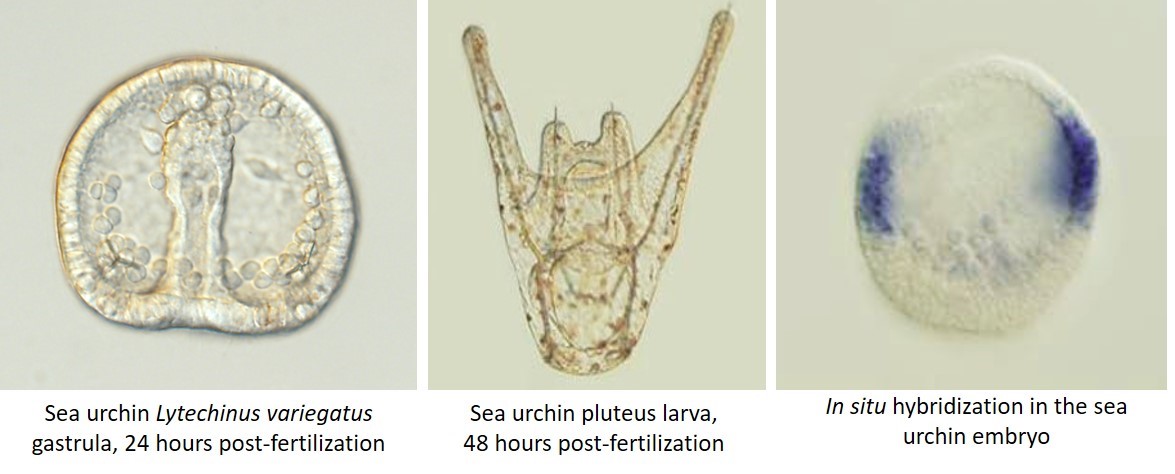
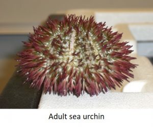
For my graduate research I studied skeleton formation in the sea urchin embryo. Most laboratories get adult sea urchins either by lab-organized diving trips if the lab is close enough to the ocean, or much more commonly by purchasing them from a supplier. When the sea urchins get to the lab, they need to be carefully acclimatized to the artificial sea water and the temperatures of lab aquaria, as any stress can make them spawn out their precious gametes prematurely. The maintenance of these aquaria and the monitoring of a new shipment of animals is an important and time-consuming job. Harvesting sea urchin sperm and eggs is as simple as injecting potassium chloride into the adults, after which they usually die (sad face!). In some species, however, adults can be shaken vigorously to make them spawn, and these animals can be used a few more times.

A major disadvantage of the sea urchin as a model system is the inability to establish stable transgenic lines or to easily create knockouts. Most analyses of development therefore depend on knockdown and overexpression experiments, which though easy to perform by microinjections, are not always clean. On the other hand, the ability to produce very large numbers of embryos from each round of fertilization gives the sea urchin a strong advantage for large-scale experiments. Since in situ hybridization and immunohistochemistry can be done on whole embryos, no time is spent sectioning embryos prior to analysis. Also, these embryos are very easy to analyze with several types of microscopy. Sea urchin embryonic development lasts only 2-4 days depending on the species, a definite benefit for moving a project along. An unexpected advantage of the sea urchin is the ability to replicate results in several closely-related species, which makes for a stronger case when putting together a story for publication. This also makes it an interesting model for evo-devo research especially when comparing multiple urchin species to other echinoderms.


Fish, fish and more fish


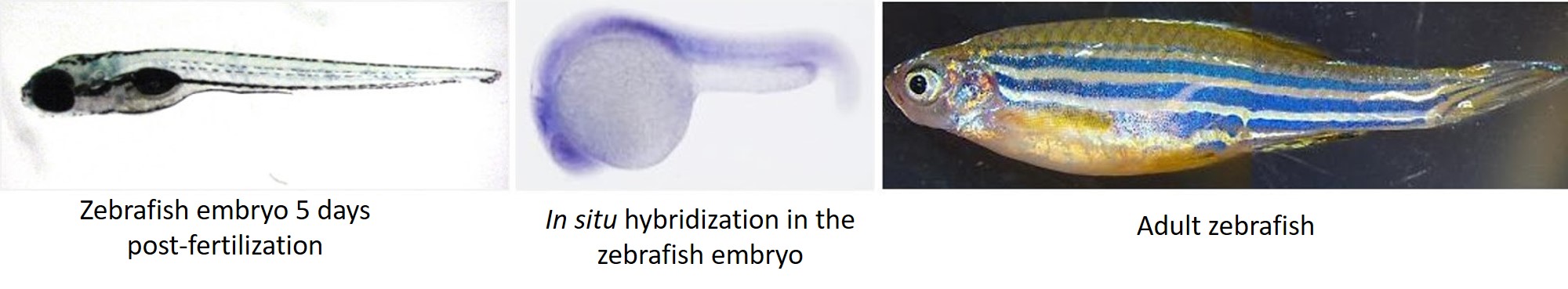
After my initial love affair with the sea urchin, I transitioned to zebrafish during my postdoc where I made zebrafish models for rare human diseases. Unlike the average sea urchin lab where adult animals are acquired from an external supplier, zebrafish labs often have several stable lines in-house, maintained by breeding adult fish and raising their offspring. While all this aquaculture can be time-consuming for a graduate student or postdoc, many institutions have fish core facilities and/or staff hired specifically for fish husbandry. Male and female zebrafish are usually kept separately, and to get them to breed, you only need to put them together in the same tank and they take over from there.

The zebrafish has many of the same advantages as the sea urchin, as it is easy to get large numbers of relatively transparent embryos. Adult animal and embryo care is similar to the sea urchin, as are methods of microinjection and analysis of results. The distinct advantage of the zebrafish (and indeed the main reason I found it attractive as a model system) is the immense power of transgenics in this system.  Stable transgenic lines and knockouts are standard in the zebrafish field, and this opens up a world of possibilities for a researcher.

Another advantage of this system is the ability to culture embryos into adulthood. This lets you study a phenotype throughout the lifespan of the fish. Moreover, since the zebrafish is a closer relative to humans, antibodies raised against human proteins work better in the fish than they do in sea urchins. Aside from learning about zebrafish morphology and how to make the best use of the transgenic toolkit, my transition from sea urchin to zebrafish was seamless.


Chicken, anyone?


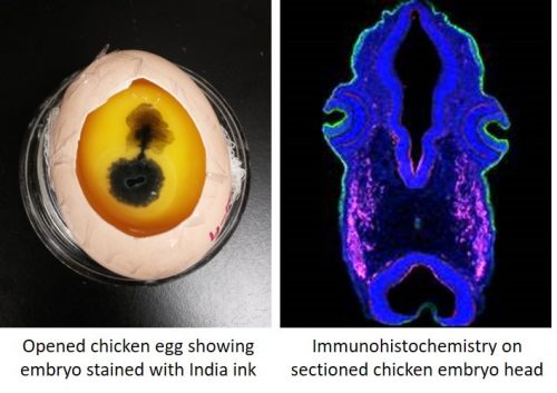
My current postdoctoral work is on neural crest and placode cells in the chicken embryo, which is distinct from the first two systems described. We get our embryos by incubating fertilized chicken eggs shipped in from a supplier, and as orders for eggs must be placed relatively far in advance, a researcher’s plan for experiments can fizzle out very quickly if egg quality is poor for a specific shipment. Also, a lot of time is spent on egg prep work, and you start out with fewer eggs, which means fewer embryos for your experiments.

Unlike aquatic embryos where you can just peek into a petri dish to tell what stage of development your embryos are, chicken eggs have to be opened and sometimes stained to check embryo development. Very early embryos especially do not take too kindly to this abuse, and may stop growing. My favorite thing about chick work is that you can stop and restart development multiple times simply by changing incubation temperature. This means that you can set a timer on an incubator and have your embryos ready at the stage at which you want them, when you want them. This cuts down on those infamous late nights in the lab.

The toolkit available to a chicken embryologist is relatively large and effective, and of the three systems described, antibodies generally work best in the chick. It is possible (but not at all common) to create stable transgenic lines of chicken, and other more common gene manipulation techniques such as morpholinos also work well. Since chicken embryos are relatively large, they often must be sectioned to observe the results of experiments.

The one thing I couldn’t escape with all three systems was the fact that embryo quality changed with the weather and the seasons.


The verdict?

Through all these transitions, I have found that studying development is a state of mind that transcends any specific model system, and the expertise you gain from one system is very easily transferable to another. However, if forced to pick a favorite, I would confess that my heart still belongs to the sea urchin.


Thumbs up (13 votes)
Loading...

Tags: , ,
Categories: Lab Life, Research