Three months ago I did the unthinkable and moved to England, a strange haggis-free land. Starting my BBSRC PhD Internship (introductory post here) at the Node having left my Scottish lab was an amazing opportunity to get more involved with the dev bio community.
My main task while here was the day to day running of the Node; twitter, jobs and asking authors of interesting papers to write for us, thank you to all the researchers that did! You can read one of my favourite posts on using scRNA-seq to understand cell fate conversion here. I also conducted our people behind the papers interviews and a particular favourite was on the fascinating puzzle of plant cell topology, I even attempted to read On Growth and Form after their interview!
The chance to read and explore such a wide variety of topics has been eye-opening and there is so much fascinating research being conducted on topics I didn’t know existed. Learning about so many different model systems has shaped my thinking about my future in research (any Cephalopod neurobiology labs needing a Postdoc soon?).
Alongside keeping the Node running, my second project focused on refurbishing the resources section on the Node. My goal here was to make use of my position as an active researcher to make the resources page a community area to find useful links for research, teaching and to encourage researchers to engage in outreach ad advocacy. This resources list is by no means comprehensive and we still need your input. Please get in touch to let us know what is useful to you, what needs to be added/updated and any way that the resources section can be improved!
This internship has been a wonderful insight into the developmental biology community and I am so grateful to Aidan and Katherine for the opportunity and their guidance throughout the three months. I encourage all researchers to write for the Node, get your research out there! I am now returning to my PhD, all that is left to say is so long and thanks for all the Zebrafish.
The story behind this study provides yet another example of where the pursuit of a few chance observations developed into an interesting project in its own right.
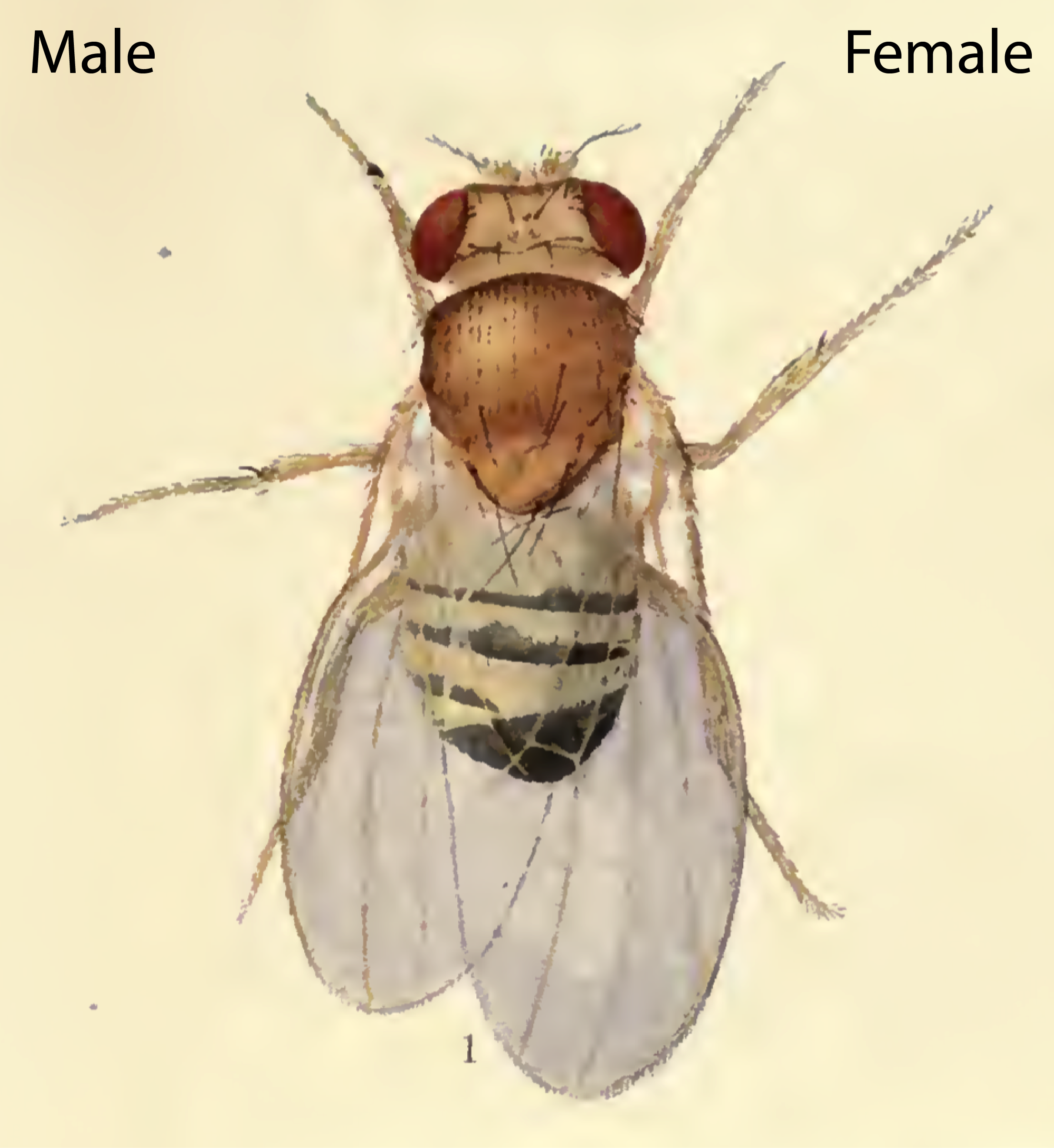
I started my postdoc in the lab of Alex Gould at The Francis Crick Institute with the objective of investigating how nutrient deprivation affects the growth of different body parts of the fruit fly larva. These experiments required precise measurements of organ sizes, and it soon became pretty clear that in order to get the most useful data, males and females had to be analysed separately. Not because the different sexes necessarily show different growth responses to starvation, but because female body parts are consistently 20-40% larger than their male counterparts. Hence, if males and females are pooled, the data have a large variance. Surprisingly, a lot of studies that have analysed larval growth seem to have ignored this sex difference in body size, officially termed sexual size dimorphism (SSD). So SSD effectively doubled my workload but it also opened up a new question: how does the sex of the larva influence its growth?
Summary of the key findings
We used two opposite angles of attack to identify the mechanism underlying larval SSD. First, in a sort of top-down approach, we asked if there were any measurable differences in growth parameters or behaviour that could give insights into underlying genetic mechanisms. Second, using a bottom-up strategy, we manipulated sex determination genes in individual cell types to see if the sex of a specific body part influenced overall body size.
For the first approach, I painstakingly weighed individual male and female larvae at different stages in order to determine their growth curves. When a larva hatches from the embryo it only weighs around 10mg, about 1/5th of the mass of a grain of salt! [1], and it grows to a final size of about ~2mg, around half the mass of a sesame seed [2], before it undergoes pupal development about 4 days later. This work established that males and females have the same size at larval hatching and that they have similar time windows of growth. Nevertheless, females begin to have a higher growth rate from about the middle of the second larval instar onwards. Interestingly, I found that the greatest difference in the fold rate of growth between the sexes occurs relatively early in larval development, during the second larval instar. This means that any pathways that control sex differences in body size should be active before or during the second larval instar.
Fig 1: Diagram of the sex determination pathway in Drosophila. In females, the presence of two X-chromosome activates the expression of the splicing factor Sex-lethal (Sxl). Sxl maintains its own expression and causes sex-specific splicing of transformer (tra), such that a functional Tra protein is only produced in females. Tra is another splicing factor that causes the sex-specific splicing of the transcription factors doublesex (dsx) and fruitless (fru), which regulate sex-specific morphologies and behaviour. Sxl also represses the translation of male-specific lethal 2 (msl-2) in females, thereby limiting assembly of the X-chromosome dosage compensation complex to males.
For the second approach, we used the amazing GAL4/UAS system of Brand and Perrimon [3] to manipulate a gene called Sex-lethal (Sxl), the master regulator of fly sex determination. Sxl is normally only expressed in females, and it directs the sex-specific splicing of downstream genes in order to specify the development of female morphology and behaviour (Fig 1). Sxl mutant females are masculinised both in terms of their morphology and their body size, demonstrating that Sxl controls SSD by promoting higher female growth.
I used RNAi to inhibit Sxl expression in different tissues and, strikingly, found that knockdown with a pan-neuronal Gal4 driver reduced the body size of females to that of males (Fig 2A). In the reverse experiment, restoring Sxl expression in neurons could partially rescue the female body size of Sxl mutants. Thus, Sxl acts in neurons in a non-cell autonomous manner to boost the growth of the female body. To pinpoint the neuronal population in which Sxl functions to promote SSD, a panel of neuronal subset-specific Gal4 drivers were then screened. We found that Sxl acts additively in at least two non-overlapping subsets of neurons: 1) the insulin producing cells (IPCs), a cluster of 7 neurosecretory cells that are known to secrete several insulin-like peptides into the circulation, and 2) cells expressing the Gad1-Gal4 driver, which is active in GABAergic neurons (although we found that the overlap with GABA-expressing neurons is only partial).
Fig 2: Effect of pan-neuronal knockdown of Sxl on sexual size dimorphism (SSD) in larval and imaginal tissues. Sxl RNAi was expressed using a pan-neuronal Gal4 driver (elav[c155]-Gal4) and the effect on SSD was measured for various body parts. Measurements were taken at the end of larval development, in wandering L3 (wL3) larvae (A-C) or in adult flies (D). The right graphs in each panel plot the female to male ratio as a quantification of SSD. Pan-neuronal Sxl RNAi abolishes SSD at the level of larval body mass (A) and the fat body nuclear diameter (B), while SSD of the wing imaginal disc remains intact (C). Pan-neuronal Sxl RNAi reduces, but does not abolish SSD of the adult wing (D).
The results of both approaches raise four tricky questions that Alex and I have been thinking about, and also investigating, with varying levels of success:
How does the new study fit with the dogma that somatic sex determination in flies is cell-autonomous?
We think the answer to this important question lies in differences between how Sxl regulates the SSD of the larval versus the adult body. On the face of it, our finding that Sxl acts in neurons to increase female body growth would seem to conflict with classic genetic studies of gynandromorphs (flies mosaic for male and female cells). These sex mosaic flies had clearly demonstrated that male/female morphologies are controlled cell-autonomously. Not only that but, in a bilateral gynandromorph, the male side is smaller than the female side [4] (Fig 3). So how can we square the classic findings with our new observations?
Fig 3: Drawing of a bilateral gynandromorph (reproduced from [4]). The drawing depicts a mosaic fly with male cells developing on the left side and female cells developing on the right side, likely as a result of a loss of one X-chromosome during early embryonic divisions. Note that the male side shows typical male morphological differentiation such as the sex-combs (dark bristled on front leg), as well as a smaller body size compared to the female side.
The first thing to note is that the presence of the cell-autonomous effects revealed by gynandromorphs does not completely rule out additional non-cell autonomous effects. In fact, such a dual SSD mechanism does seem to operate in mammals. For example, a recent mouse study suggests that gonadal hormones and direct effects of sex chromosomes combine to produce overall differences in growth and metabolism between males and females [5, 6]. Furthermore, we and others [7, 8] have found that manipulation of Sxl or its downstream target transformer does indeed have cell-autonomous effects on growth, although they are too small to account for the full extent of SSD.
Secondly, we realised that the gynandromorph studies only looked at the external structures of the adult fly. These are derived from small groups of diploid cells called imaginal discs, which grow within the larval body and only transform into adult structures during metamorphosis. In contrast, I had measured SSD for the larval body itself, the bulk of which is composed of large polyploid cells, which are degraded during metamorphosis and so do not make it to the final fly. This realization led to the testable hypothesis that SSD is regulated differently in the larval polyploid tissues versus the diploid imaginal tissues. Unlike many of our crazy hypotheses, this one turned out to stand the test of time as neuronal knockdown of Sxl abolished SSD in the fat body (a larval tissue), yet it had no effect on SSD in the wing imaginal disc (the adult wing precursor) (Fig 2B and 2C). Thus, neuronal Sxl in females specifically boosts the growth of larval not imaginal tissues during the period of juvenile development. Nevertheless, by the time the imaginal discs have transformed into the mature external structures of the adult fly, I did observe a moderate effect of neuronal Sxl knockdown on SSD (Fig 2D). To account for this, we propose that, by promoting larval tissue growth, neuronal Sxl also indirectly boosts the growth of imaginal tissues, because a larger larval body confers increased pupal resources (nutrients and/or growth factors) to sustain the growth of imaginal structures during pupal stages. An alternative explanation, which we cannot rule out for now, is that neuronal Sxl also acts during pupal stages to directly influence the size of imaginal structures.
Is there a role for insulin signalling in SSD?
We think the overall answer to this question is “yes but only for imaginal tissues” and here’s our logic for why this may be the case. Sxl functions in insulin producing cells and it is known that insulin secretion from these cells is a key regulator for growth, so an attractive hypothesis is that Sxl promotes insulin secretion in females, thereby boosting female growth. Interestingly, SSD in mammals is driven in part by gonadal hormones causing sex differences in insulin-like growth factor 1 (IGF-1) release. However, our data suggest that SSD of the larval body (i.e. the target of neuronal Sxl) is established independently of sex differences in insulin signalling. This is because, at the early stages when the sex difference in growth rates is maximal (the second instar larva), no sex differences in insulin secretion or insulin signalling could be detected. In addition, several manipulations aimed at changing insulin secretion from IPCs also had no effect on larval body SSD. And finally, mutant larvae that lack one or all of the insulin-like peptides (Ilps) produced in the insulin producing cells are very small yet their SSD remains intact. These findings suggest to us that Ilps are not required for the establishment of sex differences in growth in the early larva.
But the twist here is that there is evidence suggesting a role for insulin signalling in SSD in older larvae and in adult flies. Thus, insulin receptor mutant adult flies show a strong reduction in SSD (measured as dry body mass) [9]. At late larval stages, it has also been reported that insulin secretion from IPCs and insulin signalling is higher in females [7]. It is therefore likely that sex differences in insulin signalling in old larvae contribute to SSD in imaginal tissues, even though at this time larval body growth has largely finished. In support of this, I found that when IPCs are genetically manipulated to decrease their size and so presumably their insulin secretion, SSD is abolished in the wing imaginal disc but it remains intact in the larval body, despite the smaller absolute body masses of both sexes. Clearly, more work is needed to understand why insulin signalling appears to regulate the SSD of larval versus imaginal tissues so differently.
What is downstream of neuronal Sxl to boost female body growth?
We wish we could answer this tricky question but, unfortunately, all we can give you right now is a list of factors that are probably NOT involved.
As mentioned above, the current evidence indicates that insulin like peptides are not strong candidates for the Sxl targets in IPCs that are relevant for establishing larval body SSD. Nevertheless, it is possible that Sxl regulates the secretion of other signalling molecule(s) into the circulation, either directly from IPCs, or from cells that the IPC neurons connect with. Notably, IPCs send axons to the ring gland [10, 11], a major hemosecretory organ, which could provide such a systemic signal. The role of Sxl in the Gad1-Gal4 neurons is even less clear.
Two direct downstream targets of Sxl that have been widely studied are transformer and msl-2 (see Fig 1). Our loss of function and rescue experiments strongly suggest that neither transformer nor msl-2 play a role in the neurons regulating larval SSD. The msl-2 data also ruled out the possibility that the reduction in female body sizes we saw in Sxl loss of function experiments were due to “sickness” caused by ectopic activation of msl-2 expression and the dosage compensation machinery. It is likely that a new Sxl target is involved, but identification of this may well require unbiased sequencing experiments or other genome-wide techniques, using brains from the relevant developmental stage.
Is increased female body growth driven by increased feeding?
We have been asked this question a lot at conferences. We did measure feeding rates in early second and early third instar larvae. In both cases, sex differences in the absolute food intake rate (i.e. intake per larva) were detected, but when these were normalised for body mass, mass-specific food intake rates (i.e intake per mg of larval tissue) appear to be the same for males and females. This suggests to us that sex differences in absolute food intake are a consequence of a larger female body mass, but not the driver of it. So why then do female larvae have a higher mass-specific (i.e. fold) growth rate than males despite both sexes having similar mass-specific feeding rates? One possible explanation here is that female larvae are more efficient at converting nutrients into tissue mass than males.
It is important to highlight that there are caveats to our conclusion that mass-specific food intake is equal in male versus female larvae. This is because we measured food intake over 20-30 minutes using a blue dye uptake method that would not be sensitive enough to detect very small differences. Some clever method for measuring food consumption over much longer periods might be necessary to detect very small sex differences in mass-specific food intake. Unfortunately, this may be tricky as larvae not only eat their food but they also burrow in it, and defecate into it. However, there is another reason for thinking that the neuronal Sxl mechanism for establishing SSD does not involve sex-specific feeding. This is because increased feeding rates in females would be predicted to increase the growth of both larval and imaginal tissues, yet we observed that imaginal tissues inside the larva are blind to neuronal Sxl regulation.
Concluding remarks
Sex determination and growth regulation have been extensively studied in Drosophila for many years. It is therefore very surprising that only a handful of studies have investigated how sex influences growth in this model organism. The neuronal Sxl relay mechanism that we have recently discovered contrasts with the text book view that somatic sexual differentiation in insects is controlled in a strictly cell-autonomous fashion. Future research into the neuronal Sxl mechanism for larval SSD will be needed to identify the relevant Sxl targets and perhaps endocrine growth regulatory mechanisms that act selectively on larval polyploid tissues rather than diploid imaginal discs. Despite this, our research has already revealed glimpses that suggest that the regulatory logic of SSD in mammals and insects may be more similar than previously thought. This encouraging news opens up the possibility of using our favourite model organism to study how sex differences in growth and metabolism impact upon disease – an important emerging area of biomedical research.
References
“Grain of salt”: Bluebulb Projects’ The Measure of Things; [02.12.2017]. Available here.
Our latest monthly trawl for developmental biology (and other cool) preprints. Let us know if we missed anything.
This month in preprint news – here at The Company of Biologists, set just outside Cambridge in the UK, we’re hiring a new ‘Preprints Community Manager’. You can find out more here.It’s a great opportunity to get in to science communication and publishing, and of course preprints!
And this month in preprints, we found platypuses and pigeons, xenotransplants and reoriented axes, CRISPR tools and liquid droplets. The preprints were hosted on bioRxiv, PeerJ, andarXiv. Use these links to get to the section you want:
Human-specific NOTCH-like genes in a region linked to neurodevelopmental disorders affect cortical neurogenesis. Ian T Fiddes, Gerrald A Lodewijk, Meghan M Mooring, Colleen M Bosworth, Adam D Ewing, Gary L Mantalas, Adam M Novak, Anouk van den Bout, Alex Bishara, Jimi L Rosenkrantz, Ryan Lorig-Roach, Andrew R Field, Maximillian Haeussler, Lotte Russo, Aparna Bhaduri, Tomasz J Nowakowski, Alex A Pollen, Max L Dougherth, Xander Nuttle, Marie-Claude Addor, Simon Zwolinski, Sol Katzman, Arnold Kreigstein, Evan E Eichler, Sofie R Salama, Frank MJ Jacobs, David Haussler
Loss of Wt1 in the murine spinal cord alters interneuron composition and locomotion. Danny Schnerwitzki, Sharn Perry, Anna Ivanova, Fabio Viegas Caixeta, Paul Cramer, Sven Guenther, Kathrin Weber, Atieh Tafreshiha, Lore Becker, Ingrid L. Vargas Panesso, Thomas Klopstock, Martin Hrabe de Angelis, Manuela Schmidt, Klas Kullander, Christoph Englert
Higher-order inter-chromosomal hubs shape 3-dimensional genome organization in the nucleus. Sofia A Quinodoz, Noah Ollikainen, Barbara Tabak, Ali Palla, Jan Marten Schmidt, Elizabeth Detmar, Mason Lai, Alexander Shishkin, Prashant Bhat, Vickie Trinh, Erik Aznauryan, Pamela Russell, Christine Cheng, Marko Jovanovic, Amy Chow, Patrick McDonel, Manuel Garber, Mitchell Guttman
The Transcriptional Logic of Mammalian Neuronal Diversity. Ken Sugino, Erin Clark, Anton Schulmann, Yasuyuki Shima, Lihua Wang, David L. Hunt, Bryan M. Hooks, Dimitri Trankner, Jayaram Chandrashekar, Serge Picard, Andrew Lemire, Nelson Spruston, Adam Hantman, Sacha B. Nelson
| Stem cells, regeneration & disease modelling
Neuroepithelial rosettes from Medelnik, et al.’s preprint
T helper cells modulate intestinal stem cell renewal and differentiation. Moshe Biton, Adam Haber, Semir Beyaz, Noga Rogel, Christopher Smillie, Karthik Shekhar, Alexandra Schnell, Zuojia Chen, Chuan Wu, Jose Ordovas-Montanes, David Alvarez, Rebecca H. Herbst, Itay Tirosh, Grace Burgin, Danielle Dionne, Michael E. Xifaras, Mei Zhang, Alex K. Shalek, Ulrich H. von Andrian, Daniel B. Graham, Orit Rozenblatt-Rosen, Hai Ning Shi, Vijay Kuchroo, Omer Yilmaz, Aviv Regev, Ramnik J. Xavier
Developmental And Genetic Regulation Of The Human Cortex Transcriptome In Schizophrenia. Andrew E Jaffe, Richard E Straub, Joo Heon Shin, Ran Tao, Yuan Gao, Leonardo Collado Torres, Tony Kam-Thong, Hualin S Xi, Jie Quan, Qiang Chen, Carlo Colantuoni, William S Ulrich, Brady J Maher, Amy Deep-Soboslay, The BrainSeq Consortium, Alan Cross, Nicholas J Brandon, Jeffrey T Leek, Thomas M Hyde, Joel E Kleinman, Daniel R Weinberger
Gene expression imputation across multiple brain regions reveals schizophrenia risk throughout development. Laura M Huckins, Amanda Dobbyn, Douglas Ruderfer, Gabriel Hoffman, Weiqing Wang, Antonio F Pardinas, Veera M Rajagopal, Thomas D Als, Hoang Tan Hoang, Kiran Girdhar, James Boocock, Panagiotis Roussos, Menachem Fromer, Robin Kramer, Enrico Domenici, Eric Gamazon, Shaun Purcell, CommonMind Consortium, Schizophrenia Working Group of the Psychiatric Gen, iPSYCH-GEMS Schizophrenia Working Group, Ditte Demontis, Anders Borglum, James Walters, Michael O’Donovan, Patrick F Sullivan, Micahel Owen, Bernie Devlin, Solveig K Sieberts, Nancy Cox, Hae Kyung Im, Pamela Sklar, Eli Ayumi Stahl
Aging and neurodegeneration are associated with increased mutations in single human neurons. Michael A Lodato, Rachel E Rodin, Craig L Bohrson, Michael E Coulter, Alison R Barton, Minseok Kwon, Maxwell A Sherman, Carl M Vitzhum, Lovelace J Luquette, Chandri Yandava, Pengwei Yang, Thomas W Chittenden, Nicole E Hatem, Steven C Ryu, Mollie B Woodworth, Peter J Park, Christopher A Walsh
Germline loss of MBD4 predisposes to leukaemia due to a mutagenic cascade driven by 5mC. Mathijs A. Sanders, Edward Chew, Christoffer Flensburg, Annelieke Zeilemaker, Sarah E. Miller, Adil al Hinai, Ashish Bajel, Bram Luiken, Melissa Rijken, Tamara Mclennan, Remco M. Hoogenboezem, François G. Kavelaars, Marnie E. Blewitt, Eric M. Bindels, Warren S. Alexander, Bob Löwenberg, Andrew W. Roberts, Peter J. M. Valk, Ian Majewski
Hemimetabolous genomes reveal molecular basis of termite eusociality. Mark C. Harrison, Evelien Jongepier, Hugh M. Robertson, Nicolas Arning, Tristan Bitard-Feildel, Hsu Chao, Christopher P. Childers, Huyen Dinh, Harshavardhan Doddapaneni, Shannon Dugan, Johannes Gowin, Carolin Greiner, Yi Han, Haofu Hu, Daniel S. T. Hughes, Ann-Kathrin Huylmans, Carsten Kemena, Lukas P. M. Kremer, Sandra L. Lee, Alberto Lopez-Ezquerra, Ludovic Mallet, Jose M. Monroy-Kuhn, Annabell Moser, Shwetha C. Murali, Donna M. Muzny, Saria Otani, Maria-Dolors Piulachs, Monica Poelchau, Jiaxin Qu, Florentine Schaub, Ayako Wada-Katsumata, Kim C. Worley, Qiaolin Xie, Guillem Ylla, Michael Poulsen, Richard A. Gibbs, Coby Schal, Stephen Richards, Xavier Belles, Judith Korb, Erich Bornberg-Bauer
Platypus sampling sites from Martin, et al.’s preprint
Genetically encoded calcium indicators from Shen, et al.’s preprint
A genetically encoded Ca2+ indicator based on circularly permutated sea anemone red fluorescent protein. Yi Shen, Hod Dana, Ahmed S. Abdelfattah, Ronak Patel, Jamien Shea, Rosana S. Molina, Bijal Rawal, Vladimir Rancic, Yu-Fen Chang, Lanshi Wu, Yingche Chen, Yong Qian, Matthew D. Wiens, Nathan Hambleton, Klaus Ballanyi, Thomas E. Hughes, Mikhail Drobizhev, Douglas S. Kim, Minoru Koyama, Eric R. Schreiter, Robert E. Campbell
A suite of transgenic driver and reporter mouse lines with enhanced brain cell type targeting and functionality. Tanya L Daigle, Linda Madisen, Travis A Hage, Matthew T Valley, Ulf Knoblich, Rylan S Larsen, Marc M Takeno, Lawrence Huang, Hong Gu, Rachael Larsen, Maya Mills, Alice Bosma-Moody, La’Akea Siverts, Miranda Walker, Lucas T Graybuck, Zizhen Yao, Olivia Fong, Emma Garren, Garreck Lenz, Mariya Chavarha, Julie Pendergraft, James Harrington, Karla E Hirokawa, Julie A Harris, Medea McGraw, Douglas R Ollerenshaw, Kimberly Smith, Baker A Baker, Jonathan T Ting, Susan M Sunkin, Jerome Lecoq, Michael Z Lin, Edward S Boyden, Gabe J Murphy, Nuno da Costa, Jack Waters, Lu Li, Bosiljka Tasic, Hongkui Zeng
Cross-Site Comparison of Ribosomal Depletion Kits for Illumina RNAseq Library Construction. Zachary T Herbert, Jamie P Kershner, Vincent L Butty, Jyothi Thimmapuram, Sulbha Choudhari, Yuriy O Alekseyev, Jun Fan, Jessica W Podnar, Edward Wilcox, Jenny Gipson, Allison Gillaspy, Kristen Jepsen, Sandra Splinter BonDurant, Krystalynne Morris, Maura Berkeley, Ashley LeClerc, Stephen D. Simpson, Gary Sommerville, Leslie Grimmett, Marie Adams, Stuart S. Levine
An interlaboratory study of complex variant detection. Stephen E Lincoln, Justin M Zook, Shimul Chowdhury, Shazia Mahamdallie, Andrew Fellowes, Eric W Klee, Rebecca Truty, Catherine Huang, Farol L Tomson, Megan H Cleveland, Peter M Vallone, Yan Ding, Sheila Seal, Wasanthi DeSilva, Russell K Garlick, Marc Salit, Nazneen Rahman, Stephen F Kingsmore, Swaroop Aradhya, Robert L Nussbaum, Matthew J Ferber, Brian H Shirts
Community-driven data analysis training for biology. Bérénice Batut, Saskia Hiltemann, Andrea Bagnacani, Dannon Baker, Vivek Bhardwaj, Clemens Blank, Anthony Bretaudeau, Loraine Guéguen, Martin Čech, John Chilton, Dave Clements, Olivia Doppelt-Azeroual, Anika Erxleben, Mallory Freeberg, Simon Gladman, Youri Hoogstrate, Hans-Rudolf Hotz, Torsten Houwaart, Pratik Jagtap, Delphine Lariviere, Gildas Le Corguillé, Thomas Manke, Fabien Mareuil, Fidel Ramírez, Devon Ryan, Florian Sigloch, Nicola Soranzo, Joachim Wolff, Pavankumar Videm, Markus Wolfien, Aisanjiang Wubuli, Dilmurat Yusuf, Rolf Backofen, Anton Nekrutenko, Björn Grüning
A 3-year PhD fellowship in evolutionary developmental biology and genomics is available in the lab of Michalis Averof, at the Institut de Génomique Fonctionnelle de Lyon (IGFL) in France. The fellowship is funded by the Marie Curie ITN programme EvoCELL.
Some animals have the ability to regenerate parts of their body (limbs, tail, internal organs) after severe injury. However the molecular and cellular mechanisms underlying the regeneration of such complex organs are still poorly understood. The project will investigate cell differentiation during leg regeneration in the crustacean Parhyale hawaiensis, an emerging model for limb regeneration. The PhD fellow will determine the spectrum of cell types present in Parhyale limbs, their molecular profiles, and whether that diversity of cell types is fully restored after regeneration. In collaboration with other teams of the EvoCELL network, s/he will compare the transcriptional profiles of diverse cells types across phyla, in an effort to identify the evolutionary origins and conserved molecular signatures of progenitor cells.
The student will be trained in various technologies to analyze limb regeneration in Parhyale and compare it to regeneration in other species. S/he will use single-cell transcriptomics to establish the exhaustive repertoire of cell types prior to, during and after regeneration. S/he will participate in all steps of the experiment: dissociate limb cells, prepare libraries and computationally analyze the data. The fellow will also be trained in transgenesis, confocal microscopy and in situ hybridization in order to validate the identity of the detected cell populations and use markers to follow the fate of those cells during regeneration. This work will involve short periods of training/research in the laboratories of other EvoCELL partners and collaborators, and participation in yearly meetings and practical courses organized by the network, to take full advantage of the wide range of expertise available in EvoCELL.
The host lab (https://averof-lab.org) is based in the IGFL – an institute co-founded by the École Normale Supérieure de Lyon and the CNRS, whose scientific focus lies at the interface of developmental biology, functional genomics and evolution (http://igfl.ens-lyon.fr/). The lab’s working language is English. The fellow will be co-supervised by Michalis Averof and Mathilde Paris.
Applicants should have a Masters degree or equivalent (e.g. a 5-year university degree) to be eligible to embark on a PhD at the École Normale Supérieure de Lyon. To be eligible for the fellowship, candidates must be within the first four years of their research career and not hold a doctoral degree. Candidates of all nationalities may apply, but they must not have resided or carried out their main activity (work, studies, etc.) in France for more than 12 months in the 3 years immediately prior to their appointment (short stays such as holidays or compulsory national service are not taken into account).
The suggested starting date for the PhD is October 1, 2018.
Applicants should send a short letter of interest and CV to michalis.averof@ens-lyon.fr, and ask two referees to send recommendation letters to the same electronic address. The closing date for applications is February 2, 2018.
One author’s perspective on the origins of this study (Gavin Clowry)
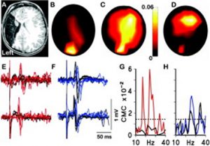
For me the origins of this research paper stretch back nearly twenty years. I was part of a research group, led by the paediatric neurologist Professor Janet Eyre, studying the neurobiology of cerebral palsy including neurophysiological studies on infants and children. A little boy with hemiplegia generated some remarkable results. He was unusual in that he had suffered a brain lesion relatively early in development compared to most CP sufferers, around mid-gestation, resulting in destruction of the sensorimotor cortex unilaterally. Transcranial magnetic stimulation, coupled with electromyographic recording, showed that muscles contralateral to the lesion were strongly responsive to stimulation of the ipsilateral motor cortex, not unusual in hemiplegics. What was unusual, though, was the strong response of the same muscles to stimulation of the intact contralateral occipital cortex. Ten more years of experiments and imaging were required to confirm these observations [1] but it was shown that in response to the lesion, visual cortex had acquired or maintained projections to motor centres in the spinal cord (Figure 1).
Figure 1 (A) T1-weighted magnetic resonance imaging of the brain at age 4 years showing left middle cerebral artery territory infarction. (B, C) Topography of corticomuscular coherence (CMC) with the electromyogram (EMG) of the right (paretic) first dorsal interosseus (FDI) (B) at 17Hz, which is centered over O1 and (C) at 24Hz, which is significant at O1 and C4. (D) Topography of CMC with the EMG of the left FDI at 24Hz, which is centered over C4. (E, F) Transcranial magnetic stimulation (TMS) motor evoked potentials (MEPs) recorded in the EMG of the right and left FDIs when L.J. was aged 14 months (upper traces) and 48 months (lower traces) evoked when the TMS coil was placed (E) over the occipital cortex (O1) and (F) over the right motor cortex (C4). Stimulation over C4 (F) led to the expected motor responses in the contralateral (left) FDI (individual responses shown in black) and also to MEPs in the ipsilateral (right/paretic) FDI (responses shown in blue). This is a pattern previously recognized in the motor supply to the paretic hand in congenital hemiplegia. (E) Finally and uniquely, stimulation over O1 produced MEPs in the paretic (right) FDI (red lines) but no MEPs in the unaffected (left) FDI (black lines). All evoked responses were of shorter latency at 48 months than at 14 months, as expected. (G) CMC spectra recorded at O1, showing significant coherence with the EMG of the right FDI (red line) but not the left FDI (black line). (H) CMC spectra recorded at C4 showing significant coherence with the EMG of the right (blue line) and left (black line) FDI. The dashed line indicates the upper 95% confidence limits for a CMC of zero for both (G) and (H). From [1] with permission.
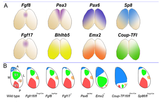
This set us speculating on the nature and plasticity of the protomap in human. Around this time strong evidence was being presented in favour of the protomap hypothesis originally proposed by Pasko Rakic [2]. The hypothesis stated that the organisation of the cerebral cortex into functional areas is determined by the co-ordinated and compartmented expression of genes in time and space at the earliest stages of its development, prior to its connection with the sensory input that could drive its maturation. Experiments in mice (Figure 2) [3, 4] were showing that altered gradients of transcription factor expression across the early cortex in mutant and transgenic mice led to an altered functional map for primary cortical areas. But does all this hold true for human development? For our next programme of research we teamed up with Prof Susan Lindsay and the Human Developmental Biology Resource to explore gene expression in the early stages of human cortex development especially to look for evidence for a human protomap.
Figure 2. A. Shows location of expression of the morphogen Fgf8 and some of its downstream effectors, all of which show high anteromedial expression (Fgf17, Pea3, Sp8) along with transcription factors expressed in an opposing gradient (Coup-TFI, Bhblb5) and Pax6 and Emx2, expressed in opposing anterolateral to posteromedial gradients. B. Summarises the effects of experiments manipulating the expression of these morphogens or transcription factors upon the size and location of primary cortical areas, usually identified and delineated in perinatal animals by expression of specific cell adhesion molecules. From [ 5 ] with permission.
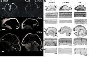
Our first success was to show that the transcription factors PAX6 and EMX2 form counter-gradients across the human cortex, just as they do in mouse, but only at the very earliest stages of cortical plate formation (Figure 3) [6]. Unlike in mouse, where expression is confined to progenitor cells, EMX2 was found to be expressed in a gradient in the post-mitotic neurons of the cortical plate as well. We also found arealised expression for other genes, by histological techniques, from quantitative measurements of mRNA expression, and in fetal cells cultured from different cortical regions [7, 8]. For instance, the layer V specific transcription factor CTIP2, along with transmembrane signalling molecules and corticospinal tract markers ROBO1 and SRGAP1, are more highly expressed in the frontal cortex between 9-12 PCW which might represent the predominance of corticofugal projection neurons in frontal sensorimotor areas.
Figure 3. (A) Expression of opposing gradients of PAX6 and EMX2 in the human forebrain between 7.5 and 9 post-conceptional weeks (PCW) by in situ hybridisation. Note that the Pax6 gradient disappears by 9 PCW. (B) Markers for corticofugal neurons, CTIP2, ROBO1 and SRGAP1 are expressed in a high anterior to low posterior gradient in the human cortex at 8-10 PCW. Ant, anterior, Pos, posterior; Med, medial; lat, lateral.
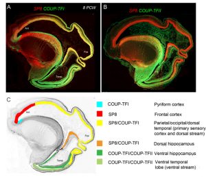
However the crucial question is whether such a developmental scheme observed in rodents is sufficient to produce all the extra areas of specialised association cortex, along with the vastly increased interconnectivity between these areas, seen in primates [9]? We believe our latest study may shed some light on this question [10]. We studied expression patterns of the arealising transcription factors COUP-TFI, COUP-TFII and SP8. What was noticeable in humans was that SP8 and COUP-TFI overlap extensively in the ventricular zone of visual, auditory and somatosensory cortex. This differs from the mouse in which COUP-TFI and SP8 show little overlap. Combinatorial expression of COUP-TFI and SP8 could maintain a common genetic identity for future primary sensory areas (visual, auditory and somatosensory) and a partially shared identity with SP8-expressing frontal motor cortex with which these sensory areas will interconnect, along with allied association cortex, via dorsal sensorimotor pathways. In mouse COUP-TFII is confined to a very small portion of the posterior cortex but in human is expressed extensively throughout the ventral temporal and ventral posterior cortex where it overlaps with COUP-TFI expression. Perhaps the expansion of cortical COUP-TFII expressing territory in human fetal brain mirrors the increased size and complexity of the association areas of the ventro-temporal cortex. An extension of this observation is that dorsal and ventral hippocampus are also differentiated by combinatorial expression of SP8/COUP-TFI and COUP-TFII/COUP-TFI respectively. Each domain has distinct functions and distinct efferent and afferent connections. It appears that the protomap for human hippocampal specialisation is laid down early in development.
Figure 4 (A) Expression of opposing gradients of SP8 and COUP-TFI in a sagittal section of human fetal telencephalon at 8 Post-conceptional weeks. (B) compartmentalised expression of SP8 and COUP-TFII in the developing cerebral cortex. (C) summarises the findings in A and B demonstrating how the progenitor zones of the cortex are subdivided into compartments by transcription factor expression that give rise to different functional areas of cortex in maturity.
It has been a 15 year journey to reach the beautiful images in Figure 4 with many interesting diversions along the way; for instance, when studying PAX6 expression we discovered a new class of progenitor cell in the subventricular zone of the cortex [11] which was subsequently shown to be a new class of radial glia [12]. We now know that humans as well as rodents have a protomap to guide cortical development, but this protomap is plastic and can be perturbed by genetic alteration, pharmacological interventions and lesions. Although more investigation is needed, it seems highly likely that the protomap has evolved in complexity as the cerebral cortex has evolved in complexity, rather than entirely new mechanisms being required to shape the cortex’s functional organisation, although, as ever, more research is needed to test this assertion. Ensuring that neurons in the cortex have the right regional and functional identity is crucial to establishing their long and short range connectivity, deficits in which are proposed to underlie neurodevelopmental orders such as autism and schizophrenia, so there is good reason to extend this journey a while longer yet.
Clowry GJ, Alzu’bi A, Harkin LF, Sarma S, Kerwin J, Lindsay S (2017) Charting the protomap of the human telencephalon. Seminars in Cell and Developmental Biology, Epub ahead of print, doi.org/10.1016/j.semcdb.2017.08.33.
Established by the British Society for Developmental Biology in 2014, The Gurdon/The Company of Biologists Summer Studentship scheme provides financial support to allow highly motivated undergraduate students an opportunity to engage in practical research during their summer vacation. Each year, ten successful applicants spend eight weeks in the research laboratories of their choices, and the feedback we receive is outstanding.
Our tenth report from the 2017 group of student awardees comes fromLiam McMulkin(student at The University of Dundee), who undertook his studentship with Dr. Marios Stavridis and Dr. David Martinat The University of Dundee.
My project’s aim was to expand British Sign Language (BSL) glossary for biology, more specifically areas relating to Developmental Biology.
British Sign Language (BSL) is a form of sign language which involves in the use of hand movements, gestures, body language and facial expressions to communicate. BSL is mainly used by deaf people. Unfortunately, BSL usually does not have signs for technical words that are not normally used in daily conversations, in biology for example, adenosine triphosphate, centriole, ectotherm, and many more which consequently lead to deaf people not having an equal access to biology compared with hearing people e.g. Interpreters don’t have signs for biological terms, which results in the use of fingerspelling. However, fingerspelling can be a lengthy process as every letter has to be spelt and is therefore not appropriate for a biology lecture, and also it is unpleasant to watch. Could you imagine a lecturer speaking out individual letter to spell out a word, more than twenty times in an hour lecture? E.g. electrophoresis and electroporation. Sometimes it can be difficult to distinguish between two words in a same lecture e.g. pluripotent, totipotent etc. especially when they are spelt out one letter at a time.
Scottish Sensory Centre has National 4 Biology BSL glossary. Unfortunately, they cannot source more funding to expand their BSL glossary for biology. Therefore, I decided to do something. I want to change this, I want deaf people to have a better access to biology with a better standard of BSL. This is a good timing to change this, as now it is an exciting time to study biology as new technologies open up novel areas of discovery e.g. genetic engineering and stem cells. Deaf people deserve to learn in their first language.
Dr. Marios Stavridis and Dr. David Martin agreed to support me to change this. They persuaded me to apply for this fantastic BSDB Gurdon Summer Studentship. After a month of waiting, I was very pleased to find out my application turned out successful. Straight away, I was given an unrestricted access to the staff in the Division of Cell and Developmental Biology at the University of Dundee, also space in the building to use as my base and meet with staff and postgraduate students to develop this glossary. In the process, I gained a first-hand experience of topics in Developmental Biology spanning the areas from Evo-Devo, live imaging of gastrulation, stem cells, neural and mesoderm development. This allowed me to get a feel for the terms beyond a dictionary definition. BSL is a very visual language and imagery is important in making good terminology. I spent a month doing this, before I ran the workshops.
The workshops were held on 8th and 10th August at University of Dundee. I sent out invitations using BSL via a video on Facebook with closed captions. The video had many shares, and I think it has shocked some people as they never knew that BSL has a limited glossary. University of Dundee also released a video about my workshops, which has reached to many people. Other news agencies such as Times Higher Education also released a news piece about my project. I am pleased that my project was well informed to the public because it raises awareness about BSL and its glossary.
Over 15 BSL users (aged from 20 and 70) attended and took part in the process of developing signs. Dr. Marios Stavridis briefly introduced what Developmental Biology is. Then, a Ph.D student provided a talk about her research work – two BSL interpreters struggled translating the talk due to lack of complex signs. Before we started developing new signs, a BSL linguistic, Gary Quinn introduced how to develop signs, and ensure they follow BSL grammar. I prepared a PowerPoint with terms we lack signs in with definitions and other helpful resources. All the participants worked together really well and developed over 70 new signs! At the end of the last workshop, the Ph.D student repeated her talk, and the participants were extremely shocked how improved the translations were from the same interpreters.
After the workshops, I spent roughly three days signing the newly developed signs. I then shared the signs online via Facebook for review. The feedbacks I received were really positive.
Now, I am at the last stage before finishing the project. The developed signs are required to be reviewed by Scottish Sensory Centre Glossary Manager, Dr. Audrey Cameron and a BSL linguistic, Gary Quinn before they are uploaded online at Scottish Sensory Centre website.
I can’t express how much I appreciated everyone’s support in this project. I millions of times thank to BSDB for selecting me to part of their studentship programme. Also, millions of times thank to The Robertson Trust for covering the costs for running the 2-day workshops at University of Dundee. I thank all the participants for all their efforts in developing signs for complex terms. Thank you to Dr. Marios Stavridis for being my supervisor. Also, for arranging an unrestricted access to the College of Life Sciences which gave me unique experience meeting world-leading scientists and observed their real work. Many thanks to Dr. David Martin for co-supervising me, and allow me to borrow his high-quality filming devices. My filming skills have improved! Finally, many thanks to Francesca Carrieri for her time observing her work and her time to come along to my workshop to deliver a talk on her research work.
In conclusion, I am very pleased I took this opportunity to improve BSL vocabulary for biology, which will improve deaf people’s access to education and science-related workplaces. Also, general science conversations using BSL. From this project, I hope more funding bodies have recognised the work of Scottish Sensory Centre and support them expanding BSL vocabulary to help deaf students in education. Finally, I really hope this project encourages Scottish Sensory Centre to add more of university-level vocabulary rather than just school-level vocabulary for help deaf higher-education students like myself.
Gaining a first-hand experience of topics in Developmental Biology at College of Life Sciences, University of Dundee. Left to Right: Anne Whittaker (communication support worker), Francesca Carrieri and myself.
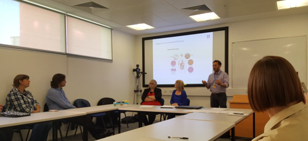
Dr Marios Stavridis introducing Developmental Biology on 8th August 2017 at the first sign development workshop at University of Dundee. Challenging for interpreters. The picture was taken by Dr David Martin.
Positions are available to study the physical principles of morphogenesis in the Mechanics of Morphogenesis / Davidson Laboratory at the University of Pittsburgh in the Department of Bioengineering. Our group focuses on studying the molecular, cellular, and tissue-scale processes that regulate mechanical properties and force-production during morphogenesis. Projects can involve quantitative cell biology, biophysics, bioengineering, and embryology.
Postdoctoral candidates will have recently completed a PhD and have a background in either bioengineering, biophysics, cell and developmental biology, or cell- and tissue- mechanics. The research environment at the University of Pittsburgh includes a dynamic community of bioengineers, developmental biologists, cell- and tissue-level biomechanics, and theoretical biologists. Nearby resources include the Peterson Institute of NanoScience and Engineering and the Pittsburgh Supercomputing Center. Contemporary Pittsburgh is a diverse vibrant city undergoing a renaissance led by world class Universities and the University of Pittsburgh Medical Center. The University of Pittsburgh is an Equal Opportunity Employer. Women and minorities are especially encouraged to apply.
Interested applicants should forward their CV and statement of research interests to:
Lance Davidson (lad43@pitt.edu)
Professor of Bioengineering
University of Pittsburgh
mechmorpho.org
The Marine Biological Laboratory seeks a Research Assistant II or III to join the laboratories of Kristin Gribble and David Mark Welch in the Josephine Bay Paul Center. The successful candidate will help develop genome editing techniques, including CRISPR/Cas9, in rotifers, a novel aquatic invertebrate model system for studies of aging, neurobiology, developmental biology, ecology, and evolution. Specific goals of the project include designing guide RNAs, optimizing microinjection methodologies, phenotyping and genotyping mutant strains, and screening genes of interest.
Qualifications:
Applicants should have a B.A., B.S., or Master’s degree in biology, cell/molecular biology, biochemistry, or a related field. This position requires proficiency and previous experience in basic molecular biology techniques, microscopy, microinjection, and CRISPR/Cas9 methodology. We are seeking an independent, organized, enthusiastic, and productive individual with robust problem solving skills. Excellent interpersonal skills, attention to detail, and a strong work ethic are essential. Position level and salary will depend upon education and experience. The ideal candidate will have working familiarity with RNAi and transgenic protocols. Proficiency in bioinformatics is a plus. Previous experience in established animal model or in non-model systems is preferred.
Established by the British Society for Developmental Biology in 2014, The Gurdon/The Company of Biologists Summer Studentship scheme provides financial support to allow highly motivated undergraduate students an opportunity to engage in practical research during their summer vacation. Each year, ten successful applicants spend eight weeks in the research laboratories of their choices, and the feedback we receive is outstanding.
Our ninth report from the 2017 group of student awardees comes fromMiguel Robles Garcia(student at The University of East Anglia), who undertook his studentship with Andrea Münsterbergat The University of East Anglia.
This summer I had the opportunity to undertake an internship at Andrea Münsterberg’s Laboratory at the University of East Anglia, where I am currently studying for a Bachelor’s Degree in Biological Sciences. Under the supervision of a PhD student, Johannes Wittig, and a postdoctoral researcher, Dr Estefanía Lozano-Velasco, I was able to spend seven weeks learning the ins and outs of everyday research. During this internship, my role was to focus on the early stages of heart development in chicks.
During vertebrate development, the heart is one of the first organs to develop. It is known that during this process many malformations can occur, which are capable of affecting correct heart function leading to potential defects or death. The developmental stages between mammals and birds are similar starting with cardiac looping and resulting in chamber formation. Due to the importance of the heart, its development is tightly regulated by different transcriptional and post transcriptional signalling pathways driven by transcription factors and microRNAs (miRs) respectively. miRs are regulators of gene expression that inhibit the translation of mRNA. During this project, my focus was to aid the postdoctoral researcher with her investigation of miR-133 function, which is thought to regulate BAF60b chromatin during heart development.
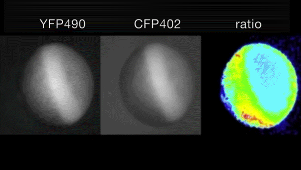
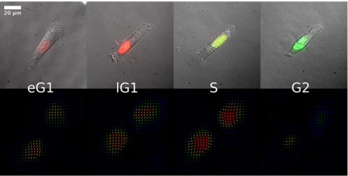
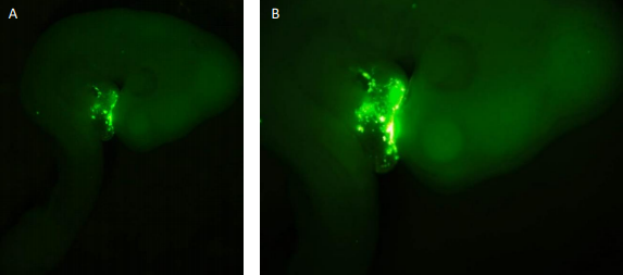
I started my time in the lab by dissecting wild type (WT) embryos from stages HH18-19 and HH23-25. The staging of the chick development follows the one described by Hamburger and Hamilton from 1951. Throughout my time in the laboratory, I became familiar with the dissection technique in which the different membranes that cover the embryo need to be removed. This is needed so that in future analyses (such as those that are mentioned) the membranes would not infer in the procedure and to make the tissue more available. My project then shifted towards the injection of embryos at stages HH 14-16 with the antagomir-133 (AM133). An antagomir is a modified oligonucleotide that binds to its specific micro RNA and inhibits its action. Therefore, this technique can be used to investigate potential effects in hearts which are deprived of a specific micro RNA. I used AM133 which is designed to inhibit miR-133. I also used scrambeled antagomiR (SCR) as a control for the procedure to see that the actual way of injecting the oligonucleotide wasn’t the one affecting the development of the heart. Both of these oligonucleotides have 5’ fluorescent label that allows the injection to be seen at first with the naked eye, but later after further incubation, with the aid of a GFP microscope (Figure 1). These antagomiRs were injected into a hollow cavity close to the heart walls while the heart is still beating without killing the embryo. After
the injection, the embryos were sealed with tape and returned to an incubator at 37°C for 24/48h depending on the intended analysis.
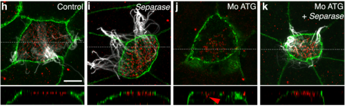
Figure 1. Fluorescence seen in embryos injected with the SCR probe. As depicted the fluorescence is seen only in certain parts of the heart and nowhere else. This shows that only in these places the inhibitor has bound to its target. Figure A shows the whole embryo while B is a close up of the same embryo. The fluorescence signal is stronger on the right-hand side of the heart.
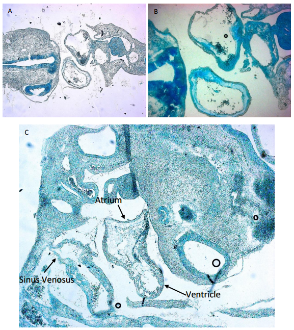
While some of the embryos were used for imaging and making sure the inhibitor bound in the correct place as shown in Figure 1, other embryos were embedded for sectioning. This involved placing the embryos in a gelatine medium that would then solidify at room temperature. Two different approaches were used for this in which one meant leaving the embedded embryos at room temperature for the gelatine to solidify slowly while the other was performed using dry ice for a rapid solidification. This allowed me to section the embryos for a closer examination of the heart. SCR embryos were compared to AM133 injected embryos to see if there is any difference in morphology. However, in order to image the slides containing the embryo section I had to stain them. For this we used an Alcian blue staining procedure (Figure 2). In this case the medium surrounding the tissues of the embryos would be discarded and only the glycosaminoglycans within the actual tissue were stained blue. This was also performed for embryos that had a double knock out of miR133 and miR1.
Figure 2. Image shows Alcian Blue staining of cryosections of embryos injected with antagomir-133 and SCR. A) Cross section of SCR injected embryo. The two hollow forms in the middle are the atria (down) and Truncus arteriosus (up) which can be seen connecting to the sinus venosus. B) Cross section of another SCR embryo showing a closer look at the heart. C) shows a sagital section. The top structure of the image is the head while the structure along the bottom is the back and tail of the embryo. The heart can be seen again in the middle of the embryo. Two different chambers are observable the top right one being the atria and ventricle while the bottom left one is the sinus venosus.
I want to thank the BSDB Gurdon Studentship for granting me the opportunity to have what I can just describe as an incredible experience. It has allowed me to develop my skills and scientific mind which I will be able to apply in my future studies and career. This opportunity is of great importance as, as a third-year student, I wasn’t sure of how daily lab teamwork alongside other scientists that are dedicated to their research felt like. I would also like to thank the Münsterberg lab for hosting me and Estefanía and Johannes for their guidance through these weeks.
A Brain Research UK-sponsored research assistant position for a highly motivated scientist is available in the laboratory of Dr. Claudia Barros at the Peninsula School of Medicine of Plymouth University, UK. The project is an opportunity to characterise novel targets and its human orthologue genes potentially involved in brain tumour initiation and growth, which have been identified in our laboratory. Molecular biology, cellular and biochemical techniques will be employed, including gene loss and gain of function assays, FACS, RT-qPCR, immunochemistry, western-blotting and confocal imaging. Work will make use of human brain tumour cell lines, tumour tissues and Drosophila brain as a model. A relevant 1st class or 2:1 (or equivalent) Bachelor degree is required and a postgraduate research degree may be preferred. You must have experience in some of the mentioned techniques, in particular cell culture and tissue work, and a suitable background knowledge. Good analytical, organisational and presentation skills, ability to multitask and collaborate with team members, and a genuine dedication to the research work are essential. Please include a cover letter detailing suitability/ experience/ interest and an academic CV with your application, in addition to ensure that 1-2 academic references are received.
Full or part time; Fixed term: initially 7 months if full time; Salary start: £24,983/ year.
 (4 votes)
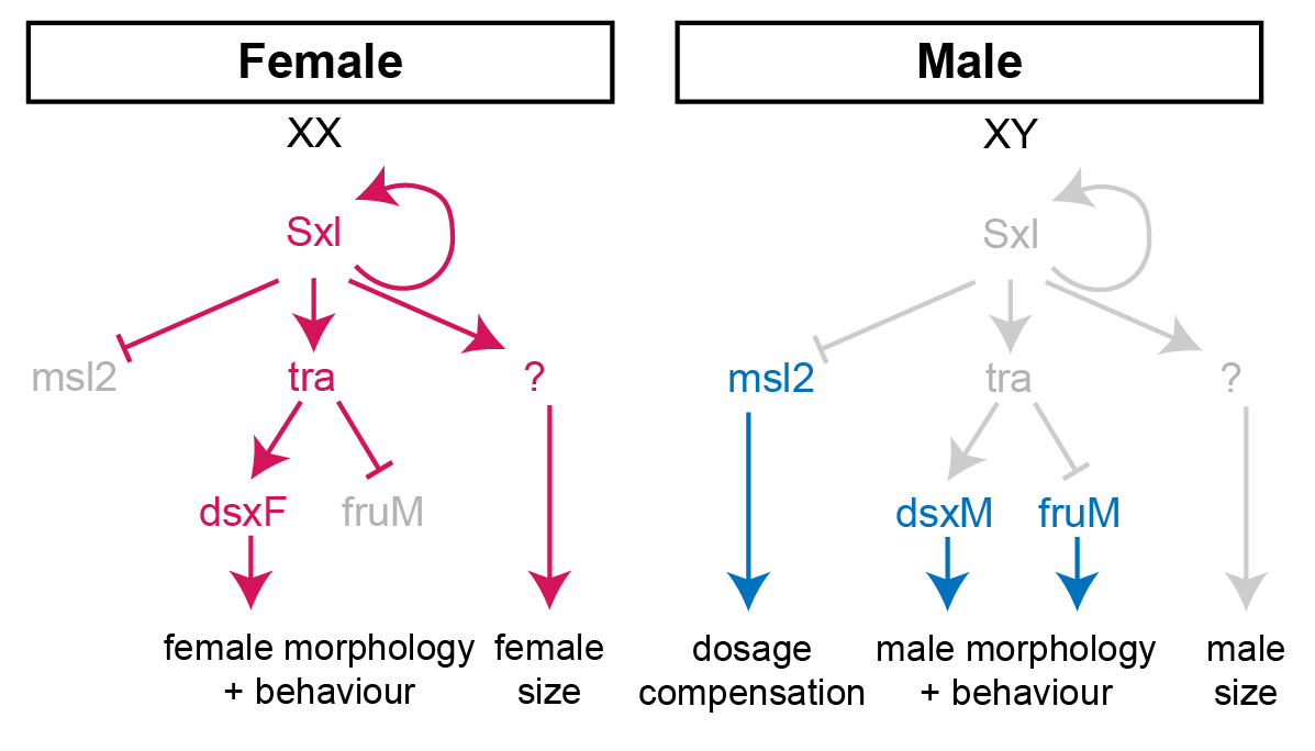

 (No Ratings Yet)