Checkpoints ensure that mouse oocytes with DNA damage arrest in meiosis I, preventing non-viable embryo formation, however the mechanisms which activate this checkpoint have so far eluded researchers. This week we feature a paper published in the latest issue of Development that reveals that the unique ability of mouse oocytes to sense DNA damage by rapid kinetochore checkpoint activation. First author Simon Lane and his PI Keith Jones of the University of Southampton, told us more.
Keith Jones and Simon Lane
Keith, please can you give us your scientific biography and the main questions your lab is trying to answer?
KJ I am Head of Biological Sciences, and Chair of Cell Biology at the University of Southampton. In the mid 1990s I worked at the Medical Research Council Experimental Embryology & Teratology Unit in London, examining the way in which sperm-driven changes in intracellular calcium at fertilization were regulated and how they affected embryo quality, helping to establish a link between early events of fertilization and long term embryo quality.
Between 1998 and 2008 I held an academic position in the Institute for Cell and Molecular Biosciences at the University of Newcastle-upon Tyne, UK. My lab has helped develop the use of Fluorescent Proteins to study the process of meiosis in real-time. This approach led to recent developments in the understanding of how the meiotic divisions are regulated. In 2008, until 2012, I moved to University of Newcastle, Australia, where Simon joined me as a PhD student. In 2012 I moved back to the UK and Simon followed me, then as a postdoc having successfully defended his thesis.
My lab is focused on understanding how the oocyte makes the transition in meiosis from a mature egg.
Simon, how did you come to join the Jones lab?
SL After receiving an interesting lecture on fertilisation in ascidian eggs I applied for a summer placement in the cell biology labs in Newcastle University. There I met Keith who was about to move his lab to Australia and was recruiting PhD students. I didn’t hesitate at the opportunity to do a PhD in a subject that was very interesting and in such an exciting part of the world!
Can you give us a brief summary of why you decided to ask the questions in your paper and the previous research that led you to this story?
KJ Along with the John Carroll lab (then UCL, now Monash) we made the original discovery of the ability of oocytes to arrest in meiosis I a few years ago.
The current work is an extension of that initial study. The first study was all about reporting the phenomenon. The present one, published in Development, is a more detailed investigation of the mechanism by which arrest is achieved.
Can you give us the key results of the paper in a paragraph?
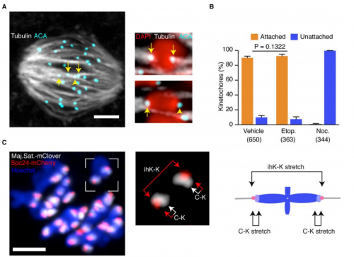
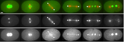
KJ In a nutshell it shows that oocytes have a profound ability to arrest in meiosis I in response to DNA damage, and that the mechanism by which this achieved is unusual. It’s a process that involves the kinetochore, rather than the sites of DNA damage, and it doesn’t involve the usual DDR kinases ATM and ATR. We also show that the response is specific to the first meiotic division, as it is absent in mature eggs.
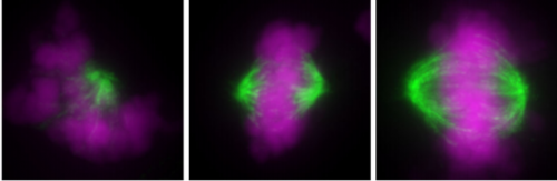
Imagining the kinetochores, from Figure 6, Jones, et al. 2017
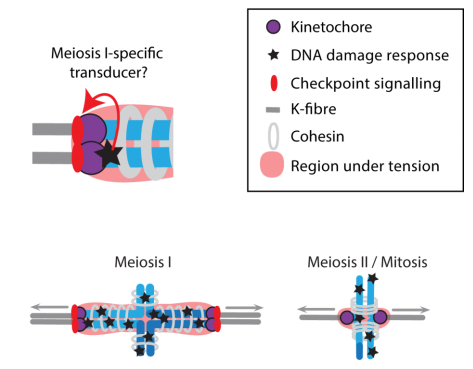
You suggest three models for MI oocyte sensitivity to DNA damage, how might you proceed to test your preferred model?
KJ The paper shows the response specific to the first meiotic division. We are therefore pursuing the role of various proteins known to play specific functions during the first but not the second meiotic division.
Possible models to explain meiosis I-specific arrest, from Figure 9, Jones, et al. 2017
In the paper you talk about potential implications for human oocytes: how well do you think the mouse model translates and do you have any plans to test this research in human cells?
KJ We already know that follicular fluid collected from the ovaries of women who have endometriosis is able to cause arrest of mouse oocytes during in vitro maturation. The mechanism we believe is that ROS levels are higher in endometriosis, a phenomenon associated with inflammation, and the increased free radicals have the ability to damage DNA.
When doing the research, did you have any particular result or eureka moment that has stuck with you?
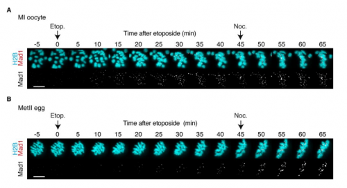
SL I like the feeling when you are working late to complete an experiment but then you see something new and interesting and you think to yourself, I might just be the first person who has ever seen this. The experiment where the Mad1 response to DNA damage is completely different between oocytes and eggs was like that.
DNA-damaged mature eggs can complete meiosis II, from Figure 8, Jones et al. 2017
And on the flipside: any moments of frustration or despair?
SL There are many of these moments (more frustration than despair), it’s a part of the process I guess. So many things have to come together at once to get each experiment working so it keeps you constantly on your toes!
What are your career plans following this work?
SL I’m currently in the process of applying for fellowships.
And what is next for the Jones lab?
KJ I’d really like to figure out how the kinetochore function in meiosis. This seems a little dull at first, yet the structure of the chromosomes and how the segregate in meiosis I are unique, with co-segregation of sister kinetochores happening only in this division. Understanding how this co-segregation is achieved and how the meiotic spindle microtubules interact with the fused sister kinetochores are probably the most fundamental unknowns in meiosis.
Finally, what do you two like to do when you are not in the lab?
KJ My partner likes to tell me that my work is my only hobby! I think, although am not certain, this is a windup. I enjoy walking, good wines (I have been really surprised at the excellent sparkling wines made in Hampshire- next to Southampton), cooking, and BBC4 podcasts-take these in any combination. My work takes me round the world so I consequently do enjoy travel.
SL I keep fit with boot-camp style training and also like to experiment with 3D printing and electronics projects.
The Reprogramming and Hematopoiesis lab is currently seeking a highly motivated postdoctoral fellow!
Reprogramming and Hematopoiesis lab
Cellular reprogramming can be achieved experimentally in different ways, including nuclear transfer, cell fusion or expression of transcription factors. We aim to uncover how hematopoietic stem cell and effector cell identity is established employing cellular reprogramming logic. Ultimately our work may allow the generation of patient-specific hematopoietic cells for regenerative medicine and immunotherapy. To explore these aims, we use a variety of approaches, including cellular reprogramming through gene transduction (Pereira et al, Cell Stem Cell, 2013) and single cell gene expression profiling during embryonic development (Pereira et al, Developmental Cell, 2016). Hematopoiesis is a core area of research at the Medical Faculty at Lund University. Within this broader research area the Division of Molecular Medicine and Gene therapy harbors an ensemble of international research groups with a focus on understanding both normal and malignant hematopoiesis and to develop new strategies for therapeutic intervention. Our lab is generously funded by the Wallenberg Centre for Molecular Medicine and the Knut and Alice Wallenberg Foundation.
Candidate Profile
The candidate should be an enthusiastic and motivated scientist willing to join a young international research group in a highly dynamic and multidisciplinary environment (with English as main language). Candidates with a passion for cell identity and epigenetics as well as immunology and/or hematopoiesis who recently completed their PhD thesis or currently finishing up are encouraged to apply. The successful candidate will join a research program at the interface between the fields of cellular reprogramming and stem cells, hematopoiesis and oncoimmunology. Excellent verbal and written communication skills in English are required.
Research at Lund University
Lund University is Scandinavia’s largest institution for education and research and consistently ranks among the world’s top 100 universities. The Lund Stem Cell Center hosts 15 research groups in experimental hematology and is one of Europe’s most prominent in the field of hematopoietic research. This environment has all facilities and equipment essential for the project including an outstanding animal facility, technical platforms for flow cytometry and cell sorting, a human ES/iPS core facility, viral vector technology and single cell genomics facility. This creates a very interactive environment with weekly seminars and annual retreats for students, postdocs and PIs.
Experimental Approaches
Key approaches will include flow cytometry, high-content automated image acquisition and analysis, single cell gene expression and chromatin profiling, cellular transplantation, Crispr/Cas9 and small molecule screening and the generation and characterization of new mouse models.
Start of Position and Application Deadline
The position start date is flexible from October 2017. Application deadline: 31st October 2017.
How to apply
Please send a letter of motivation, your curriculum vitae, and the contacts for three references to:
Pereira, C.F.**; Chang, B.; Gomes, A.; Bernitz, B.; Papatsenko, D.; Niu, X.; Swiers, G.; Azzoni, E.; Brujin M.F.T.R.; Schaniel, C.; Lemischka, I.R.; Moore, K.A. Hematopoietic Reprogramming In Vitro Informs In Vivo Identification of Hemogenic Precursors to Definitive Hematopoietic Stem Cells. Developmental Cell 2016, 36 (5), 525-39. **corresponding author.
Pereira, C. F. **; Chang, B.; Qiu, J.; Niu, X.; Papatsenko, D.; Hendry, C. E.; Clark, N. R.; Nomura-Kitabayashi, A.; Kovacic, J. C.; Ma’ayan, A.; Schaniel, C.; Lemischka, I. R.; Moore, K., Induction of a hemogenic program in mouse fibroblasts. Cell Stem Cell 2013, 13 (2), 205-18. **corresponding author.
A new study carried out by the University of Oxford has used flat worms to look at the role of migrating stem cells in cancer
Researchers from the Aboobaker lab in the Department of Zoology used the worms (planarians) which are known for their ability to regenerate their tissues and organs repeatedly. This process is enabled by their stem cells, which constantly divide to make new cells.
Cell migration – or the movement of cells from one part of the body to another – is a key function of cells in our bodies. New stem cells are constantly required to maintain tissue and organs functions, and they are expected to migrate to where they are needed. However, control of these movements can fail, and cancers can form when these cells migrate to places they aren’t supposed to be.
By understanding how stem cells are programmed to move, what activates them and how they follow a correct path, researchers may be able to design new treatments for cancer.
‘We already knew that these worm stem cells have a lot in common with our own stem cells, but we knew nothing about how they migrate and if this process relates to how our cells migrate,’ says Dr Prasad Abnave, first author of the study, published in Development.
‘We wanted to establish if the same mechanisms had been evolutionary conserved or not, we hoped that they would be, as this would make an excellent model for studying all aspects of stem cell migration.’
However, before the team could start working with the worms, they had to overcome a small problem. ‘Perhaps a little counterintuitively, the sheer abundance of stem cells in planarians makes it difficult to study migration,’ said Professor Aziz Aboobaker.
‘In order to trace the movement of cells you need to create a field for them to move into so you can be sure of the direction and speed at which their moving, but if the cells you are interested in are already everywhere that is difficult to do.’
Luckily the team were able to draw on over 100 years of previous work. In one particular experiment that used x-rays to kill planarian stem cells, it was found that the animals survived the treatment if part of the worm was kept under a lead shield, as ‘presumably the stem cells under the lead shield migrate to the rest of the animal and everything is fine.’ said Abnave.
With the help of Dr Mark Hill at Oxford’s Department of Oncology the group were able to design an apparatus that allowed them to use X-rays to leave behind a thin strip of stem cells. These cells could then be observed as they migrated through the rest of the organism to where the original stem cells had been killed.
‘This collaboration gave us a great opportunity to apply previous experience gained in studying cancer cells to a study involving cells in a whole organism. It will provide a useful tool to improve our understanding of stem cells, and their potential role in cancer,’ said Mark.
‘It sounds simple, but it took a long time to design an apparatus and techniques with which we could study many worms at once. That was key in being able to study how migration was controlled and for performing high quality experiments that could really generate reproducible results,’ said Aboobaker.
Professor Gillies McKenna, the Director of the CRUK / MRC Institute for Radiation Oncology and Biology commented: ‘This project is an example of why Oxford is such a rewarding place to do research. People from different departments and disciplines bringing their expertise together to tackle a problem neither could do alone but together shedding new light on both fundamental biology and also on cancer.’
By studying how the worms respond to injury, the team found that stem cells migrated very precisely to the affected area. However in the absence of damaged tissue the cells sat still and did not migrate.
Using a technique called RNA interference the team were then able to remove the function of regulatory genes already known to be important in cell migration (and to play a role in human cancers) and found that they were all also required for migration of planarians stem cells. These genes included proteins known as transcription factors that are important because they act as ON/OFF switches for hundreds of other genes.
‘This was a very satisfying result as it confirmed our suspicion that our simple worms will be very useful for understanding stem cell migration, now we have proven the system we can look intensely for new mechanisms that control or interact with cell migration and have a real expectation that we find will also be true for our migrating cells” said Abnave. One advantage of our worms is that they are easy to work with and we can make rapid progress.’
Next the team hopes to look for new genes that control stem cell migration using the system they have developed.
Our latest monthly trawl for developmental biology (and other cool) preprints. See last year’s introductory post for background, and let us know if we missed anything.
This month features butterfly eyespots, brain development in vivo and in silico, lots on cell commitment in embryos and dishes, a diverse selection of modelling preprints, and, right at the end in our ‘Why not…’ section, some algorithmic science art inspired by sand-bubbler crabs!
Gene neighbourhood integrity disrupted by CTCF loss in vivo. Dominic Lee, Wilson Tan, George Anene, Peter Li, Tuan Danh, Zenia Tiang, Shi Ling Ng, Motakis Efthymios, Matias Autio, Jianming Jiang, Melissa Fullwood, Shyam Prabhakar, Roger Foo
High-Resolution Dissection of Conducive Reprogramming Trajectory to Ground State Pluripotency. Asaf Zviran, Nofar Mor, Yoach Rais, Hila Gingold, Shani Peles, Elad Chomsky, Sergey Viukov, Jason D. Buenrostro, Leehee Weinberger, Yair S. Manor, Vladislav Krupalnik, Mirie Zerbib, Hadas Hezroni, Diego Adhemar Jaitin, David Larastiaso, Shlomit Gilad, Sima Benjamin, Awni Mousa, Muneef Ayyash, Daoud Sheban, Jonathan Bayerl, Alejandro Aguilera Castrejon, Rada Massarwa, Itay Maza, Suhair Hanna, Ido Amit, Yonatan Stelzer, Igor Ulitsky, William J. Greenleaf, Yitzhak Pilpel, Noa Novershtern, Jacob H. Hanna
Loss of MECP2 leads to induction of p53 and cell senescence. William E Lowry, Minori Ohashi, Peiyee Lee, Kai Fu, Benni Vargas, Denise E. Allen, Elena Korsakova, Jessica K Cinkornpumin, Carlos Salas, Jennifer C Park, Igal Germanguz, Konstantinos Chronis, Edward Kuoy, Stephen Tran, Xinshu Xiao, Matteo Pellegrini, Kathrin Plath
Pan-arthropod analysis reveals somatic piRNAs as an ancestral TE defence. Samuel H. Lewis, Kaycee A. Quarles, Yujing Yang, Melanie Tanguy, Lise Frezal, Stephen A. Smith, Prashant P. Sharma, Richard Cordaux, Clement Gilbert, Isabelle Giraud, David H. Collins, Phillip D. Zamore, Eric A. Miska, Peter Sarkies, Francis M. Jiggins
Repeat associated mechanisms of genome evolution and function revealed by the Mus caroli and Mus pahari genomes. David Thybert, Maša Roller, Fábio C. P. Navarro, Ian Fiddes, Ian Streeter, Christine Feig, David Martin-Galvez, Mikhail Kolmogorov, Václav Janoušek, Wasiu Akanni, Bronwen Aken, Sarah Aldridge, Varshith Chakrapani, William Chow, Laura Clarke, Carla Cummins, Anthony Doran, Matthew Dunn, Leo Goodstadt, Kerstin Howe, Matthew Howell, Ambre-Aurore Josselin, Robert C. Karn, Christina M. Laukaitis, Lilue Jingtao, Fergal Martin, Matthieu Muffato, Michael A. Quail, Cristina Sisu, Mario Stanke, Klara Stefflova, Cock Van Oosterhout, Frederic Veyrunes, Ben Ward, Fengtang Yang, Golbahar Yazdanifar, Amonida Zadissa, David Adams, Alvis Brazma, Mark Gerstein, Benedict Paten, Son Pham, Thomas Keane, Duncan T. Odom, Paul Flicek
ATR is a multifunctional regulator of male mouse meiosis. Alexander Widger, Shantha K Mahadevaiah, Julian Lange, Elias ElInati, Jasmin Zohren, Takayuki Hirota, Marcello Stanzione, Obah Ojarikre, Valdone Maciulyte, Dirk de Rooij, Attila Toth, Scott Keeney, James MA Turner
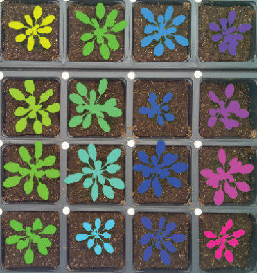
PlantCV v2.0: Image analysis software for high-throughput plant phenotyping. Malia A Gehan, Noah Fahlgren, Arash Abbasi, Jeffrey C Berry, Steven T Callen, Leonardo Chavez, Andrew N Doust, Max J Feldman, Kerrigan B Gilbert, John G Hodge, J Steen Hoyer, Andy Lin, Suxing Liu, César Lizárraga, Argelia Lorence, Michael Miller, Eric Platon, Monica Tessman, Tony Sax
Segmented plants with help from Rasberry Pi, from Tovar, et al.’s prperint
Raspberry Pi Powered Imaging for Plant Phenotyping. Jose Tovar, John Steen Hoyer, Andy Lin, Allison Tielking, Steven Callen, Elizabeth Castillo, Michael Miller, Monica Tessman, Noah Fahlgren, James Carrington, Dmitri Nusinow, Malia A. Gehan
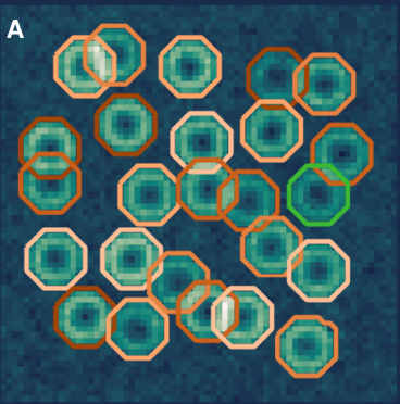
Segmenting Calcium flashes in Reynolds, et al.’s preprint
Mapping nonapoptotic caspase activity with a transgenic reporter in mice. Peter Nicholls, Thomas Pack, Nikhil Urs, Sunil Kumar, Gabor Turu, Evan Calabrese, Wendy Roberts, Ping Fan, Valeriy Ostapchenko, Monica Guzman, Flavio Beraldo, Vania Prado, Marco Prado, Ivan Spasojevic, Joshua Snyder, Kafui Dzirasa, G. Allan Johnson, Marc Caron
Multi-platform discovery of haplotype-resolved structural variation in human genomes. Mark J.P. Chaisson, Ashley D. Sanders, Xuefang Zhao, Ankit Malhotra, David Porubsky, Tobias Rausch, Eugene J. Gardner, Oscar Rodriguez, Li Guo, Ryan L. Collins, Xian Fan, Jia Wen, Robert E. Handsaker, Susan Fairley, Zev N. Kronenberg, Xiangmeng Kong, Fereydoun Hormozdiari, Dillon Lee, Aaron M. Wenger, Alex Hastie, Danny Antaki, Peter Audano, Harrison Brand, Stuart Cantsilieris, Han Cao, Eliza Cerveira, Chong Chen, Xintong Chen, Chen-Shan Chin, Zechen Chong, Nelson T. Chuang, Deanna M. Church, Laura Clarke, Andrew Farrell, Joey Flores, Timur Galeev, Gorkin David, Madhusudan Gujral, Victor Guryev, William Haynes-Heaton, Jonas Korlach, Sushant Kumar, Jee Young Kwon, Jong Eun Lee, Joyce Lee, Wan-Ping Lee, Sau Peng Lee, Patrick Marks, Karine Valud-Martinez, Sascha Meiers, Katherine M. Munson, Fabio Navarro, Bradley J. Nelson, Conor Nodzak, Amina Noor, Sofia Kyriazopoulou-Panagiotopoulou, Andy Pang, Yunjiang Qiu, Gabriel Rosanio, Mallory Ryan, Adrian Stutz, Diana C.J. Spierings, Alistair Ward, AnneMarie E. Welsch, Ming Xiao, Wei Xu, Chengsheng Zhang, Qihui Zhu, Xiangqun Zheng-Bradley, Goo Jun, Li Ding, Chong Lek Koh, Bing Ren, Paul Flicek, Ken Chen, Mark B. Gerstein, Pui-Yan Kwok, Peter M. Lansdorp, Gabor Marth, Jonathan Sebat, Xinghua Shi, Ali Bashir, Kai Ye, Scott E. Devine, Michael Talkowski, Ryan E. Mills, Tobias Marschall, Jan Korbel, Evan E. Eichler, Charles Lee
Assessment of the impact of shared data on the scientific literature. Michael Milham, Cameron Craddock, Michael Fleischmann, Jake Son, Jon Clucas, Helen Xu, Bonhwang Koo, Anirudh Krishnakumar, Bharat Biswal, Francisco Castellanos, Stan Colcombe, Adriana Di Martino, Xi-Nian Zuo, Arno Klein
A persistent lack of International representation on editorial boards in environmental biology. Johanna Espin, Sebastian Palmas-Perez,Farah Carrasco-Rueda, Kristina Riemer, Pablo Allen, Nathan Berkebile, Kirsten Hecht, Renita Kay Kastner-Wilcox, Mauricio Nunez-Regueiro, Candice Prince, Maria Constanza Rios-Marin, Erica P. Ross, Bhagatveer Sangha, Tia Tyler, Judit Ungvari-Martin, Mariana Villegas, Tara Cataldo, Emilio Bruna
Here are the highlights from the current issue of Development:
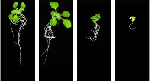
Making a move: EMT holds the key to planarian regeneration
During development and wound healing, progenitor cells are required to migrate to different locations before they can differentiate into terminal tissue types. This cell migration often involves epithelial-to-mesenchymal transition (EMT), a process by which cells delaminate from an epithelium and become motile. On page 3440, Aziz Aboobaker and colleagues investigate how neoblasts, the adult stem cell population present in planarians, are able to migrate to sites of damage in order to regenerate tissue after irradiation. Using a shielded X-ray irradiation assay, they show that neoblasts require β-integrin and the activity of a matrix metalloproteinase to interact with the extracellular matrix and move through the tissue, just as in EMT. In addition, they show that migration requires EMT-associated transcription factor orthologs, such as snail-1, snail-2 and zeb-1. Strikingly, the differentiation status of cells also affects their ability to migrate. Finally the authors report that, even in the absence of wounding, a notum-dependent signal from the brain, which normally lacks resident stem cells, draws in migrating neoblasts to maintain tissue homeostasis. Together, these results suggest that EMT-related mechanisms controlling cell migration are conserved among bilaterians and provide insights into how progenitor populations move to a site of wounding before regeneration begins.
How mouse oocytes give DNA damage the SAC
Cells in embryos and adult tissues have mechanisms that allow them to identify and respond to DNA damage, thereby ensuring that deleterious mutations cannot arise and persist in individuals. On page 3475, Keith Jones and colleagues investigate the mechanism by which mouse oocytes arrest upon DNA damage. This response involves activation of the spindle assembly checkpoint (SAC), which normally prevents the onset of anaphase until all chromosomes are correctly attached to the spindle. In this study, the authors find that, within minutes of DNA damage, SAC-associated proteins are not recruited to the sites of damage along chromosome arms, but instead become concentrated at the chromosome kinetochores, which act as a platform to generate the SAC signal. SAC activation is dependent on the activity of aurora kinase and MPS1 kinase but, interestingly, does not rely on PI3K-related kinases important for the DNA damage response in other systems. Furthermore, the authors show that the arrest response is unique to oocytes in meiosis I and does not occur in oocytes undertaking meiosis II. These results uncover a new mechanism by which DNA damage is dealt with in oocytes and provide clues into how the formation of genetically abnormal embryos is prevented.
Imp and Syp call time on Drosophila neuroblasts
Drosophila neurons are born from progenitors, known as neuroblasts, in a temporally controlled manner. Given that the timing of birth affects the type of neuron that is generated, this process must be tightly regulated over time so that a diverse array of neuronal progeny is produced. The RNA-binding proteins IGF-II mRNA-binding protein (Imp) and 15 Syncrip/hnRNPQ (Syp) are known to exhibit temporally graded expression patterns in neuroblasts, and have thus been shown to regulate the process of neuronal fate specification. Now, on page 3454, Tzumin Lee and colleagues uncover a role for Imp and Syp in neuroblast decommissioning, as well as in neuron differentiation. ‘Decommissioning’ is the process by which neuroblasts shrink and exit the self-renewing progenitor state before forming terminally differentiated neurons. The authors find that Imp and Syp are crucial for this two-stage ‘decommissioning’ process. Imp regulates shrinkage of the neuroblast so that this event does not occur prematurely, while Syp acts subsequently to promote the accumulation of Prospero in the nucleus, leading to cell-cycle exit. Together, these results provide a mechanism by which neuroblast decommissioning occurs in the Drosophila brain and enhance our understanding of how neural stem cells are controlled during development.
PLUS:
An interview with Jayaraj Rajagopal
Jayaraj (Jay) Rajagopal is a Principal Investigator at the Center for Regenerative Medicine at Massachusetts General Hospital and an Associate Professor of Medicine at Harvard Medical School. His lab works on the development and regeneration of the lung, using stem cell and animal models to develop novel insights that hopefully will provide inspiration for therapies to help treat human lung disease. In 2017, he was awarded the Dr Susan Lim Award for Outstanding Young Investigator at the International Society for Stem Cell Research (ISSCR) meeting in Boston (MA,USA), where we met him to talk about how a fish tank started a life-long fascination with the lung, the transition to running his own lab, and his optimism for the future of both basic stem cell research and its clinical translation. Read the Spotlight article on p. 3389.
On the evolution of bilaterality
Bilaterality – the possession of two orthogonal body axes – is the name-giving trait of all bilaterian animals. These body axes are established during early embryogenesis and serve as a three-dimensional coordinate system that provides crucial spatial cues for developing cells, tissues, organs and appendages. How bilaterality evolved and whether it evolved once or several times independently is a fundamental issue in evolutionary developmental biology. Recent findings from non-bilaterian animals, in particular from Cnidaria, the sister group to Bilateria, have shed new light into the evolutionary origin of bilaterality. In their Hypothesis article, Grigory Genikhovich andUlrich Technau compare the molecular control of body axes in radially and bilaterally symmetric cnidarians and bilaterians, identify the minimal set of traits common for Bilateria, and evaluate whether bilaterality arose once or more than once during evolution.
The PAR proteins: from molecular circuits to dynamic self-stabilizing cell polarity
PAR proteins constitute a highly conserved network of scaffolding proteins, adaptors and enzymes that form and stabilize cortical asymmetries in response to diverse inputs. They function throughout development and across the metazoa to regulate cell polarity. In recent years, traditional approaches to identifying and characterizing molecular players and interactions in the PAR network have begun to merge with biophysical, theoretical and computational efforts to understand the network as a pattern-forming biochemical circuit. In their Review article, Charles Lang andEdwin Munro summarize recent progress in the field, focusing on recent studies that have characterized the core molecular circuitry, circuit design and spatiotemporal dynamics.
Can injured adult CNS axons regenerate by recapitulating development?
In the adult mammalian central nervous system (CNS), neurons typically fail to regenerate their axons after injury. During development, by contrast, neurons extend axons effectively. A variety of intracellular mechanisms mediate this difference, including changes in gene expression, the ability to form a growth cone, differences in mitochondrial function/axonal transport and the efficacy of synaptic transmission. In turn, these intracellular processes are linked to extracellular differences between the developing and adult CNS. During development, the extracellular environment directs axon growth and circuit formation. In adulthood, by contrast, extracellular factors, such as myelin and the extracellular matrix, restrict axon growth. In their Review article, Brett Hilton andFrank Bradke, we discuss whether the reactivation of developmental processes can elicit axon regeneration in the injured CNS.
The new Center for Stem Cell & Organoid Medicine (CuSTOM) at Cincinnati Children’s Hospital Medical Center (CCHMC) is launching a major new initiative to recruit outstanding tenure-track or tenured faculty at the Assistant to Associate Professor level.
CuSTOM (https://www.cincinnatichildrens.org/research/divisions/c/custom) is a multi-disciplinary center of excellence integrating developmental and stem cell biologists, clinicians, bioengineers and entrepreneurs with the common goal of accelerating discovery and facilitating bench-to-bedside translation of organoid technology and regenerative medicine. Faculty in CuSTOM benefit from the unique environment and resources here to accelerate their studies of human development, disease and regenerative medicine using organoid platforms.
CCHMC is a leader in organoid biology and one of the top ranked pediatric research centers in the world, providing a unique environment for basic and translational research. Among pediatric institutions CCHMC is the third-highest ranking recipient of research grants from the National Institutes of Health. CCHMC continues to make major investments in research supporting discovery with 1.4 million square feet of research space and subsidized state-of-the-art core facilities including a human pluripotent stem cell facility, genome editing, high-throughput DNA analysis, biomedical informatics, a Nikon Center of Excellence imaging core and much more.
We invite applications from innovative and collaborative investigators focused on basic or translational research in human development and/or disease using stem cells or organoid models. Successful candidates must hold the PhD, MD, or MD/PhD degrees, and will have a vibrant research program with an outstanding publication record.
Applicants should submit their curriculum vitae, two to three page research statement focused on future plans, and contact information for three people who will provide letters of recommendation to CuSTOM@cchmc.org. Applications must be submitted by January 5, 2018.
The Cincinnati Children’s Hospital Medical Center, and the University of Cincinnati are Affirmative Action/Equal Opportunity Employers. Qualified women and minority candidates are especially encouraged to apply.
We are inviting applications for a post-doctoral scientist to join the Waltzer team in the “Genetic Development and Reproduction” (GReD) research unit in Clermont-Ferrand (France).
Our group investigates how blood cell development is controlled during normal and pathological situations. Using mainly Drosophila as a model system, we aim at deciphering the gene regulatory network that regulates blood cell progenitor maintenance and differentiation. We are particularly interested in the transcriptional and post-transcriptional regulation of blood cell fate 1,2 and in the development of fly models for leukemia 3.
Following the recent relocation of our team to the GReD, we are now seeking to recruit a talented and motivated post-doc to expand our work into the epigenetic and epitranscriptomic fields. The proposed project will focus on characterizing the molecular mechanism of action of a conserved enzyme involved in hematopoiesis and leukemia, using a combination of genetic, developmental & molecular approaches in Drosophila.
Qualifications & skills
The candidate must hold a PhD in Life Sciences, in the field of molecular biology, developmental biology and/or genetics. Candidates with previous experience with fly handling, NGS analysis and/or proteomics are particularly encouraged to apply.
Fellowship
The position is available immediately and is funded for three years. Salary scale for the post is in the range from 35.000 and 46.000€ p.a. inclusive, depending on experience.
Environment
The Genetics, Reproduction and Development laboratory (https:www.gred-clermont.fr) is a multi-disciplinary CNRS-INSERM-University research institute on the medical campus of the University Clermont-Auvergne (35,000 students) the main university in central France, localised in the lively city of Clermont-Ferrand, with attractive living conditions.
The GReD consists of 14 groups with research interests ranging from genome dynamics and epigenetic control, development and stem cell biology to endocrinology and cancer. It offers state-of-the-art equipments and all technological facilities required for the project and constitutes an excellent environment for high quality and intensive scientific life and training.
To apply:
Please send your C.V., a cover letter with statement of research interest, and contact details of at least 2 referees to lucas.waltzer@uca.fr
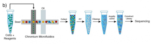
Cell polarisation is crucial for normal development and controlled by complex molecular interactions in the cytoplasm and at the membrane. Today we feature a paper recently published in Developmental Cell that describes a single-cell biochemistry technique and its insights into polarity protein dynamics the developing wormembryo. We caught up with first author Dan Dickinson, who carried out the work as a postdoc in Bob Goldstein‘s lab in UNC Chapel Hill, and has recently started his own lab in the University of Texas.
Looking back, did you always want to be a scientist?
Well, math and science were always my favorite school subjects, even going back to my elementary school years. I was most interested in astronomy early on – I wanted to be an astronaut in second grade, and most of my early science fair projects involved the solar system or telescopes in some form. I got excited about biology in 7th grade, when we were introduced to Mendelian genetics in a science unit. It was my first introduction to the idea that living things behaved according to a set of knowable rules, and I was hooked almost immediately.
And can you tell us your scientific biography up until now?
My first “real” science experience was the year after I graduated from high school. I was younger than most of my classmates, and so I decided to take a year off before college. I got a job as a tech with a small startup pharmaceutical company that was trying to develop generic versions of several chemotherapeutic drugs. In return for washing dishes half-time, they let me do some small research projects trying to optimize drug yields. It was a fun year and a great window into how corporate research works.
I went to college at Iowa State and joined Gloria Culver’s lab halfway through my freshman year. The lab studied the biochemistry of ribosome assembly in E. coli, and I worked on a couple of different projects related to how one particular protein, called S15, binds and alters the structure of the ribosomal RNA.
During my senior year, I got invited to apply for a Fulbright scholarship, and received an award to go to Switzerland for a year and work in a research lab. I got connected with Martin Pruschy at University Hospital in Zurich, who was studying the signalling pathways that are activated in response to ionizing radiation in cancer cells. My project in that lab was trying to identify novel proteases that were activated by radiation. It was pretty high-risk and the project never really went anywhere, but it was my first time working full-time in an academic lab and I learned a lot about how science really works. It was great preparation for grad school.
I got my Ph.D. at Stanford, where I was jointly advised by James Nelson (a cell biologist) and Bill Weis (a crystallographer and biochemist). I proposed a new project for the two labs, studying the evolution of cell-cell adhesion in the slime mold Dictyostelium. The projected started out because I got curious about evolution, started doing blast searches, and found out that Dicty has homologs of beta-catenin and alpha-catenin, two cell-cell adhesion proteins that we had previous assumed were only present in animals. I wanted to figure out what Dicty was doing with these proteins, and we went in with the hypothesis that it might have something to do with adhesion during the multicellular phase of its life cycle. We were surprised to learn that in fact, during the multicellular phase, Dicty forms a tissue that looks very much like an animal epithelium, and this tissue required the catenins for its structure and polarity. This was a big deal because it suggested that the basic organizational principles behind animal multicellularity might be much more ancient than anyone thought.
I wanted to move into an animal model system as a postdoc, and picked the Goldstein lab after interviewing in several worm and fly labs. I was attracted to the simplicity of C. elegans embryos and the ability to study development at single-cell resolution.
It’s funny – I can see why someone would ask that after having read my papers (especially from my postdoc). But I don’t think of myself as a “methods guy” at all. I didn’t start working on methods until the second year of my postdoc, and it was only because I got frustrated by the lack of available tools for the experiments I wanted to do. In hindsight, I guess I do enjoy the challenge of working out new techniques, but I’ve always been motivated by the biology.
What do in vivo biochemical methods like the one described in your paper promise to tell us about development?
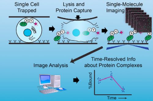
Well, at the risk of over-generalizing, I’d say that most developmental problems have historically been inaccessible to biochemistry. There are exceptions, of course – Xenopus egg extracts come to mind as a beautiful biochemical system with developmental relevance – but broadly speaking, it’s been hard to get pure populations of cells or tissue from in vivo models in sufficient quantity for biochemistry. The sc-SiMPull approach we developed, combining nanoscale microfluidic lysis and single-molecule biochemistry, represents one solution to that challenge.
The graphical abstract describing single-cell biochemistry in vivo. Dickinson, et al. 2017, Dev Cell.
Can you give us the key results of the paper in a paragraph?
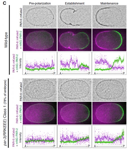
There were two really key findings. The first was a demonstration that technically, it is possible to get dynamic information about protein-protein interactions in single C. elegans zygotes by lysing them and performing single-molecule pull-down. Second, by applying this approach, we found that the PAR complex – a critical cell polarity determinant in many systems – dynamically oligomerizes during polarity establishment. We studied the significance of that transient oligomerization using targeted mutants and live imaging, and found that PAR complex oligomerization is essential for PAR proteins to get transported to the anterior of the cell by the cortical flows that establish polarity. We also found that a cell cycle kinase, PLK-1, controlled PAR complex oligomerization by directly phosphorylating one of the complex members (a protein called PAR-3). This was exciting because prior to our work, I don’t think anyone really understood how the timing of polarity establishment was controlled.
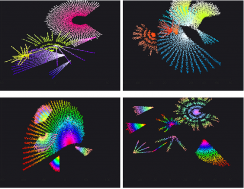
Polarising embryos and single molecule images derived from them, from Figure 2, Dickinson, et al. 2017. Dev Cell.
How applicable do you see sc-SiMPull being to other systems?
I think it should be straightforward to apply to other systems, and in fact that’s one of the things we’re going to work on in my own lab (which just started this past month, at the University of Texas at Austin). Now that the basic approach is worked out, applying it to other systems should be as simple as adjusting the design of the microfluidic chips to accommodate whatever sample we want to study.
I was fortunate to be in touch with the PIs on those other papers, Nate Goehring and Fumio Motegi, before any of us submitted, and we communicated periodically during the review process. I think all three of us had a genuine desire to see everyone get credit for their work – which I believe is the right attitude to have. All three papers reached a similar conclusion about the role of PAR complex oligomerization in polarity establishment, but there’s actually very little overlap in terms of the experiments each group did. That’s an ideal situation, because the papers reinforce each other and increase our confidence that the conclusions are correct.
Improper embryonic polarity following blocked PAR-3 oligomerization, from Figure 5, Dickinson, et al. 2017. Dev Cell.
When doing the research, did you have any particular result or eureka moment that has stuck with you?
The first time I saw single molecules that I’d pulled out of an embryo, that was a big moment when I knew this was going to work. Also, when I first put the oligomerization-blocking mutant embryos on the microscope and saw that they had an interesting polarity defect, that was a big day when it became clear we’d actually discovered something interesting.
And what about the flipside: any moments of frustration or despair?
I actually starting working on sc-SiMPull almost 5 years ago – before CRISPR – and the CRISPR work was kind of a detour along the way. In fact, the whole reason I started doing CRISPR in the first place was because I thought that endogenous gene tagging was a pre-requisite for the kind of quantitative biochemistry I envisioned (we could have counted complexes of overexpressed proteins, but what would we really have learned?). CRISPR turned out to be a much longer detour than I’d expected. In hindsight, it was worth it, of course, but there were some struggles along the way and times when I wondered whether I’d ever get back to the work I actually wanted to be doing.
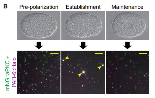
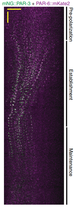
Kymograph of cortical PAR-3 and PAR-6 during the first cell cycle, from Figure 3, Dickinson, et al. 2017. Dev Cell.
Austin is great. It’s a really fun place to live, it’s relatively affordable, and from a scientific standpoint I think the department is phenomenal. I’ve felt very welcome here and am excited to be getting the lab up and running. This is an exciting time because I feel like we finally have the tools for the kind of science I want to do, and I’m looking forward to deconstructing the PAR polarity system at a level of mechanistic understanding that hasn’t been possible previously. We’ll keep working on worms, but I want to expand into some other systems too.
Finally, what do you get up to when you are outside of the lab?
I have two little boys, ages 3 and 5, who are just tremendous fun. I love watching the reactions they get when they tell their friends and teachers that their dad smashes worms for a living. Since we moved here I’ve been spending most of my weekends building various backyard climbing structures for them to mess around on. I also love to cook (and eat), and I compensate for all the cooking and eating by road cycling about 100 miles a week. One of my favorite things about Austin so far is the cycling culture – there are bike lanes everywhere, and they are widely used even in the heat of the summer. It’s been a lot of fun to explore the city and surrounding area that way.
An NIH-funded postdoctoral fellow position is open at UC Davis School of Medicine.
The postdoc opening is in the Knoepfler Lab. The focus of research will be on stem cell epigenomics, particularly as it relates to pluripotency, neural differentiation, and oncogenic transformation. Approaches will include functional genomics assays such as ChIP-Seq, chromatin conformation capture, CRISPR genetics work, and cell biological research. The Knoepfler lab is also part of the UC Davis Genome Center, the Comprehensive Cancer Center, and the Institute for Regenerative Cures.
Qualifications:
Applicants must have a PhD, an MD, or both. Preference will be given to applicants who have a strong track record as students of biological research including published work and ideally one or more published first author papers. Genomics, next generation sequencing, bioinformatics, stem cell or cancer biology would be a plus for candidates. Excellent written and oral communication skills are required.
To apply: E-mail knoepfler@ucdavis.edu a 1-page overview of research experience and career goals, a CV, and contact information for 3 references.
UC Davis School of Medicine is an outstanding research and teaching institution committed to diversity, and located in beautiful Northern California.
From the spots of a leopard and stripes on a zebra to the pigmentation of sea shells and arrangement of sand dunes in a desert, repeating patterns are present at vastly different scales throughout the natural world. During embryonic development, repeating patterns are also prevalent and lead to diverse structures such as the digits of the limb, intestinal villi, cartilaginous rings of the trachea and rugae of the palate. But how do these patterns form in the first place?
A common feature with repeating patterns is that they arise through a process of self-organisation from local interactions within the system. The ability to self-organize is nowhere more apparent than in an embryo, where a single fertilised cell can develop into a complex organism with many different structures. To the mathematician and famous war-time code breaker Alan Turing, this emergence of a body plan (morphogenesis) was an example of symmetry breaking; where homogenous or ‘symmetric’ tissue, in terms of it being developmentally equivalent, transitions from a uniform to an organised heterogeneous state. This phenomenon inspired Turing to publish his seminal 1952 work ‘The chemical basis of morphogenesis’[1]. In his paper, Turing theorised that a pair of interacting chemical molecules (which he termed ‘morphogens’) that are both diffusible but at different rates, when acting across an entire field with local fluctuations can break symmetry and produce a regularly spaced pattern. This concept is now commonly referred to as the Turing reaction-diffusion model.
Some 20 years later two German scientists, Hans Meinhardt and Alfred Gierer, provided a general mechanism for such pattern formation [2]. Meinhardt and Gierer showed that a system only requires a network where a self-enhancing molecule with a short diffusible range known as the ‘activator’ stimulates the production of its own ‘inhibitor’ molecule that operates over a larger spatial range. Simply put, for a pattern to form, there needs only be local self-enhancing activation coupled with long-range inhibition.
Although this Meinhardt-Gierer model originally described biological molecules, the fundamental interactions of the network can explain the formation of non-biological patterns. Indeed, a nice example to introduce their model is during the formation of sand dunes. In this system, grains of sand are blown across a desert by the prevailing wind. Tiny random fluctuations in the system (such as a small rock making a bump) will cause sand grains from the wind to be deposited and soon a small mound will begin to form. This deposition of sand is the activator. Now as the wind continues to blow, this new mound will trap more sand and consequently will increase in size. This can be thought of as local self-activation. However, this increasing deposition of sand at the growing mound means that the number of sand grains remaining immediately downwind is vastly depleted. As a result, no sand is deposited in the area immediately behind the mound, generating a space until the next dune can form. This is the long-range inhibition in the system.
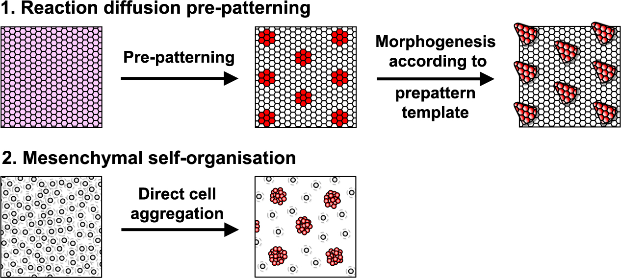
Reaction-diffusion systems are signal driven processes based on diffusing elements (e.g extracellular proteins, such as growth factors) that lead to a spatially patterned change in cell state usually resulting in altered gene expression. This arrangement of cell states then acts as a template or ‘prepattern’ from which anatomical structures are produced by inducing local cell aggregation, growth or survival (Fig. 1). However, importantly, these models are not the only type of mechanism that can produce pattern. A second class of model relies upon the ability of cells to self-organise directly, requiring no prepattern. In these systems periodic focal points of high cell density are created through chemotaxis, mechanical deformation of the environment, or cell-cell adhesion (Fig. 1). Although no prepattern is required, this type of pattern formation shares the same constraints as the first class of reaction-diffusion systems (local activation coupled with long range inhibition), but in these systems local cell clustering provides the activation whereas the inhibition results from depletion of cells around the emerging aggregates. Thus, these two classes of patterning mechanism are distinguished by the entity that moves to break symmetry – whether diffusible signals (reaction-diffusion) or cells (mesenchymal self-organisation).
Figure 1. Different processes capable of producing periodic patterns. 1 – A signal driven system where diffusible elements produce a prepattern template from which morphogenesis is guided. 2 – Cell driven mesenchymal self-organisation where cells organise themselves directly into aggregates without instruction from a prepattern. Figure adapted from Glover et al., 2017 [3].
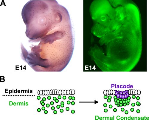
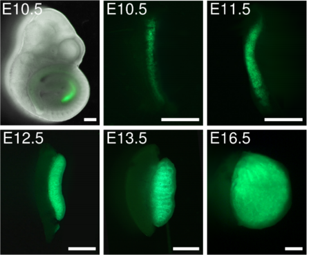
The developing skin is a good model in which to study periodic pattern formation as it is structurally quite simple and readily produces easily appreciated repeating patterns. At embryonic day 13, mouse skin is composed simply of an epithelial sheet (epidermis) lying atop a loose mesenchyme (dermis). As far as we can tell no pre-programmed information relevant to defining hair follicle positions is in there at the start and so the tissue can be deemed symmetric at this stage. Yet, only a day later (either in vivo or in culture) the skin has partitioned itself into a dotted pattern of altered gene expression and cell aggregations that form the hair follicle primordia (Fig. 2). This model system allows us to investigate pattern formation in a tightly framed way as we do not need to worry about the (admittedly interesting) earlier problem of how the skin develops to that stage, nor the (also interesting) question of how the primordia go on to make fully functional hair follicles and the intervening space becomes mature skin. So how is the symmetry of the homogeneous tissue broken to give a repeating spatial pattern of cell clusters and divergent cell fates? More specifically, instead of concerning ourselves with determining how the pattern produces an exact density and size of spots, we wanted to understand how the tissue symmetry is broken to give a spatially ordered arrangement.
Figure 2. The primary hair follicle array in mouse. A) In embryonic day 14 (E14) mouse embryos a periodic pattern of hair follicles can be observed through detection of the early placode marker Dkk4 (left panel) or by visualisation of dermal cell organisation using a fluorescent reporter (right panel). B) Between E13 and E14 mouse hair follicle formation occurs, consisting of a compaction and invagination of epidermal cells to form the placode, which sits directly above an aggregation of mesenchymal cells called the dermal condensate. Figure adapted from Glover et al., 2017 [3].
A more complete reaction-diffusion system
As several signalling pathways have been shown to be essential for early hair primordium formation, we were interested in the integration between them and whether this integration might constitute a pattern forming reaction-diffusion system. We restricted our consideration to molecules that are members of three major pathways (BMP, FGF and WNT) known to be involved in hair follicle development [4-6], and to those mRNAs that have short half-lives and so can undergo rapid regulation at the timescale of the primary hair pattern formation. We assessed the transcriptional regulatory interactions between these molecules to define the network of interactions between them. To test whether our experimentally derived network was capable of creating a periodic pattern we sought the expertise of Dr Vaclav Klika (Czech Technical University, Prague). Vaclav’s mathematical analysis showed that this multiple species reaction-diffusion system is capable of breaking symmetry to produce a periodic pattern. From this it is plausible that interactions between WNT, FGF and BMP pathways are sufficient to generate the hair follicle pattern.
More than signals alone
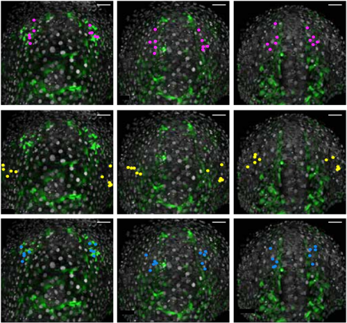
Because complete hair follicle development also requires the formation of dermal condensates that underlie each placode, we wanted to explore how these mesenchymal cells aggregate and how this is regulated by the pathways and molecules we identified in our gene regulatory network. To answer this question we enlisted the help of Dr Richard Mort (based at the University of Lancaster) to track dermal cell movement during condensate formation. Using live cell imaging we found that the mesenchymal cells that form the dermal condensate are those located in its immediate vicinity, suggesting that local cues arising during patterning guide the dermal cells. As it was likely that any such signals would come from the epidermis we examined the timing between the epidermal signal driven pattern and corresponding dermal cell rearrangement. By analysing the gene expression of early placode markers and comparing this with the cellular organisation we found the existence of a molecular prepattern in the epidermis that precedes condensate formation. Further investigation revealed that this prepattern provides a template of local FGF sources that attract dermal cells ultimately leading to condensate formation.
Making everything a hair follicle
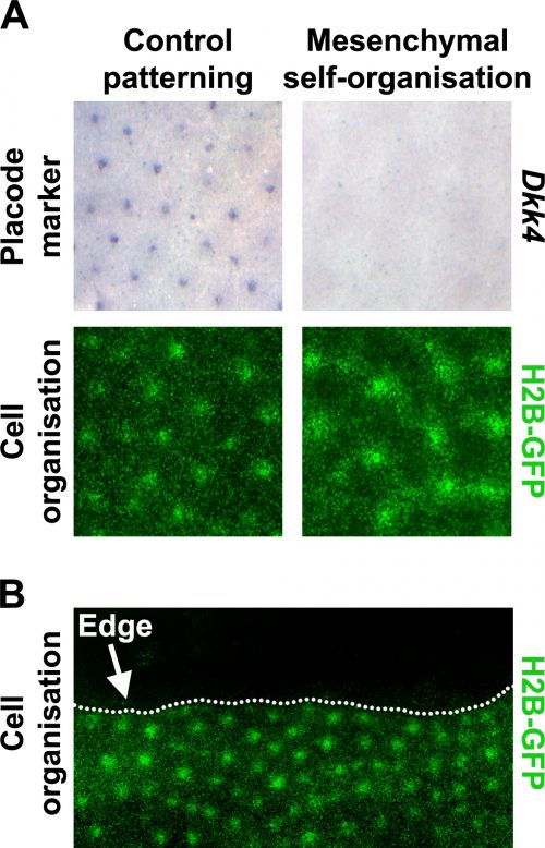
Prior literature indicated that hair follicle primordia are distinguished by their high FGF and low BMP activity [5, 7]. We wondered what would happen if we imposed these conditions across the entire skin. Using a pharmacological inhibitor of BMP signalling and a recombinant FGF protein, we treated unpatterned skins to achieve this effect. The resulting pattern of the skins cultured in these conditions looked rather similar to control skins, based on dermal cell aggregation, but we found that expression of epidermal placode marker genes was absent in these skins (Fig. 3A). We realised that this condition had revealed a previously unrecognised developmental potential in the skin; that the dermis has the capability to pattern by itself without instruction from of an epidermal prepattern.
To analyse the formation of these mesenchyme-only patterns we enlisted the help of Dr Franziska Matthäus (FIAS in Frankfurt) who specializes in methods to analyse cell motility. Through particle image velocimetry (PIV) analysis we began to identify distinct differences, such as far greater cell movement and incorporation into condensates, between the mesenchyme-only and the unperturbed patterning processes.
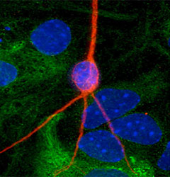
Figure 3. A) Under conditions that recreate the environment of hair follicle primordium, mesenchymal cells (visualised using TCF/Lef::H2B-GFP reporter mice) can self-organise in the absence of epidermal instructions (placode marker – Dkk4). B) In reaction-diffusion signal based systems patterns form at the cut edge. This behaviour at the tissue edge can be used to determine the type of patterning mechanism.
Living on the edge
To work out whether the mesenchymal cell only patterns arise through a fundamentally different patterning system we used the fact that although the skin appears developmentally homogeneous, it is unavoidable that it has edges where dissected away from the embryo to put into culture. The behaviour of patterns at these tissue edges can be very informative when trying to distinguish the underlying mechanism of a pattern’s generation (Fig. 3B).
Patterning systems relying on active inhibitory signals will form a row of foci close to the edge as the inhibitor diffuses off the edge, thereby giving a competitive advantage to those cells at the perimeter. Conversely, in cell driven patterning systems the pattern will stay away from the edge as cell number becomes limiting for the nucleation of new cell aggregates, and at the edge there are simply fewer cells available to recruit. By inserting cuts into skins cultured in both control and in our conditions recreating the hair follicle primordium, we see that the mesenchymal only patterning is characteristic of a cell driven mechanism rather than a signal driven reaction-diffusion system as is observed for the control experiments. This suggested that these two systems rely on fundamentally different mechanisms.
We then searched for a molecular mechanism for mesenchyme-only patterning. We found that disruption of TGFβ signalling abolished mesenchyme-only patterning, but that these perturbations had only modest effects on normal patterning, which would fit the idea that these patterning processes occur through divergent mechanisms with a different reliance on TGFβ signalling. In addition, we found that local sources of TGFβ2 attract mouse dermal cells, revealing that TGFβ2 serves as a widely expressed attractant that draws mesenchymal cells together.
Finally, having determined that restricted TGFβ signalling was critical for mesenchyme-only patterning we wanted to determine its role in the interplay between normal and mesenchymal patterning mechanisms during hair development. We found that TGFβ2 treatment substantially enhances dermal cell attraction to FGF sources and that when TGFβ signalling is inhibited dermal cells migrate very poorly towards to local FGF sources. Thus, in normal development TGFβ signalling creates an environment conducive to cell recruitment to the local epidermal FGF sources. These sources, defined by the signal driven network as the prepattern, create restricted microenvironments that provide the conditions for mesenchyme-only patterning to occur such that a dermal condensate is formed. This demonstrates that fundamentally distinct patterning systems can operate together during embryonic organ formation, but in this case a hierarchy exists wherein one system guides the other.
Conclusions
Research into biological pattern formation has been enjoying a resurgence in recent years. Examples of Turing systems underpinning the formation of the limb digits [8], villi of the intestine [9] or the transition of colour spots on lizard skin [10] have been described. In addition to signal focused patterning mechanisms, during the weeks following the release of our work, two new papers [11, 12] have described cell processes driving chicken feather patterning and mouse hair follicle assembly, highlighting the ability of mesenchymal cells to self-organise. Studying the interplay between cell and signal driven processes during embryogenesis promises to be an exciting field of investigation in the future, which could provide fresh insight in the fundamental areas of developmental biology.
This work would not have been possible with the input of many collaborators. The mathematical guidance, simulations and models provided to us by Kevin Painter, Vaclav Klika and Franziska Matthäus has allowed us to confirm our preliminary laboratory findings and provide a more comprehensive answer to the initial question we asked. The imaging experiments and tools for cell tracking designed by Richard Mort enabled us to accurately follow dermal cells during hair follicle formation making it possible for us reject a hypothesis of pre-determined cell sorting, highlighting the utility of live cell imaging when studying developmental processes. Collaborations such as these, where scientists specialising in a range of different fields and from different locations work together on a single problem is the basis for outstanding research and I strongly encourage establishing similar relationships to help advance your own research.
This work was funded by the BBSRC and carried primarily in Dr Denis Headon’s group at The Roslin Institute near Edinburgh. You can discover the full story, findings and experiments in our paper:
Turing, A.M., The Chemical Basis of Morphogenesis. Philosophical Transactions of the Royal Society of London Series B-Biological Sciences., 1952. 237(641): p. 37-72.
Gierer, A. and H. Meinhardt, A theory of biological pattern formation. Biological Cybernetics, 1972. 12(1): p. 30-39.
Glover, J.D., et al., Hierarchical patterning modes orchestrate hair follicle morphogenesis. PLoS Biol, 2017. 15(7): p. e2002117.
Mou, C., et al., Generation of the primary hair follicle pattern. Proc Natl Acad Sci U S A, 2006. 103(24): p. 9075-80.
Huh, S.H., et al., Fgf20 governs formation of primary and secondary dermal condensations in developing hair follicles. Genes Dev, 2013. 27(4): p. 450-8.
Andl, T., et al., WNT signals are required for the initiation of hair follicle development. Dev Cell, 2002. 2(5): p. 643-53.
Botchkarev, V.A., et al., Noggin is a mesenchymally derived stimulator of hair-follicle induction. Nat Cell Biol, 1999. 1(3): p. 158-64.
Raspopovic, J., et al., Modeling digits. Digit patterning is controlled by a Bmp-Sox9-Wnt Turing network modulated by morphogen gradients. Science, 2014. 345(6196): p. 566-70.
Walton, K.D., et al., Villification in the mouse: Bmp signals control intestinal villus patterning. Development, 2016. 143(3): p. 427-36.
Manukyan, L., et al., A living mesoscopic cellular automaton made of skin scales. Nature, 2017. 544(7649): p. 173-179.
Shyer, A.E., et al., Emergent cellular self-organization and mechanosensation initiate follicle pattern in the avian skin. Science, 2017. 357(6353): p. 811-815.
Lei, M., et al., Self-organization process in newborn skin organoid formation inspires strategy to restore hair regeneration of adult cells. Proceedings of the National Academy of Sciences, 2017. 114(34): p. E7101-E7110.
 (1 votes)
 (No Ratings Yet)
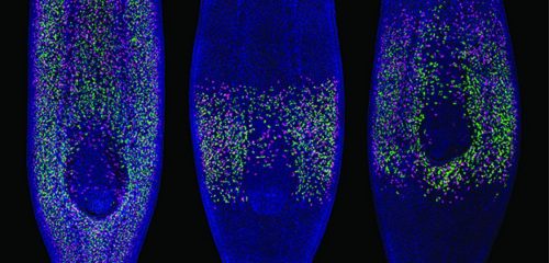


 Jayaraj (Jay) Rajagopal is a Principal Investigator at the Center for Regenerative Medicine at Massachusetts General Hospital and an Associate Professor of Medicine at Harvard Medical School. His lab works on the development and regeneration of the lung, using stem cell and animal models to develop novel insights that hopefully will provide inspiration for therapies to help treat human lung disease. In 2017, he was awarded the Dr Susan Lim Award for Outstanding Young Investigator at the International Society for Stem Cell Research (ISSCR) meeting in Boston (MA,USA), where we met him to talk about how a fish tank started a life-long fascination with the lung, the transition to running his own lab, and his optimism for the future of both basic stem cell research and its clinical translation. Read the Spotlight article on p.
  Bilaterality – the possession of two orthogonal body axes – is the name-giving trait of all bilaterian animals. These body axes are established during early embryogenesis and serve as a three-dimensional coordinate system that provides crucial spatial cues for developing cells, tissues, organs and appendages. How bilaterality evolved and whether it evolved once or several times independently is a fundamental issue in evolutionary developmental biology. Recent findings from non-bilaterian animals, in particular from Cnidaria, the sister group to Bilateria, have shed new light into the evolutionary origin of bilaterality. In their
  PAR proteins constitute a highly conserved network of scaffolding proteins, adaptors and enzymes that form and stabilize cortical asymmetries in response to diverse inputs. They function throughout development and across the metazoa to regulate cell polarity. In recent years, traditional approaches to identifying and characterizing molecular players and interactions in the PAR network have begun to merge with biophysical, theoretical and computational efforts to understand the network as a pattern-forming biochemical circuit. In their
  In the adult mammalian central nervous system (CNS), neurons typically fail to regenerate their axons after injury. During development, by contrast, neurons extend axons effectively. A variety of intracellular mechanisms mediate this difference, including changes in gene expression, the ability to form a growth cone, differences in mitochondrial function/axonal transport and the efficacy of synaptic transmission. In turn, these intracellular processes are linked to extracellular differences between the developing and adult CNS. During development, the extracellular environment directs axon growth and circuit formation. In adulthood, by contrast, extracellular factors, such as myelin and the extracellular matrix, restrict axon growth. In their
 (6 votes)