Here we highlight some developmental biology related content from other journals published by The Company of Biologists.
JCS kicked off 2017 with a Special Issue relevant to many developmental biologists: 3D cell biology. It’s packed full of commentaries, interviews, research articles and techniques, and well worth a browse.
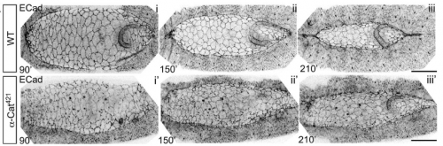
Nicole Gorfinkiel and colleagues showed that α- Catenin stabilises actomyosin foci and E-Cadherin to promote apical contraction in the Drosophila amnioserosa.
A position (#122764) is available immediately for a Research Technician/Faculty Specialist to contribute to our studies in neural crest and placodes. The Technician will conduct research, assist in the training of students, and take part in the management of the laboratory of Dr. Lisa Taneyhill at the University of Maryland. Laboratory skills should include the ability to perform various molecular biology and biochemical assays, such as recombinant DNA/cloning; immunoprecipitation and immunoblotting; and/or immunohistochemistry. Experience with microscopy, chick embryology, and tissue culture is desirable. For more information on the lab, please see http://www.ansc.umd.edu/people/lisa-taneyhill. A Bachelor’s degree (B.A. or B.S.) in a related field and prior laboratory research experience is essential. Fluency in spoken and written English is required. Salaries are highly competitive, negotiable and commensurate with qualifications. Fringe benefits offered. Applicants must apply through eTerp at https://ejobs.umd.edu. Applications will be accepted until a suitable candidate is identified.
Our latest monthly trawl for developmental biology (and other cool) preprints. See June’s introductory post for background, and let us know if we missed anything
2017 started where 2016 had left off, with an number of preprints covering most corners of developmental biology, plus more relevant work from related fields. Looking at the list below, it’s clear that a mix of young and established labs are using preprints. Following the trends of last year, they were predominantly found on bioRxiv, with some also on arXiv and PeerJ Preprints.
This month features regeneration in mouse digits and whole ascidians, how body axes form in gastruloids, the link between metabolism and signalling in mice, and developmental plasticity in Arabidopsis. There also were a whole bunch of fly papers, and new tools including a new ImageJ and ‘WikiGenomes’. Happy preprinting!
Molecular and functional variation in iPSC-derived sensory neurons. Jeremy Schwartzentruber, Stefanie Foskolou, Helena Kilpinen, Julia Rodrigues, Kaur Alasoo, Andrew J Knights, Minal Patel, Angela Goncalves, Rita Ferreira, Caroline L Benn, Anna Wilbrey, Magda Bictash, Emma Impey, Lishuang Cao, Sergio Lainez, Alexandre J Loucif, Paul J Whiting, HIPSCI Consortium, Alex Gutteridge,Daniel J Gaffney
Stem cell differentiation is a stochastic process with memory. Patrick S. Stumpf, Rosanna C. G. Smith, Michael Lenz, Andreas Schuppert, Franz-Josef Müller, Ann Babtie, Thalia E. Chan, Michael P. H. Stumpf, Colin P. Please, Sam D. Howison, Fumio Arai, Ben D. MacArthur
Distinguishing Mechanisms Underlying EMT Tristability. Dongya Jia, Mohit Kumar Jolly, Satyendra Chandra Tripathi, Petra Den Hollander, Bin Huang, Mingyang Lu, Muge Celiktas, Esmeralda Ramirez-Pena, Eshel Ben-Jacob, Jose N. Onuchic, Samir M. Hanash, Sendurai A. Mani, Herbert Levine
It′s okay to be green: Draft genome of the North American Bullfrog (Rana [Lithobates] catesbeiana). S Austin Hammond, René L Warren, Benjamin P Vandervalk, Erdi Kucuk, Hamza Khan, Ewan A Gibb, Pawan Pandoh, Heather Kirk, Yongjun Zhao, Martin Jones, Andrew J Mungall, Robin Coope, Stephen Pleasance, Richard A Moore, Robert A Holt, Jessica M Round, Sara Ohora, Nik Veldhoen, Caren C Helbing, Inanc Birol
Progress Towards a Public Chemogenomic Set for Protein Kinases and a Call for Contributions. David H Drewry, Carrow I Wells, David M Andrews, Richard Angell, Hassan Al-Ali, Alison D Axtman, Stephen J Capuzzi, Jonathan M Elkins, Peter Ettmayer, Mathias Frederiksen, Opher Gileadi, Nathanael Gray, Alice Hooper, Stefan Knapp, Stefan Laufer, Ulrich Luecking, Susanne Muller, Eugene Muratov, R. Aldrin Denny, Kumar S Saikatendu, Daniel K Treiber, William J Zuercher, Timothy M Willson
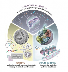
The 4D Nucleome Project. Job Dekker, Andrew S Belmont, Mitchell Guttman, Victor O Leshyk, John T Lis, Stavros Lomvardas, Leonid A Mirny, Clodagh C O’Shea, Peter J Park, Bing Ren, Joan C Ritland, Jay Shendure, Sheng Zhong, The 4D Nucleome Network
Genome Graphs. Adam M Novak, Glenn Hickey, Erik Garrison, Sean Blum, Abram Connelly, Alexander Dilthey, Jordan Eizenga, M. A. Saleh Elmohamed, Sally Guthrie, André Kahles, Stephen Keenan, Jerome Kelleher, Deniz Kural, Heng Li, Michael F Lin, Karen Miga, Nancy Ouyang, Goran Rakocevic, Maciek Smuga-Otto, Alexander Wait Zaranek, Richard Durbin, Gil McVean, David Haussler, Benedict Paten
Biocuration as an undergraduate training experience: Improving the annotation of the insect vector of Citrus greening disease.Surya Saha, Prashant S Hosmani, Krystal Villalobos-Ayala, Sherry Miller, Teresa Shippy, Andrew Rosendale, Chris Cordola, Tracey Bell, Hannah Mann, Gabe DeAvila, Daniel DeAvila, Zachary Moore, Kyle Buller, Kathryn Ciolkevich, Samantha Nandyal, Robert Mahoney, Joshua Von Voorhis, Megan Dunlevy, David Farrow, David Hunter, Taylar Morgan, Kayla Shore, Victoria Guzman, Allison Izsak, Danielle E Dixon, Liliana Cano, Andrew Cridge, Shannon Johnson, Brandi L Cantarel, Stephen Richardson, Adam English, Nan Leng, Xiaolong Cao, Haobo Jiang, Chris Childers, Mei-Ju Chen, Mirella Flores, Wayne Hunter, Michelle Cilia, Lukas A Mueller, Monica Munoz-Torres, David Nelson, Monica F Poelchau, Josh Benoit, Helen Wiersma-Koch, Tom D’elia, Susan J Brown
Recommended by Bob Goldstein, University of North Carolina at Chapel Hill
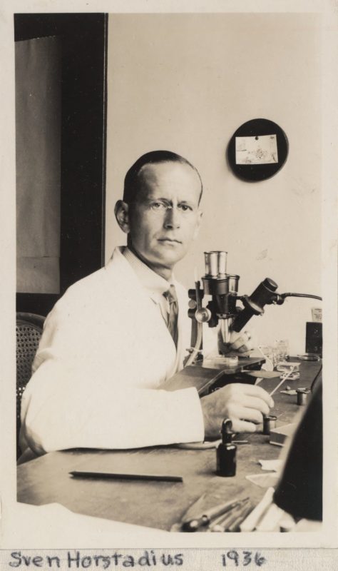
Sven Hörstadius stands alongside the likes of Boveri, Spemann, Mangold and Driesch as a giant of experimental embryology in the first half of the twentieth century. While his 1950 treatise on the neural crest in vertebrate head development became an early bible for the field, he is probably best known for his work on sea urchins. His 1939 manuscript summarises a whole suite of work from himself and others dealing with various kinds of determination: of the axes of the embryo, of cell fate by instruction or position, and of ‘species character’ by the cytoplasm or the nucleus.
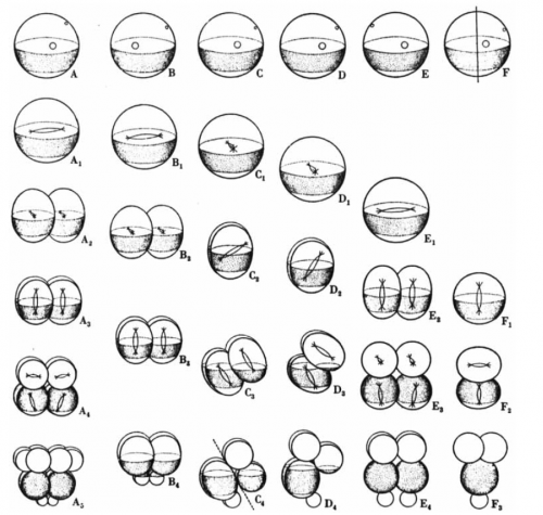
The focus is on the purple sea urchin, Paracentrotus lividus, which Hörstadius studied during trips to the Stazione Zoologica in Naples, once a haunt of Driesch and Boveri and still an active centre of research. P. lividus was a particularly useful urchin species because the eggs and early embryos have a vegetal pigment band, allowing polarity to be easily traced. In previous decades the basics of itsdevelopment had been sketched out, including where successive cleavage planes lie in the cell, the movements of gastrulation, and fate of some blastomeres. However, while fundamental rules of development could be inferred from observation alone, Hörstadius was an experimentalist, and intervened in development in a variety of creative ways to investigate determination. Like Rosa Beddington, subject of the previous post in this series, he was known as an expert dissector: he could isolate blastomeres with fine glass needles, and create chimeras by fusing part of one animal to part of another and placing a small glass ball on top “to give the necessary pressure”. His 1939 paper is full of this sort of cut and paste embryology, and the outcomes are often unexpected (“The following is a very strange phenomenon…”).
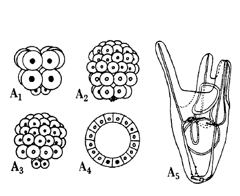
Stages of early Paracentrotus development, from uncleaved egg to 64-cell stage, from Figure 1, Hörstadius, 1939. Reproduced with permission of Wiley†.
The paper also devotes a lot of time accounting for contradictory results from other researchers. In particular, there seems to be a long standing beef with one Leopold von Ubisch, with some wonderfully formal put-downs:
“von Ubisch does not admit the possibility that halves of equatorial and subequatorial eggs are different. This is strange…” (p162)
“von Ubisch concludes that the cytoplasm has no influence on the species character of the skeleton. I do not find this conclusion convincing” (p169)
“The diagram of Fig. 9 has been criticized by von Ubisch. But his objections were, it seems to me, anticipated in the original paper” (p158)
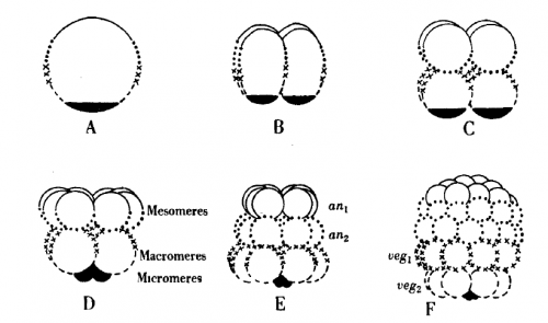
Among the flood of experiments and ideas that Hörstadius describes, some stand out. The first results section addresses the control of spindle position and orientation in the divisions that make the 16 cell embryo with its characteristic micromeres at the vegetal pole. These divisions can be delayed by shaking or adding diluted sea water; once this delay is relieved, the embryo starts dividing again. However, instead of picking up where they left off, the embryos would often skip a cell division such that, for instance, a delayed 4 cell embryo would divide to give micromeres (column B in the figure below).
Cleavage in Paracentrotus in normal (A) and delayed conditions (B-F). From Figure 2, Hörstadius, 1939. Reproduced with permission of Wiley†.
Hörstadius proposed that the micromere division is determined by the ‘activation’ of material in the vegetal cytoplasm, and that this activation occurs independently of how many cell divisions had occurred. After a certain amount of time, the embryo wants to make micromeres, irrespective of the number of divisions completed, and changes spindle position to do so. There is a kind of ‘cleavage clock’ which sets the type of cell division that occurs next.
The next three sections concern the embryonic axes. Hörstadius began his career under the supervision of John Runnström, who proposed that the sea urchin egg held two gradients, one emanating from the animal and one from the vegetal pole. The gradients “interact mutually and are partially hostile to each other”, and a cell’s fate is dependent on the relative strength of each of the gradients in the cytoplasm it inherits. Before the molecular biology revolution, before experimental and what was then called ‘chemical’ embryology were united, the cytoplasm had “qualities” or “forces” that influenced development.
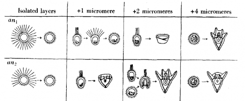
The development of isolated an1 and an2 layers, with or without added micromeres. From Figure 9, Hörstadius, 1939. Reproduced with permission of Wiley†.
One of the ways Hörstadius investigated the double gradient hypothesis was to add micromeres (the inheritors of the most vegetal, and hence most ‘active’ cytoplasm) to layers of animal cells. These layers, if isolated and left alone, go on to form useless ciliated balls. Remarkably, add enough micromeres and you could rescue normal development, and gradually increasing the micromere number led to progressively ‘better’ development. Along with the results of various other experiments involving adding bits of one embryo to bits of another, this implied that the amount of animal and vegetal material you start with is crucial to the outcome. To explain these results, Hörstadius
“…assume[d] an animal and a vegetative gradient, both reaching the opposite pole and progressively diminishing. The animal and the vegetative qualities or forces have to interact in order to bring about normal differentiations, e.g. vegetative influences are necessary for the formation of ciliated band and stomodaeum, animal ones for gastrulation and skeleton formation, and so on. The differentiation depends – within wide limits – upon the relative amounts of animal or vegetative material present.” (p173)
While Lewis Wolpert used these experiments to inform his models of positional information, half a century later the picture had changed as Eric Davidson and others challenged the idea of a double gradient. In the updated model, specification is conditional, and mediated by successive interactions between micromeres and blastomeres, the micromeres having been initially autonomously specified by maternally supplied factors. The model combines modern data on signalling pathways and gene expression with a reinterpretation of Hörstadius’ classic experiments and their insight that cell interactions could direct development.
A heterosperm merogone formed from Paracentrotus lividus cytoplasm and Psammechinus microtuberculatus nucleus, from Figure 12, Hörstadius, 1939. Reproduced with permission of Wiley†.
The paper ends with the question of the role of the nucleus in inheritance. Hörstadius took up work originally started by Boveri half a century earlier, removing the nucleus from eggs of one species and fertilising them with the sperm of another to make ‘heterosperm merogones’. The nucleus coming from one species, and the cytoplasm the other, he could ask which species the larva ended up looking like, and hence where ‘character’ was determined. In one combination, the merogone followed the characteristics of the species from which the nucleus was taken, but due to high variability in other combinations, Hörstadius had to be cautious in his intepretation:
“…the nucleus obviously has a positive effect on the species character. It is to be regretted that the characters in question are not defined sharply enough to permit of a conclusion as to the possible role of the cytoplasm.” (p172)
Even treading this lightly, the results are consistent with the work of Boveri, Spemann, Waddington, Wilson and Stevens, all of which put the nucleus, and the mysterious substance contained within it, in the driving seat of development and heredity.
The paper is testament to the power of cut and paste embryology, and the rich potential of the sea urchin embryo as a model. We can leave it Carl Olaf Jacobsen, one of only two graduate students trained by Hörstadius during his long time as a Professor in Uppsala, to summarise his supervisor’s legacy:
“…his experiments on sea urchin larvae shed light on a couple of the most central findings in developmental biology, namely that the uneven distribution of the egg-cell contents give rise to early embryo cells with shifting qualities, and that communication between these cells has an essential role in the differentiation process.”
This paper’s a fun read because it’s dense with simple and clever experiments. And many of the experiments built a foundation for our current understanding of how animal development works. But I love it most of all because it’s a great example of old literature that includes questions that we, as a field, forgot were fundamental questions. How does Hörstadius’ cleavage clock work? I think we still don’t know.
Hörstadius’ work was the inspiration for much of what I have done over the past 30 years. The following information was circulated over the years first by my mentor who had met Hörstadius, and more stories were circulated years ago when there was a festscrhriften thrown in honor of Hörstadius’ life in Stockholm. Unfortunately it was years after his death. Apparently until after his death the Swedes didn’t realize how well known Hörstadius actually was.
The post mentions that Hörstadius worked under Runnström. That was true and it was the cause for several things in his life. The Swedish system had very few professors so Horstadius worked under Runnström for most, if not his entire career. For that reason the Swedes never considered Hörstadius as particularly famous because he was not in charge. Ruunström was also involved in Hörstadius’ science so I was told. Runnström was a major proponent of morphogenetic gradients, especially double gradients. Consequently, the double gradients that are part of Hörstadius’ sea urchin work fit the ideas of Runnström – or perhaps were strongly suggested by him. Perhaps it was only gossip, but I heard from several senior scientists that Hörstadius never really believed that double gradient nonsense but since he was in Runnström’s lab that was part of the cost. He was later admitted to the Swedish Academy and to the Royal Academy largely for his work on neural crest, but the sea urchin work has stood the test of time with a greater presence than the neural crest work.
Höstadius was praised for his microsurgical abilities. He was the only one in the world who could do those remarkable dissections of the tiny sea urchin embryos and the cut and paste experiments were considered amazing feats. That was how I was initially taught about his work. He taught himself to dissect on the stage of a compound microscope. That meant he had to teach himself to move his needles in the opposite direction relative to what was seeing through the objective. His dissection tray was a piece of photographic film on the bottom of a shallow petri dish. That was because the photographic film had a coating of gelatin on it which was just thick enough to score a trough in which his embryos would be placed. Then, all of his dissections were done by hand. By contrast, I do all the same dissections but in my case I have major help from micro manipulators, a dissection microscope which shows movements in the same direction as what I can see though the objective, and I can record everything with a camera whereas he had to draw everything. As I indicated, he had great hands in addition to a perceptive brain.
† Further usage of any Wiley content that appears on this website is strictly prohibited without permission from John Wiley & Sons, Inc. Please contact Wiley’s Permissions Department either via email: permissions@wiley.com or use the RightsLink service by clicking on the ‘Request Permission’ link accompanying this article on Wiley Online Library (www.onlinelibrary.wiley.com)”
Aidan Maartens
This post is part of a series on forgotten classics of developmental biology. You can read the introduction to the series here and read other posts in this series here. We also would love to hear suggestions for future Forgotten Classics – let us know in the comments box.
Faculty of Biology, Medicine and Health, The University of Manchester, Manchester, Uk
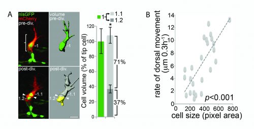
Collective cell migration is involved in many biological processes. In particular it is required to build new tissues during morphogenesis and to repair them during wound healing. Cancer cells however also exploit it during invasion of other tissues. In order for a group of cells to migrate together as a coordinated group they must establish a hierarchy of cellular identities, generally thought of as “leader” and “follower” cells (Friedl & Gilmour 2009). How this hierarchy is established and robustly maintained is key to understanding the process of collective cell migration. To examine this, in our recent study we explored the collective movement of endothelial cells undergoing angiogenesis (the generation of new blood vessels from existing ones) in zebrafish embryos. We were particularly interested in how these collectively migrating cells managed to maintain their organisation while undergoing divisions.
During sprouting angiogenesis the collectively migrating cells take on the roles of either a leading “tip cell” or a following “stalk cell” (Herbert & Stainier, 2011). The tip cell is the first cell to leave the existing vessel and is highly motile. This is then followed by the stalk cells, which are less motile and go on to form the main trunk of the developing vessel. Previous work has established that this hierarchy is driven by differing levels of Vascular endothelial growth factor (VEGF) signalling, with the tip cells having high levels compared to the stalk cells. This is thought to be established via a Notch/Delta controlled lateral inhibition, whereby tip cells induce a reduction in VEGF signalling in the stalk cells (Herbert & Stainier, 2011).
Sprouting angiogenesis in a zebrafish embryo. The “tip cell” (far left) first sprouts from the existing blood vessel and is followed by the “stalk cells”. Blue is DNA and red is the cell membranes
During angiogenic sprouting migrating endothelial cells are required to undergo mitosis (Schoors et al. 2015). This presents the cells with a problem, as the two daughter cells must acquire two different migratory profiles, dependent on their resultant positions. The more distal cell must take on the tip cell identity, while the more proximal cell becomes the trailing stalk cell. However, division will partition the components of the VEGF signalling and Notch pathway components, which if equal will presumably induce competition for tip cell identity between the two daughter cells. This would disrupt the migration of the group of cells and ultimately impede angiogenesis. However, far from this, tip/stalk cell identities are actually established almost instantaneously after division, much faster than notch/delta mediated lateral inhibition is thought to take (upwards of 5 hours) (Matsuda et al. 2015; Bentley et al. 2014).
After division cells immediately display distinct tip and stalk cell behaviours. Costa et al., (2016)
The question – How are the tip/stalk identities of collectively migrating endothelial cells re-established so quickly after mitosis?
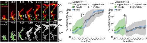
Computer modelling suggested that the answer to this question could be that these cells undergo a form of asymmetric cell division and produce daughters of different sizes. The model predicted that a larger daughter cell would inherit more of the VEGF signalling machinery, giving it higher levels of VEGF signalling and thereby establishing it as the tip cell. Live imaging of zebrafish angiogenic endothelial cells revealed that indeed the most distal daughter cell (the tip cell position) is on average 1.8-1.9 times larger than the more proximal daughter cell (the stalk cell position). Furthermore, the size of each daughter cell was shown to be proportional to its migratory speed, meaning that sister cells that had the biggest difference in their size also have the biggest difference in their speeds. Furthermore, in vitro work also demonstrated that larger cells inherit a larger proportion of VEGF receptor mRNA, as well as having higher levels of VEGF signalling. In order to demonstrate that these differing levels of VEGF signalling were necessary to define post-mitotic tip/stalk cell identities, zebrafish embryos were treated with low levels of a drug that blocks VEGF signalling. This low dose didn’t inhibit signalling altogether but prevented any cell from signalling at the levels necessary to be a tip cell. Under these conditions both daughter cells assumed stalk-like identities after mitosis.
A) The divisions of zebrafish angiogenic endothelial cells are asymmetric; one cell (the tip cell, cell 1.1) is larger than the other (the stalk cell, cell 1.2) B) The size of a cell is proportional to its speed of migration. Costa et al., (2016)
How might the asymmetry in daughter cell size be achieved?
Preliminary data suggests that the size asymmetry seen in angiogenic endothelial cells is (at least in part) generated by the positioning of the mitotic spindle towards the proximal pole of the cell (Costa et al. 2016). Thereby shifting the division plane away from the volumetric centre of the cell. Other classical asymmetric cell divisions also result in cells of different sizes, for example Drosophila neuroblasts and one-cell C. elegans embryos, though the role of this asymmetry has not been extensively explored (Cabernard et al. 2010; McNally 2013). However some clues may be gleaned as to how angiogenic endothelial cells manage to position their mitotic spindles such that two differently sized daughters are produced. A canonical set of proteins is known to generate the membrane associated pulling force that acts upon mitotic spindles. Partner of Inscuteable (Pins) (LGN in Drosophila and GPR1/2 in C.elegans), anchored at the membrane by Gαi (GOA-1 and GPA-16 in C.elegans), binds to Nuclear mitotic apparatus (NuMA) (Mud in Drosophila and LIN-5 in C.elegans), which in turn binds to the dynein/dynactin complex, this then pulls on the plus ends of astral microtubules (Bergstralh et al. 2013). Asymmetric enrichment of this complex can cause the spindle to be pulled towards one side of the cell, such as in the one-cell C.elegans embryo (Kiyomitsu, 2015). However, it remains to be seen whether the spindle orienting machinery is involved in positioning the mitotic spindle of angiogenic endothelial cells.
Downward movement of the mitotic spindle positions the plane of division such that 2 differently sized daughter cells are produced. Costa et al., (2016)
Asymmetric cell division is a well-described phenomenon traditionally thought of as a process employed by cells to enable them to generate cells different from themselves. An asymmetric inheritance of fate determinants or a position dependent asymmetry in external cues normally results in daughter cells becoming two different cells types. Size asymmetry (and a resultant asymmetry in signalling strengths) between daughter cells offers a simple way of introducing subtle heterogeneity into a population of a single cell type. Furthermore, if it is controlled so that the larger and smaller cells are positioned specifically, patterns (such as leading and following cells) can be produced. Further work is needed to elucidate the mechanism behind this new form of asymmetric division and it will be interesting to see whether other collectively migrating systems, or indeed any other cell types, undergo similar divisions.
References
Bentley, K., Harrington, K.I. & Regan, E.R., Can active perception generate bistability? Heterogeneous collective dynamics and vascular patterning. ALIFEhttp://dx.doi.org/10.7551/978-0-262-32621-6-ch053 (2014)
We are currently seeking applications for the role of Reviews Editor at Development. This is a temporary position (maternity cover) anticipated to last up to 12 months, starting June 2017.
Joining an experienced and successful team, this is an exciting opportunity to make a significant contribution to one of the major journals in the field of developmental biology. Development publishes primary research articles, reviews and other front section content across the breadth of the developmental biology and stem cell fields.
Applicants will hold a PhD in developmental or stem cell biology. Post-doctoral and/or previous editorial experience is desirable, although we will provide full training.
Core responsibilities:
Commissioning, handling peer review and developmental editing of material for the front section of the journal
Representing the journal at international conferences and within the wider scientific community
Writing press releases, article highlights and material for Development’s community website ‘the Node’
Creative involvement in the journal’s development
The successful candidate will have a broad interest in science, the scientific community and publishing. Excellent interpersonal and literary skills, enthusiasm and commitment are also essential requirements for the position.
The Reviews Editor will work alongside an experienced in-house team, including the Executive Editor and current Reviews Editor, as well as with our international team of academic editors.
This maternity cover position provides an excellent opportunity to gain experience on a highly successful life-science journal, and offers an attractive salary and benefits. The position is full-time and will be based in The Company of Biologists’ attractive modern office on the outskirts of Cambridge, UK.
The Company of Biologists (biologists.com) exists to support biologists and inspire advances in biology. At the heart of what we do are our five specialist journals – Development, Journal of Cell Science, Journal of Experimental Biology, Disease Models & Mechanisms and Biology Open – two of them fully open access. All are edited by expert researchers in the field, and all articles are subjected to rigorous peer review. We take great pride in the experience of our editorial team and the quality of the work we publish. We believe that the profits from publishing the hard work of biologists should support scientific discovery and help develop future scientists. Our grants help support societies, meetings and individuals. Our workshops and meetings give the opportunity to network and collaborate.
Applicants should send a CV to recruitment@biologists.com, along with a covering letter that summarises their relevant experience, why they are enthusiastic about this opportunity, and their current salary level. Applicants should be eligible to work in the UK and should be able to travel internationally.
Applications should be received by February 28th 2017, though we may be able to consider later applications.
The brown alga Ectocarpus has emerged as a model system for the evolution of muticellularity. Today’s paper, from the current issue of Development, investigates the role and evolutionary history of a gene implicated in Ectocarpus development. We caught up with first author Nicolas Macaisne and supervisor J. Mark Cock of the Station Biologique de Roscoff in Brittany.
Mark, can you tell us your scientific biography and the general focus of your lab?
JMC I am a molecular biologist that worked for many years on various aspects of land plant biology, including nitrogen fixation and self-incompatibility. In 2002 I moved to the Station Biologique de Roscoff, a marine laboratory on the north-west coast of France, with the aim of setting up a laboratory to work on brown algal developmental biology. Working with Akira Peters, we evaluated several brown algae for their potential as model systems and eventually selected the filamentous brown alga Ectocarpus (Peters et al., 2004). The choice of Ectocarpus was based on its amenability to genetic analyses and on earlier work, particularly by Dieter Müller in Constance, that had demonstrated the merits of this species as a laboratory system. The main objective of the laboratory has been to understand the molecular mechanisms that regulate life cycle progression in this brown alga. Ectocarpus has a haploid-diploid life cycle involving an alternation between two multicellular organisms, the sporophyte and the gametophyte. We have been using genetic approaches to identify both key life cycle regulators and regulators of downstream processes during the development of the two generations (Coelho et al., 2011; Peters et al., 2008). In addition, a great deal of effort has been put into generating tools for Ectocarpus, including the sequencing and analysis of its genome in collaboration with the Genoscope in Paris and the VIB in Ghent (Cock et al., 2010). More recently, the laboratory has also become interested in sex determination in the brown algae, a project led by my co-PI Susana Coelho (Ahmed et al., 2014; Lipinska et al., 2015; Luthringer et al., 2015).
And Nicolas, how did you come to work with Mark in Roscoff?
NM I joined Mark’s laboratory in 2011, just after obtaining my Ph.D. under the mentorship of Raphaël Mercier in the National Institute for Agronomic Research of Versailles, France. During my thesis, I studied the genetic regulation of meiotic DNA repair in Arabidopsis, a land plant which is a very well established model for genetics. Thus, my previous research and the model I was employing were quite far removed from Mark’s research on the biology of algae. There are very few labs that study algae, but the unusual life cycle of Ectocarpus, especially the fact that the sporophyte and the gametophyte generations are independent from one another, intrigued me. I realized that this system offered a unique opportunity to study these developmental stages independently. I didn’t appreciate how much I would learn about basic plant biology from this beautiful organism.
I also recognized Mark’s lab as a place where I could challenge myself to learn new techniques and approaches. Working with a model system for which the genome was still not completely assembled and only has a partial genetic map was daunting, but I now feel that I have gained an appreciation for how to set up a new system and look beyond our traditional models for unique insights. This experience was also incredibly valuable for allowing me to learn genome and transcriptome sequencing and analysis. These techniques are absolutely invaluable for the analysis of genes involved in all aspects of developmental biology. Mark’s expertise and the great scientific environment at the Station Biologique de Roscoff are the best assets I could have hoped for in order to achieve my goals.
What makes brown algae interesting models for developmental and evolutionary biologists?
JCM & NM Although many eukaryote groups have evolved simple multicellular forms, we generally consider that only five major groups have evolved complex multicellularity, i.e. possess macroscopic bodyplans with multiple cell types orchestrated by developmental programs (Cock and Collén, 2015). These groups are animals, land plants, brown and red algae and fungi. Complex multicellularity evolved independently in each of these five lineages and comparisons between lineages could therefore provide us with important information about the molecular principals that underlie this key evolutionary transition. Unfortunately, whilst we know a great deal about developmental mechanisms in animals and land plants, the three remaining groups remain almost completely uncharacterised. Of these latter groups, the brown algae are particularly interesting because they include species that rival land plants in terms of their developmentally complexity. We believe that it is important to extend the scope of developmental biology beyond the animal and land plant lineages to effectively address questions about the evolutionary origins of developmental processes associated with complex multicellularity.
“It is important to extend the scope of developmental biology beyond the animal and land plant lineages to effectively address questions about the evolutionary origins of developmental processes associated with complex multicellularity”
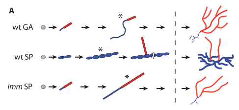
The brown algae are also interesting models to address a wide range of more specific developmental questions. Haploid-diploid life cycles, such as that of Ectocarpus, raise the question of how a single genome directs the implementation of two different developmental programs to produce either a sporophyte or a gametophyte at the appropriate stage of the life cycle. Clearly, epigenetic processes must play an important role in this alternation but at present we have no information about how this might be mediated and the epigenetic aspect will be an important avenue for future research. Historically, brown algae have made important contributions to our understanding of early embryogenesis, particularly the establishment of polarity and the asymmetric division of the initial cell (Bouget et al., 1998; Brownlee and Bouget, 1998; Goodner and Quatrano, 1993). In this context, one of the most surprising features of the Ectocarpus life cycle is the alternation between symmetrical and asymmetrical initial cell divisions during the sporophyte and gametophyte generations, respectively. If we look more broadly across the brown algae, these organisms exhibit a bewildering variety of different types of life cycle (Cock et al., 2014) and sexual systems (Luthringer et al., 2014). For example, Fucus species have diploid life cycles and the diecious members of this genus are thought to have diploid phase sex-determination systems (presumably XY or ZW sex chromosome systems) associated with oogamy, whereas the haploid-diploid life cycle of Ectocarpus is associated with a haploid-phase sex-determination system (UV chromosomes) and near-isogamy. Moreover, life cycle and sex-determination systems appear to have varied considerably during the evolution of this group providing an excellent context to link the different types of system with developmental (size, growth habit, reproductive strategy, etc.) and ecological (habitat, biotic interactions, etc.) parameters.
Brown algae also have many interesting features associated with adaptation to the extreme conditions of the coastal environment. These include their unusual cell walls that provide both strength and flexibility, and mechanisms providing resistance to both abiotic (variations in light, heat, desiccation, salinity, etc.) and biotic stresses. Their evolutionary past, which included a secondary endosymbiosis event that gave rise to the stramenopile plastid, provided brown algae with a mosaic of genetic information from various sources, including that of the red algal endosymbiont. Developmental biologists can therefore expect to discover unusual features at the molecular level and the identification of the IMM gene is probably just a foretaste of the novelties to come.
Schematic representation of the early development of the wild-type gametophyte (wt GA), the wild-type sporophyte (wt SP) and the imm mutant sporophyte (imm SP), from Figure 2, Macaisne, et al. 2017. Development.
And you appear to have quite an extensive toolkit for Ectocarpus in particular?
JMC & NMEctocarpus has a long history as a research organism stretching back to the 19th century (Charrier et al., 2008; Coelho et al., 2012). One of our priorities after selecting Ectocarpus as a model organism was to generate a genome sequence and associated genomic and genetic tools including a transcriptomic database, genetic maps and methodologies for investigating gene function. Tool generation is an ongoing process, for example we have recently re-annotated the genome sequence, adding extensive new information and significantly improving the quality of the existing information (Cormier et al., 2016). The Ectocarpus genome is currently one of the highest quality reference sequences available for the stramenopiles. The generation of tools to analyse gene function has been a more complicated process and we still lack one key tool: a robust protocol for genetic transformation. Importantly, the recent development of an RNAi approach, initially for Fucus (Farnham et al., 2013) and subsequently for Ectocarpus (Macaisne et al., 2016), has provided an alternative for reverse genetics and the recent demonstration that forward genetic approaches can be applied in Ectocarpus also represents a powerful tool to analyse gene function. We hope that our demonstration that forward genetic approaches can be applied to brown algal systems will encourage other groups to investigate developmental mechanisms in this group, leading to the emergence of macroalgal developmental biology as a field in its own right.
Can you give us the key results of your paper in a paragraph?
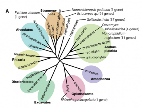
JMC & NM In most brown algae, the initial cell divides to produce two cells with basal and apical identity respectively. This is also the case for the Ectocarpus gametophyte but the sporophyte is unusual because it first establishes an extensive, filamentous basal system before deploying the apical tissues (upright filaments). The first cell division of the sporophyte is therefore developmentally symmetrical, producing the two germ tubes of a symmetrical basal filament (Peters et al., 2008). Sporophytes carrying a mutation in the IMM gene fail to produce the extensive basal system. Instead the initial cell divides asymmetrically to produce a basal rhizoid and an apical upright filament. The mutant sporophyte therefore resembles a gametophyte and the mutation was initially interpreted as causing a partial conversion of the sporophyte into a gametophyte (Peters et al., 2008). In this study, however, analysis of transcriptomic data indicated that the imm sporophyte retains sporophyte identity and we propose an alternative interpretation: that the phenotype of the imm sporophyte represents a more primitive state of the sporophyte, before it evolved the capacity to deploy the extensive basal system. Positional cloning of the IMM locus showed that it encodes a novel protein of unknown function with a repeated cysteine-rich motif that is also found in a gene from the Ectocarpus virus EsV-1. Interestingly, IMM is a member of a large family of 91 genes in Ectocarpus, which we have called EsV-1-7 domain genes after the viral gene that contains this repeated domain. Other stramenopiles either lack EsV-1-7 domain genes or possess only a single EsV-1-7 domain gene suggesting that there has been a massive expansion of this gene family in the brown lineage, perhaps associated with the transition to complex multicellularity (brown algae are the only stramenopile group that exhibit complex multicellularity). A broader search for EsV-1-7 domain genes across the eukaryotic tree of life found a very unusual distribution. In addition to the stramenopile EsV-1-7 domain genes, members of this family were only found in one cryptophyte (Guillardia theta), two chlorophyte green algae (Cocomyxa subellipsoidea and Monorapidium neglectum) and perhaps in one fungal genome (although this latter gene may be a contaminant). It is difficult to reconcile this distribution with vertical inheritance of the gene family. Interestingly, EsV-1-7 domain genes were also found in the genomes of three diverse viruses, suggesting a possible alternative explanation: viral-mediated horizontal gene transfer. This latter hypothesis may be difficult to confirm because horizontal transfer events may have been quite ancient (this appears to have been the case in the stramenopiles for example) but more complete genome sampling for the lineages in question will undoubtedly shed some light on the evolutionary history of this family.
Partheno-sporophyte germlings after introduction with siRNAs targeting imm (B) and controls (C), from Figure 2, Macaisne, et al. 2017. Development
Although IMM seems functionally restricted to the generation of the sporophyte, it is also expressed in the gametophyte: do you think it is doing anything there?
JMC & NM Gametophytes carrying the imm mutation appear to be completely normal indicating that the gene does not have any function during that generation. Of course, we cannot rule out the possibility that there is a subtle or conditional gametophyte phenotype that we have not detected, but the current data indicates that IMM has a sporophyte-specific function. Therefore, in Ectocarpus at least, IMM appears to be specifically associated with the pathway that directs the development of an extensive basal system and delays deployment of the apical system. Nonetheless, the presence of IMM transcripts during the gametophyte generation remains something of a mystery but presumably, if transcription of IMM during the gametophyte generation does not result in any observable phenotype, then there may not have been any selective pressure to prevent IMM transcription during this part of the life cycle.
The IMM gene is conserved in other brown algae, including species that do not exhibit delayed deployment of the apical system. It will be interesting in the future to investigate the role of these IMM orthologues, during both the sporophyte and the gametophyte generations of these other species.
Schematic tree of the eukaryotes showing the phylogenetic position of species that possess EsV-1-7 domain genes, from Figure 5, Macaisne, et al. 2017. Development
Do you want to hazard a guess at what EsV-1-7 domain proteins are doing?
JMC & NM At present we only have functional information of one member of this family: IMM. The phenotype of the imm mutant suggests this protein has a regulatory function because loss of function leads to changes in the expression levels of a large number of genes. The protein sequence doesn’t tell us very much more about its function except that the repeated cysteine-rich motif in the C-terminal part of the protein (the EsV-1-7 repeat) is reminiscent of a zinc finger motif. Our current guess would therefore be that these proteins contain a novel version of the zinc finger motif that allows them to act as regulatory molecules, perhaps as transcription factors for example. We are currently trying to test this hypothesis by assaying for DNA-binding activity and by attempting to obtain three-dimensional structural information for the protein.
If EsV-1-7 domain proteins have a regulatory role, the next question is what are they regulating? The only information we have at present is their expression patterns in transcriptomic data corresponding to various life cycle stages and conditions. These data, which indicate that the members of the gene family have very diverse expression patterns, suggest that the different members of the family may be involved in diverse processes but additional functional analyses will be necessary to confirm this and to explore the functions of each member of the family.
IMM mutants appear to revert to a more ancestral mode of development, and EsV-1-7 repeat genes may have got into Ectocarpus through horizontal gene transfer. So do you think viral transfer facilitated a new mode of development? It’s a very striking idea!
This would seem to be a reasonable explanation based on the unusual phylogenetic distribution of EsV-1-7 domain genes across the eukaryotes, but it is important to underline that the process would have occurred over a very long time-scale. The presence of EsV-1-7 domain genes in stramenopiles, a cryptophyte, two chlorophytes and perhaps one fungal species, together with the presence of related genes in three viral genomes does strongly suggest horizontal transfer, perhaps mediated by viruses. However, the acquisition of EsV-1-7 domain genes by the stramenopile group appears to be ancient because gene family members were also found in oomycete and eustigmatophyte genomes. Several hundred million years would therefore have separated this ancient horizontal transfer event from the evolution of the IMM gene and the associated novel mode of development in the brown algae.
Nonetheless, we agree that the idea that an important brown algal developmental regulator may have originally been acquired from a viral genome is very interesting. We also believe that the identification of IMM underlines the importance of forward genetic screens and the use of diverse model organisms in developmental biology. None of the classical animal, plant or fungal model organisms possesses EsV-1-7 domain genes, so it would not have been possible to identify this family using these systems.
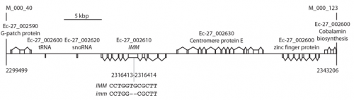
Schematic representation of the 43,708 bp interval on chromosome 27 between the closest recombining markers to the IMM locus, from Figure 5, Macaisne, et al. 2017. Development
When doing the research, did you have any particular result or eureka moment that has stuck with you?
Not really a “eureka” moment, but with the hindsight, I realize how lucky I may have been with some experiments I have undertaken. For example, when I was mapping the mutation causing the imm phenotype, after having scanned the genome of 1699 individuals with more than a hundred molecular markers, I was stuck with a relatively large genomic region containing several annotated genes (and possibly more which were not annotated yet) and no easy way to know which of these genes IMM was. So I decided to adapt a whole-genome sequencing approach to sequence specifically this region of the genome of Ectocarpus. Although this approach was successful in identifying the causal mutation, to my knowledge it had never been performed previously, and I was doing that on an alga. So with hindsight, I think I was a bit overly confident, or maybe a bit naive, in thinking that my approach would work. Thankfully it did, and now I am glad I dared to use it.
“With hindsight, I think I was a bit overly confident, or maybe a bit naive, in thinking that my approach would work. Thankfully it did”
And what about the flipside: any moments of frustration or despair?
NM To be honest, I was a bit afraid when I realized the enormity of data that was obtained from the RNA-sequencing, especially considering the fact I had, at that time, no experience in bioinformatics. However, our team and the bioinformaticians of the Station Biologique were extremely supportive; it has really been a team effort.
More generally, my major frustration is that we cannot yet genetically transform Ectocarpus, preventing us from generating another mutant allele of IMM. This would have made the validation of this gene much easier. However, given the successful use of RNAi for this paper, I am confident that transformation tools like CRISPR/Cas9 targeted mutagenesis will be implemented soon for this model organism. Such tools will have a tremendous impact in the field, hastening considerably the understanding of algal biology.
What about your plans for the future following this paper?
NM Working on a relatively young model organism, with a team that is directly involved in the development of genetic and genomic tools to study it, has been very enriching. I have now joined the laboratory of Judith L. Yanowitz at the Magee-Womens Research Institute in Pittsburgh, PA, where I went back to my primary field of expertise, the regulation of DNA repair during meiosis. I now use the nematode Caenorhabditis elegans as a model organism. Now that I have worked on three different organisms, I realized that having a multi-organism approach is highly beneficial for research as we can take advantage of the similarities and the specificities of each organism to better understand a biological process. The next step for me would be to become a principal investigator to continue my research on meiosis using C. elegans as a model and branching out to less well-developed organisms.
“So far we have only scratched the surface and there is still a great deal to be learnt about brown algal developmental biology”
And where next for the lab?
JMC So far we have only scratched the surface and there is still a great deal to be learnt about brown algal developmental biology, including the identification of the major regulators of the diverse processes discussed above. We are still a long way from having any detailed understanding of developmental processes in brown algae. Our laboratory will continue to focus on life cycle regulation and sex determination, using genetic approaches to dissect the underlying pathways but also developing new approaches, for example to investigate the role of epigenetic processes in life cycle control. We are also very interested in the evolutionary perspective and a recently launched project, in collaboration with Genoscope, aimed at obtaining genome sequences for a broad range of brown algal species will provide the genomic data necessary to place brown algal developmental processes in an evolutionary context.
Ramkumar N, Omelchenko T, Silva-Gagliardi NF, McGlade CJ, Wijnholds J, Anderson KV.
Nat Cell Biol. 2016 Dec;18(12):1281-1291
“It is not birth, marriage, or death, but gastrulation, which is truly the most important time in your life.”
Lewis Wolpert (1986)
Aptly stated by Wolpert, gastrulation is the fundamental process that shapes the morphogenesis of the embryo. It involves the formation of the three germ layers, namely the endoderm, mesoderm and ectoderm, from a single layer of epithelial cells called the epiblast. These germ layers then proceed to give rise to the different tissues and organs in the embryo. Gastrulation involves large scale cell movements, rearrangements and changes in gene expression leading to transformation of cell fates. Among them is a process called epithelial-to-mesenchymal transition (the EMT), wherein epithelial cells lose their apical-basal polarity and adopt migratory mesenchymal behavior. In the mouse embryo, this process happens at the posterior pole of the embryo in a structure called the primitive streak, which is also a convergence point for many signaling pathways in the embryo1. The sheer complexity of gastrulation and its efficiency of execution fascinated me and motivated me to work on it during my PhD.
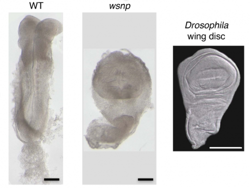
The bulk of our current knowledge and understanding of the signaling pathways involved in gastrulation comes from the analysis of mutants identified from genetic screens mainly in Drosophila and recently in other animals. Likewise, my journey to the world of Crumbs and its role in gastrulation began with a mouse mutant called wsnp(wing shaped neural plate). wsnp mutants have very little mesoderm-derived tissues and their neural plate resembles the Drosophila wing disc.
Figure 1: The wing shaped neural plate (wsnp) phenotype. In comparison to their wild-type litter mates, wsnp embryos had a shortened anterior-posterior axis, lacked visible somites and had a completely open neural plate at embryo day 8.5. At this stage, the wsnp embryos resembled a Drosophila wing disc (C), hence the name. Dorsal views, Anterior up (A, B). Scale bar- 150 µm. Image in (C) is from http://www.pbs.org/wgbh/nova/genes/fate-07.html
With the help of deep sequencing and genetic complementation analysis we identified wsnp to be a null allele of Poglut1 (Protein O-glucosyltransferase) which encodes an enzyme that adds O-glucose to the EGF repeats of proteins. At the time we discovered this, Drosophila Notch was shown to be modified by this enzyme2. While I worked on studying the role of Notch glycosylation during mammalian gastrulation, the severity of the wsnp phenotype compared to that of Notch pathway mutants led us to explore the possibility of alternate targets of the enzyme during gastrulation. Among them, the Crumbs family of proteins stood out as likely candidates. In the lab, we had previously isolated a mutant lulu, a null allele of Epb4.1l5, which had a phenotype similar to wsnp at gastrulation, and biochemical experiments had showed a direct interaction of Epb4.1l5 with mammalian Crumbs3,4. With this as our only inkling, we took a leap of faith and decided to explore the role of Crumbs in gastrulation. At that time, there was no precedent for the role of any Crumbs proteins in mouse development, let alone gastrulation. We decided on Crumbs2 because it had a long extracellular domain with EGF repeats that could be glycosylated and the conserved intracellular domain. We waited for about a year to get the conditional Crumbs2 knock out mice and the results made our wait worthwhile. We were really excited that the phenotypes of wsnp and Crb2-/- were identical. We went on to show that sugar modification of Crumbs2 is essential for its membrane localization and that in turn is essential for its function during gastrulation5.
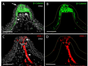
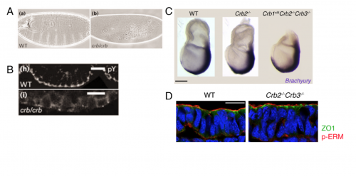
We then focused our attention to our central question – What is the function of Crumbs2 during mammalian gastrulation? We had always known Crumbs as a well-established apical determinant in Drosophila as its role in cell polarity has been well explored there6,7. We were surprised to find that the epiblast of Crumbs2-/- mutants did not have any polarity defects. To rule out the involvement of other mammalian Crumbs proteins in setting up this polarity, we generated triple mutants lacking all known mammalian Crumbs proteins. Any mouse geneticist will tell you – generating triple mutants is not fun! To our surprise, we found that mammalian Crumbs proteins were not required to establish the polarity of the epiblast epithelium. This was in contrast to the case in Drosophila, where Crumbs is essential to set up polarity of the embryonic epithelium. Additionally, in the Crumbs2-/- mouse embryos we found an accumulation of cells expressing E-cadherin and Sox2+at the streak, suggesting that these cells were still epithelial. This was contrary to what we were expecting: as a canonical polarity protein, one would imagine that a loss of Crumbs2 would lead to more cells delaminating i.e. making more mesoderm cells, but we saw the opposite. This suggested to us that the functions of mammalian Crumbs2 could be different from that of Drosophila Crumbs, and got me further engrossed into understanding how mammalian Crumbs2 functions during gastrulation.
Figure 2: Comparing mouse Crumbs null phenotype to Drosophila crb phenotype. (A) Cuticle preparation of wild-type and homozygous crb embryos showing the failure to form a continuous cuticle in crb mutants (anterior is left and ventral down). (B) The ventral epidermis of a stage 12 wild-type embryo and crb mutant embryo stained with an anti-phosphotyrosine (pY) antibody, which labels the site of adherens junctions, shows continuous junctions in wild-type which is completely lost in crb embryos (Apical is down). (C)Wild type, Crumbs2 mutants and Crumbs triple mutants (Crumbs1rd8, Crumbs2-/-, Crumbs3-/-) at E7.5 probed for expression of Brachyury, a marker of the primitive streak. Crumbs triple mutant embryos do not have a more severe gastrulation phenotype than Crumbs2 single mutants (Lateral view, anterior to left, distal down). (D) Transverse section through the epiblast of E7.75 wild type and Crumbs2-/- Crumbs3-/- double mutant embryos immunostained for ZO1 (tight junctions) and pERM (apical domain). The double mutants do not have defects in epiblast polarity (Apical is up). Scale bars in C, 50μm; D, 21μm. The extraembryonic portion of the triple mutant was removed for genotyping. Figures A and B adapted from Ansgar and Knust, 2000.
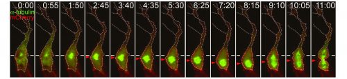
Our attention was drawn to the cells stuck at the streak. Although we were able to visualize these cells using SEM and X-linked GFP, these were static, 2-dimensional images. Cell delamination from an epithelium is a dynamic process requiring constant neighbor exchange and happens in 3-dimensional epithelium. In order to understand exactly when the cells fail during this process, we had to watch this process live in mouse embryos. While it seemed like the most obvious experiment, it was clearly the most challenging aspect of the project at many levels. At that time, there were hardly any labs that had done live imaging with fluorescent reporters in mouse embryos. While cell delamination had been extensively studied, and imaged in in vitro culture systems, only recently with the advent of better imaging technology and improved reporters scientists have started exploring it in live tissues and embryos. For us, it was a herculean task as we had to work out every aspect of the experiment including the mouse strains to use, then determine the imaging conditions to use while keeping the embryo stable. Additionally, we had to define the temporal dynamics of this process in wild-type embryos before we begin to compare it with our mutant.
As mouse embryos are very sensitive to laser light and the primitive streak is a densely packed epithelium, it is important for the reporter to be very bright to get reasonable data. Initially we tried with reporters expressed ubiquitously in the epithelium, as it would reduce the number of crosses and increase the probability of the desired genotype. However, at that time there weren’t good algorithms to segment these images making their analysis very difficult and unfruitful. To bypass this, we decided to express the fluorescent protein in a random pattern, which helps to create better 3-dimensional reconstructions of individual cells and help in their subsequent analysis. However, this would involve additional crosses and also decrease the probability of getting our desired genotype. After screening many different reporter and CRE lines that we and others at Sloan Kettering had in their labs, we began to lose hope in this experiment, as we weren’t getting the right combination of the two. It was around this time that Kathryn attended a conference where Ann Sutherland presented her lab’s work on imaging with the mT-mG reporter and the EIIA-CRE. They found that this combination gave them a good random pattern and the membrane-GFP (mGFP) was a bright reporter at this stage8. Having seen her beautiful movies with the mGFP, we decided to get these mice and eventually used them for our experiment. Despite our optimization of the imaging, there were many factors in this experiment that were beyond our control. For instance, the probability of getting a mutant with both the reporter and the CRE was low and the percentage of randomness of the CRE was unpredictable. Nevertheless, after many unsuccessful attempts, we managed to get beautiful movies of the cells in wild-type and the mutant streak and were happy to see that the cells in the mutants shrink their apical surface but fail to leave the epithelium. While we focused mainly on shape changes and time to delamination for the purpose of this paper, these movies are a gold mine of data. It is amazing how so many processes are happening simultaneously at the streak. There are cell divisions, tissue level movements, neighbor exchanges to just name a few. With the advent of light sheet microscope, there is undoubtedly lots to learn from just watching these cells live in the epithelium.
GFP positive streak cell translocates from the apical to the basal side of the epiblast in wild type embryo. The epiblast cells in the mouse embryo randomly express mGFP at E7.5 and are imaged from the primitive streak side (posterior). A 3D rendered surface (yellow) was built with Imaris for easy visualization of the individual cell shape changes. Ingressing cells have basal protrusions, translocate their cell body basally and leave the epiblast in less than 2h. In addition to the shape changes we discuss in the delaminating cell, the other cells in this region of the epithelium (streak), which are poised to delaminate are also highly dynamic. Time is h:min. Scale bar,10 µm.
The big debate- Polarity?
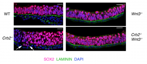
Since Crumbs was as a polarity protein, we had to determine whether the defect we saw was specific to gastrulation at the streak or that Crumbs plays a role in the epithelia by virtue of which it affects the streak. My lab meetings would always end on this debate, followed by heated arguments supporting both hypothesis. The only way to test this was how Drosophila geneticists had always done before. We had to prevent gastrulation from happening using Wnt3 mutants (with and without Crumbs2) and see whether the tissue was normal or not. Fortunately for us, we had the right tools out there to do this experiment. Since both outcomes were equally likely and exciting, irrespective of the result, it was one of the most exciting dissections ever. It would answer the question I had since my first lab meeting. I remember discussing the Wnt3 results at my lab meeting: finally, we concluded without any argument that it is the streak, and not the epiblast epithelium, that requires Crumbs2, after all.
Figure 3: Crumbs2 is required at the streak during gastrulation. Transverse section through the anterior epiblast/presumptive neural epithelium of wild type and Crumbs2 mutants, epithelia of Wnt3-/- and Crumbs2-/- Wnt3-/- double mutant embryos immunostained for SOX2 and LAMININ. Crumbs2-/- mutants have an increase in the thickness of the neural epithelium and SOX2+ve cells are present below the basement membrane (arrows) in the mutants at E8.0. Wnt3-/- and Crumbs2-/- Wnt3-/- double mutant embryos are morphologically identical. The epithelium of Wnt3-/- and Wnt3-/- Crumbs2-/- embryos do not have breaks in laminin expression or SOX2+ nuclei below the basement membrane. Scale bars: 21 μm.
Perspective makes a difference
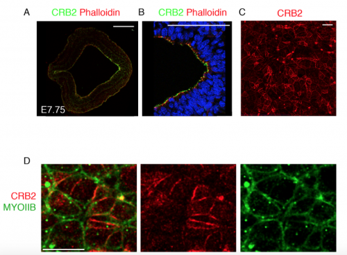
Traditionally, protein localization in the primitive streak epithelium was observed by immunostaining transverse sections of the epithelium. We too followed suit to look at Crumbs2 expression and were happy to see it localized apically in the cells of the streak. During my trials with live imaging, I began to appreciate the importance of the 3-dimensional nature of the epithelium and thought it was essential to see where Crumbs2 would fit in this 3-D context. The apical surface of the primitive streak epithelium faces the interior of the embryo, making it difficult to access and therefore not many people have looked at polarity proteins en face, in contrast to studies in Drosophila and cell culture. While learning to do en face imaging on neural epithelium, as a fun experiment I decided to look at Crumbs2 localization en face at the primitive streak. Although it took me many trials to get the streak epithelium flat, I was surprised to see that Crumbs2 localization was anisotropic. At first, I thought it could be an artefact of my sample processing. However, after repeated attempts with different approaches to sample processing and appropriate controls I was finally able to convince myself about the anisotropic pattern. Nevertheless, we still didn’t understand what it meant! I had tried co-staining with many other polarity proteins, but none of them had this pattern. I had immunostained for MyosinIIB separately, but it wasn’t until the Roper paper on Drosophila Crumbs in the salivary gland that we decided to probe for MyosinIIB and Crumbs2 together. That’s when it came together. The complementary nature of their pattern was striking and steered us to think of possible mechanisms of Crumbs2 action.
Figure 4: CRUMBS2 localisation at the primitive streak epithelium Transverse section of the primitive streak of wild-type embryo at E7.75 immunostained for CRB2 (green) and Phalloidin (red) showing the expression of CRB2 in both the anterior presumptive neural epithelium and the epiblast and its enrichment in the posterior epiblast. The streak region is magnified in (B). Extended projection of en face view of the apical surface of the epiblast layer of the primitive streak of wild type at E8.5 immunostained for CRB2 (C), showing its anisotropic distribution. The double immunostaining of the streak for CRB2 (red) and MYOSIN IIB (green), showing the reciprocal enrichment of CRB2 and Myosin IIB on different cell edges. Proximal is up. Scale bars: A, B, 100 μm; C,D 10 μm.
We hypothesize that the unequal levels of Crumbs2 in the epithelium must be an important part of the mechanism to determine which cells delaminate first. It’s a very interesting problem which hasn’t received its due. The streak region as defined by the basement membrane breakdown is easily around 4-5 cells wide and runs from the node to the distal tip of the embryo. Despite that, cells delaminate individually and leave the epithelium, suggesting that this process must be highly coordinated and tightly regulated at both cell and tissue levels. We know little about how the cells establish the complementary pattern of Crumbs2 and MyosinIIB, how they define the order in which they delaminate and how they coordinate this with their neighbors. These questions can be addressed by live imaging with appropriate reporters and will offer a wealth of information.
My journey to Crumbs emphasizes the power of genetic screens in identifying new players in gastrulation, and my paper offers a peak into the unexplored territories of mammalian gastrulation. There is a lot to learn by just observing this beautiful process in wild-type and mutant embryos. Hopefully, my work will inspire people to explore the dynamics of this process in greater detail.
References
1 Ramkumar, N. & Anderson, K. V. SnapShot: mouse primitive streak. Cell 146, 488-488 e482, doi:10.1016/j.cell.2011.07.028 (2011).
Here are the highlights from the current issue of Development:
Evolving an atypical developmental programme with IMM
The brown alga Ectocarpus has alternating haploid (gametophyte) and diploid (sporophyte) generations. Morphologically, these are distinguished by a more complex system of basal filaments in the sporophyte, initiated via symmetric divisions before the apical-basal axis is defined and the upright filaments form. This mode of development is unusual – in most brown algae, and in the Ectocarpus gametophyte generation, the first division is asymmetric to establish the apical-basal axis. The immediate upright (imm) mutation of Ectocarpus displays an asymmetric first division, and was initially thought to represent a partial switch from the sporophyte to the gametophyte developmental programme. Here (p. 409), Mark Cock and colleagues identify the gene responsible for this phenotype and provide a detailed analysis of its evolutionary history. The IMM gene is a member of the large, rapidly evolving, EsV-1-7 domain family, which exhibits an unusual distribution across eukaryotic lineages – potentially as a result of horizontal gene transfer. Transcriptional profiling suggests that, rather than imm causing a switch from the sporophyte to the gametophyte programme, the mutation blocks the extensive development of the basal filament system, such that the mutant displays a more canonical mode of sporophyte development. While the molecular and cellular function of IMM has yet to be determined, this gene appears to represent an evolutionary innovation in the Ectocarpus lineage that altered early sporophyte development.
Defining the right MOMent for cell fate decisions
The invariant lineage of C. elegans led to an early assumption that cell fate decisions are largely made cell-autonomously. However, it has subsequently become clear that inductive interactions between cells are essential for fate determination in this system. Moreover, these inductive interactions can be highly complex. The 8-cell stage blastomere, MS, gives rise to a number of body wall muscles. Their appropriate differentiation relies first upon an inhibitory signal from the ABp lineage at early stages, and subsequently upon an activating interaction from the ABa lineage a couple of cell cycles later. It has long been known that the activating interaction depends on Notch activity, but the nature of the signals, and the reason for this complex mechanism of cell fate determination, have remained unclear. Rueyling Lin and colleagues now identify zygotic MOM-2 (a Wnt ligand) as the Notch-dependent signal responsible for both the inhibitory and activating interaction (p. 419). Moreover, they provide evidence that this two-step mechanism is important because early inhibition is required to prevent precocious lineage restriction during this rapid phase of development. These data highlight the complex intercellular interactions, and the robust mechanisms, underlying cell fate determination in even a seemingly simple embryo like that of the worm.
Nan: neomorphic effects in neonatal anaemia
The KLF1 transcriptional regulator is essential for normal red blood cell differentiation, and a particular mutation in this factor is associated with congenital dyserythropoietic anaemia. The Nan mutant mouse, in which the same amino acid is mutated and which displays a semi-dominant phenotype, serves as a valuable model for this disorder. It is known that the mutant protein, Nan-KLF1, can only bind a subset of target sites as compared with the wild type, and this leads to changes in downstream gene expression in heterozygous animals – due at least partly to effects of Nan-KLF1 on the target genes whose sites it can no longer bind. James Bieker and colleagues have recently discovered that the Nan-KLF1 variant can also bind a target sequence not recognised by wild-type KLF1. Now (p. 430), they investigate the phenotypic consequences of this. Importantly, they observe neomorphic expression of a number of genes in Nan/+ heterozygotes, which appears to be due to ectopic binding of Nan-KLF1 to these new target sequences. The downstream genes affected include a number of secreted factors, such as hepcidin – a regulator of cellular iron use – and interferon regulatory factor 7 (IRF7). Thus, Nan heterozygosity in the erythropoietic lineage can confer systemic effects via the inappropriate expression of secreted factors.
PLUS:
Trends in tissue repair and regeneration
The 6th EMBO conference on the Molecular and Cellular Basis of Regeneration and Tissue Repair took place in Paestum (Italy) in September, 2016. As summarised in the Meeting Review by Galliot, Crescenzi, Jacinto, and Tajbakhsh, the scientists who attended discussed the importance of plasticity, biophysical aspects of regeneration, injury-induced immune responses, strategies to reactivate regeneration, links between regeneration and ageing, and the impact of non-mammalian models on regenerative medicine.
Formative pluripotency: the executive phase in a developmental continuum
The regulative capability of single cells to give rise to all primary embryonic lineages is termed pluripotency. Two phases of pluripotency, called naïve and primed, have previously been described. In his Hypothesis article, Austin Smith describes a third phase, called formative pluripotency, that is proposed to exist as part of a developmental continuum between the naïve and primed phases.
Cellular and molecular mechanisms of tooth root development
The tooth root is an integral, functionally important part of our dentition, and understanding how roots develop and how they can be bioengineered is of great interest in the field of regenerative medicine. In their Review article, Yang Chai and colleagues discuss recent advances in understanding the cellular and molecular mechanisms underlying tooth root formation.
“Nothing in biology makes sense except in the light of evolution” (Dobzansky, 1973).
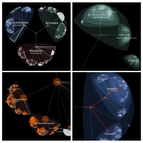
Our knowledge of the evolutionary relationships between all known organisms, the so-called Tree of Life (ToL), is crucial in all fields of biology. Many researchers in evolutionary biology are working on improving the quality and the comprehensiveness of this ToL. This implies better species sampling, more sequencing, improved evolutionary models and new algorithms for phylogenetic tree reconstruction. But displaying this information in a clear and simple way that is also convenient and useful to biologists outside the field of evolutionary biology has been largely underexplored.
In a paper published last December in PLOS Biology (de Vienne, 2016), I described an online tool, called Lifemap, for exploring the entire Tree of Life (or any large tree) in a zoomable interface (Figure 1). This tool solves many issues that previous methods had, such as the possibility to display nodes with more than two descendants (multifurcations) and the possibility to explore smoothly very large trees of more than 2 million species.
Lifemap relies on tools that were developed for cartography, especially in the context of the OpenStreetMap project, and proposes a new way of representing hierarchical structures. The exploration of the ToL in Lifemap is similar to the exploration of a geographic map: by zooming and panning. It is also possible to click on every node and tip of the tree to have access to additional information (description, picture, link to external resources, etc.), and to search for “routes” in the tree of Life (Figure 1). Finally, an efficient search engine allows for an immediate retrieval of any node or tip by its Latin or its common name.
Figure 1. Different views of Lifemap. Top-left: the three domains of the Tree of Life; Top-right: inside Bacteria; Bottom-left: orange bubbles represent the number of genomes fully sequenced; Bottom-right: a “route” from Homo sapiens to Mus musculus.
Lifemap comes in three versions, depending on the tree that is displayed and the information that is associated to nodes and tips when clicking. A first version displays the tree proposed by the Open Tree of Life (OTOL) project, a second one displays the whole NCBI taxonomy, and a third one displays a simplified version of the taxonomy with pictures and descriptions when clicking on the nodes (this last version is also available as anApp for phones and tablets on the Android market).
In the near future, I would like the NCBI and the OTOL versions of Lifemap to be proper “hubs” giving access to a multitude of information of relevance to researchers in various fields. Because I personally work on comparative genomics, I started adding information on the number of genomes that are fully sequenced according to the NCBI database (Figure 1, bottom-left). This information can be displayed directly on the interactive tree. It is useful for easily identifying species, close to our species of interest, whose genome is available. I also added links on every node and tip to the corresponding web page on the NCBI web site and on other taxonomy web resources. These examples show how flexible Lifemap is at welcoming information from various sources.
I hope that developmental biology will be a field for which information could be displayed on Lifemap as well? I will be happy to consider such options if some interest is declared!

 JCS kicked off 2017 with a Special Issue relevant to many developmental biologists: 3D cell biology. It’s packed full of commentaries, interviews, research articles and techniques, and well worth a browse.

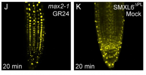 Ottoline Leyser and colleagues investigate how the plant hormone strigolactone regulates shoot development.


 (No Ratings Yet)
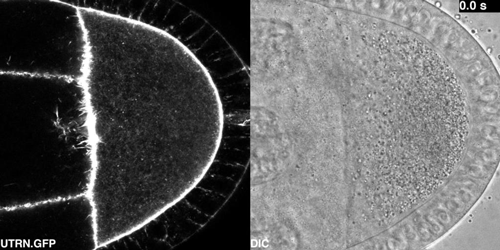
 (8 votes)
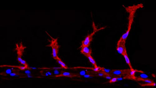
 (2 votes)


 The 6th EMBO conference on the Molecular and Cellular Basis of Regeneration and Tissue Repair took place in Paestum (Italy) in September, 2016. As summarised in the
  The regulative capability of single cells to give rise to all primary embryonic lineages is termed pluripotency. Two phases of pluripotency, called naïve and primed, have previously been described. In his
  The tooth root is an integral, functionally important part of our dentition, and understanding how roots develop and how they can be bioengineered is of great interest in the field of regenerative medicine. In their