Established by the British Society for Developmental Biology (BSDB) in 2014, the Gurdon/The Company of Biologists Summer Studentship scheme provides financial support to allow highly motivated undergraduate students an opportunity to engage in practical research during their summer vacation. Each year, ten successful applicants spend eight weeks in the research laboratories of their choices, and the feedback we receive is outstanding.
We will hear four stories from the 2016 class over the next four days, starting with Ji Hye Moon, who undertook a project in Richard Wingate’s lab in King’s College, London. You can read all the previous reports here.
The effects of transient gestational hypothyroidism on the development of foetal cerebellar nuclei
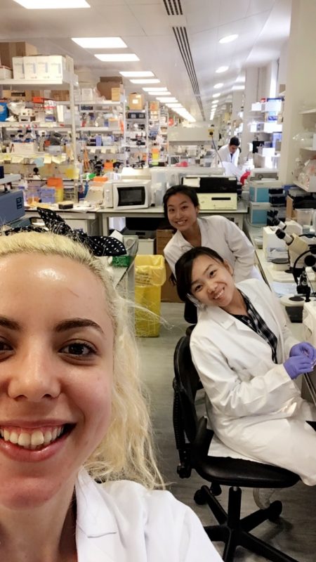
Me (Ji Hye Moon) at the far back with my colleagues from the Wingate Lab
As a graduate medical student with a previous degree in Middle Eastern Studies and Politics, I had not previously been exposed to scientific laboratory work. Progressing through the two years of pre-clinical studies, I surprised myself on just how much I enjoyed learning the science that underpins medicine and I became very aware of my lack of experience in research. I felt that I would benefit enormously from gaining exposure to this field, both in terms of my future competency as a clinician, as well as on my ability to pursue research further down in my career. As such, I felt very privileged to be able to start building up my skills and experience in lab-based research through the summer studentship this year.
The BSDB Gurdon Summer Studentship gave me the opportunity to spend 8 weeks this summer in the lab of Dr Richard Wingate in the Department of Developmental Neurobiology at King’s College London. I joined a project which aimed to examine the effects of transient gestational hypothyroidism on the development of the deep cerebellar nuclei in the foetal brain using mouse and chick models. I found this project particularly interesting as the effects of gestational hypothyroidism in the earlier stages of pregnancies are currently quite poorly understood, yet have been linked to postnatal cognitive deficits in the absence of gross malformations of the foetal brain.
The project used the drug methimazole, which is used to treat hyperthyroidism, in order to induce a transient hypothyroidism in pregnant mouse mothers to study its effect on the development of the foetal cerebellum at various embryonic and postnatal ages. All the brain samples were harvested before I started my studentship, so there were a lot of brain samples ready to be studied using immunohistochemistry (IHC) and in-situ hybridization (ISH) methods.
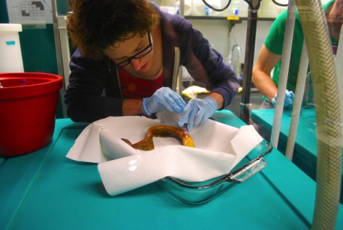
For the first part of the studentship I helped produce the wax and cryosections of the harvested brains necessary for IHC and ISH experiments. For this part of the work, I learned and honed the basic histology skills hands-on, through wax embedding, wax sectioning and cryosectioning of the mouse brain samples. Although this was quite repetitive work, I actually I found it really enjoyable and experienced a lot of satisfaction when I produced my first slide of two perfectly aligned ribbons each containing five slices of tiny, miniscule embryonic mouse brains.
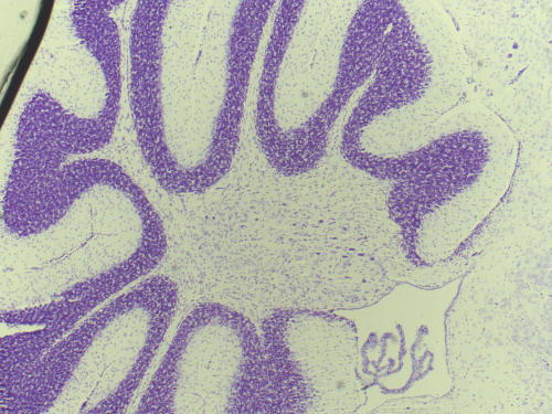
A section through the mouse cerebellum (Nissl stained) at postnatal age P23. The cerebellum is an exquisite structure with its distinct layers folded into folia.
Finally, the last few weeks of my studentship were spent producing nissl stains of the control and the experimental juvenile mice brains, which I then carefully studied to look for any subtle structural differences. I noted a potential decrease in the density of the Purkinje Cell Layer in the cerebella of the experimental group, but unfortunately was unable to complete a systematic comparison as my studentship came to an end.
I enjoyed the 8 weeks at the Wingate Lab and have benefited enormously from it. From an academic point of view, my science literacy level has improved immensely. Being exposed for the first time to the fields of mice and chick embryology and neuroanatomy as well as learning and practicing a wide range of experimental methods such as IHC and in-ovo electroporation, provided a steep learning curve that kept me focused and busy for the entire length of the studentship.
After spending two years almost exclusively in the lecture theatre, I really appreciated the hands-on learning experience and the stimulating environment. I learned a huge amount from attending the weekly lab meetings, talks and seminars from leading experts as well as interacting with my colleagues at the lab. Most of all, nothing could beat the depth of learning I gained from carrying out the experiments myself. It was fantastic to see the science come alive in front of my eyes and I found it a powerful way to learn and understand complex concepts. I was very lucky in that Dr Wingate encouraged me to tryout a variety of experimental techniques and always made time in his busy schedule for my questions.
The summer studentship was a rewarding experience on a personal level as well. It gave me a taste of what a career in research is like and an insight into the delights and the difficulties of research work. Ultimately, I’ve come to have a greater appreciation and admiration for research scientists and the enormous contribution they make to modern medicine and to the innovations that I will put into practice throughout my medical career.
I am very grateful to Dr Wingate, Dr Wilson, Margarita, Deviana, Tristan & Flo for being great hosts and teachers to me. A huge thank you in particular to Richard and the BSDB for making this opportunity possible for me!
The group of Chen Luxenburg at the Faculty of Medicine, Tel Aviv University invites applications for PhD student position.
Our laboratory is looking for excellent and highly motivated PhD students to study the role of the actomyosin cytoskeleton in skin development. Our goal is to understand how cytoskeleton derived signals regulate stem cell ability to create the skin epidermis during development, maintain it in the adult and repair it upon wounding. On top of molecular biology, tissue culture, advanced microscopy and mouse work we utilize state of the art technology that allows us to rapidly manipulate the function of any gene of interest in epidermal stem cells in utero. Several exciting projects are available for the successful candidates.
We offer state of the art laboratory with dynamic and international atmosphere and full financial support (tuition and stipend)
Candidates should hold Master’s degree in Biology/Life-sciences or related field. Preference will be given to candidates familiar with tissue culture techniques, microscopy and mouse work.
Interested candidates should email their CV and a brief paragraph describing their research experience and career plans to Dr. Luxenburg (lux@post.tau.ac.il)
Despite an overwhelming amount of carefully curated data, such as the International Shark Attack File, which indicates that your chances of being bitten by a shark are vanishingly small, humans have had a long and often macabre obsession with the impressive and sometimes daunting dentition of these stealthy marine predators. The diversity in form and function of the dentition of both living and extinct chondrichthyans (the lineage of cartilaginous fishes which includes sharks, skates, rays and chimaeras) is truly striking (some examples here). The gargantuan extinct selachian Charcharodon megalodon (literally “big tooth”) is known to science only from fossils of its immense triangular serrated teeth measuring up to 7 inches long, dwarfing the 3 inch teeth of its extant but comparatively diminutive cousin the “great” white shark C. carcharias which can nonetheless handily take down a 200kg cape fur seal (see here). In stark contrast to the sharp, often multi-cuspid, grasping and tearing tooth arrays of pelagic sharks, batoids (skates and rays) have assemblages of flat “pavement” teeth for crushing hard-shelled benthic prey (see here) while the quirky holocephalans (chimaeras/ghost sharks/rabbitfishes) have forsaken the tooth array entirely and evolved a highly derived dentition comprising relatively few large crushing tooth-plates.
That tooth looks like it could do some damage…but wait is it even a tooth? This alizarin-red stained sample is actually a denticle from the catshark S. canicula.
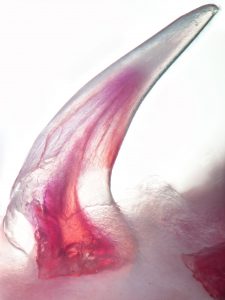
This alizarin-red stained sample from S. canicula is, in fact, a tooth (a whole tooth, and nothing but a tooth…)
Without understating the intrinsic and scientific value of this impressive morphological diversity, to an evolutionary developmental biologist the elasmobranch dentition is possessed of an even more impressive trait. This trait, if properly understood, is also one which has potentially broad developmental and biomedical applications. Unlike humans who normally produce a limited quota of exactly two generations of teeth and subsequently suffer with the consequences of tooth decay, injury, or loss, the teeth of sharks are capable of lifelong whole-organ regeneration. What’s more, sharks don’t wait for a tooth to be lost or damaged to initiate the regeneration of a replacement, but rather perpetually regenerate multiple teeth which lie in wait embedded within the jaw forming what has been endearingly referred to as the ‘conveyor-belt’ dentition. As it turns out, the capacity for endless tooth regeneration, also known as polyphyodonty, is the primitive condition of gnathostomes (jawed vertebrates) but has been lost secondarily in multiple lineages including eutherian mammals. Frustratingly for most developmental biologists, polyphyodonty has actually been lost from every major ‘classical’ model vertebrate normally studied in the lab including chick, frog, mouse and zebrafish, leaving this impressive developmental spectacle shrouded in mystery. In order to begin unravelling the secrets of polyphyodonty, we have therefore turned to the emerging model world (or simply “nature” as an evolutionary biologist would call it) and are working alongside a small but dedicated community towards turning the unassuming but satisfactorily dentate catshark, Scyliorhinus canicula, into a first-class model system. This humble but abundant shark, native to coastal waters of Europe and North Africa, was first pioneered as a model for Evo-Devo research nearly a decade ago and has since found its way into a number of Evo-Devo labs around the world including Gareth Fraser’s research group at the University of Sheffield where we maintain both developing embryos and young animals in temperature controlled artificial seawater tanks.
Embryos of S. canicula cultured in artificial seawater in the laboratory. After external gill eruption and the initiation of pharyngeal respiration embryos can easily be sortied from their eggcases, manipulated, and returned. Video courtesy of Dr. Andrew Gillis at the University of Cambridge.
As a postdoc in the Fraser group, I share two overarching research goals with the rest of the team. First, I believe that if we could understand the molecular developmental mechanisms regulating the perpetual regeneration of teeth in sharks and other polyphyodonts, and discover why and how this ability has been attenuated during the evolution of other lineages including our own, it may be possible to rescue this ability and stimulate tooth regeneration in humans. Second, as chondrichthyans diverged from the lineage leading to modern osteichthyans (bony fishes, including tetrapods) some 450 million years ago, they offer a deep comparative reference point as close to last common gnathostome ancestor and the ultimate evolutionary origin of teeth as one can get by comparing extant species. By comparing the expression of developmental genes in shark’s teeth with those of their bony cousins including us, we have already been able to show that many key components of the gene regulatory network used to build teeth are conserved between these groups, reinforcing the prevailing view of the biological homology of oral teeth. However, as to the ultimate evolutionary origin of the oral dentition the field has long suffered from a surfeit of theories and a deficit in data…until now.
Perhaps counterintuitively, the evolutionary history of teeth probably did not begin in the mouth. Palaeontologists and comparative anatomists have long recognized that teeth, comprised of an enamel/enameloid exterior layer, dentine core, and a bony root, are just one variety of a larger family of epithelial organs comprised of exactly the same components called odontodes. Long before there were even jaws which could conceivably house teeth, other types of odontodes made up a significant component of the defensive armour of many lineages of agnathan (jawless) vertebrates. It has therefore long been suspected that teeth originated though either an ‘expansion’ or ‘migration’ of external odontodes (known as denticles in elasmobranchs) into the mouth, after the origin of jaws. While Wolf-Ernst Reif correctly pointed out over 35 years ago that there is no histological or developmental gradation between the external denticles comprising the shark squamation and the oral dentition, this model can be resurrected in the light of modern network-based evolutionary developmental theory. In contemporary parlance, we could hypothesize the ‘co-option’ of the developmental mechanisms for producing odontodes (originally evolved in the external body armour at least according to the “outside-in” flavour of this model) into the oral cavity. According to this hypothesis it would only require the switching on of one or a few key master regulatory genes for odontode development in new competent cells of a different epithelium (of either endodermal or ectodermal origin) to initiate the entire developmental cascade for odontode production. Indeed, as we describe in our most recent paper, we found support for this model in that both denticles and teeth share equivalent expression patterns of many core developmental genes. That is, in addition to their equivalent anatomical components, they share a core conserved odontode gene regulatory network (oGRN) and should be considered as deeply homologous sensu Shubin et al. 2009. Individual teeth and denticles both develop via reciprocal signalling between an epithelial placode and underlying mesenchymal condensation, progress through comparable bud, papilla and morphogenesis stages during development, and are almost indistinguishable one from the other even at the molecular level. What then is a tooth except for an odontode in the mouth specialized for feeding?
Reconstruction of an XRM (X-Ray Microscopy) scan of a recently hatched specimen of S. canicula. The dentine/enameloid cusps of both teeth and denticles are shown in red, while the bony roots are rendered in green. The superficial skin is shown in blue and rendered semitransparent. Within the mouth the taste buds are visible as small blue mounds. Thanks to the amazing team at the London NHM Imaging and Analysis Centre for carrying out this work with us!
As we show in our paper, the answer to this question takes us full circle back to the most impressive property of the elasmobranch dentition: regeneration. While teeth regenerate rapidly and continuously in highly organized family rows well ahead of function, denticles do not. Using a combination of developmental gene expression analyses, cell proliferation and label-retaining analyses with the nucleoside analog BrdU, and lineage tracing with the vital dye DiI, we were able to show, at least in part, why. To make a long story (…catsharks can take upwards of 8 months to develop…) short it turns out that despite a largely conserved oGRN between teeth and denticles, the well-known stem cell factor, sox2, is only expressed in teeth. We traced the expression of sox2 from the earliest initiation of teeth through at least 5 generations and have shown that it is always retained in a small but crucial subset of epithelial cells embedded within the dental lamina immediately adjacent to the site of new tooth formation. No such pocket of set-aside stem cells could be found in denticles. Perhaps more revealing however was that the same field of sox2+ progenitors from whence the dentition emerges also gives rise to the anterior most taste-bud fields in both the upper and lower jaws. Previously, sox2 had been described as a marker of the odontogenic band, or OB, in mice and it is known to mark the future site of tooth formation in several osteichthyans along with other markers such as shh and bmp4. However, while mammals have secondarily disassociated their dentition from their gustatory faculties, many other vertebrates including sharks and teleost fishes retain a close functional and as we now know, developmental, association between teeth and taste buds. Therefore, in sharks both teeth and the anterior-most taste bud fields arise from a common sox2+ progenitor field which we have termed the odontogustatory band, or OGB, to reflect its dual developmental fate. Once this dual fate is shown definitively in an osteichthyan this will strengthen the case that this represents the ancestral gnathostome condition and reinforce the view that mammals are highly derived in this character.
Immunofluorescence reveals the location of Sox2-expressing stem cells (magenta) in the shark dentition. Here a new tooth undergoing morphogenesis can be seen immediately adjacent to one of the stem-cell niches in the shark dentition (the successional lamina). Adapted from Martin et al. 2016.
In the vertebrate lineage, taste buds are comparatively ancient epithelial sensory organs, and their origin predates that of both teeth and jaws as evidenced by their presence in living petromyzontifomes (lampreys). For as long as taste buds have existed it also appears that they have been capable of regeneration, a phenomenon which unlike polyphyodonty has actually been conserved in mammals, and is known to rely deeply on the stem cell factor sox2. Taken together these data point towards an intriguing and unexpected evolutionary scenario, namely that teeth evolved through a co-option of the oGRN from non-oral odontodes within the sox2+ progenitors of the oral cavity previously directed towards forming an entirely different type of epithelial organ – taste buds. This may not have been as grand a leap for these cells to take as it might appear. In fact, many epithelial organs utilize many of the same developmental genetic blueprints to coordinate their development. There would be far fewer evolutionary steps to take in order to modify a pre-existing morphogenetic field competent to produce one type of epithelial organ (taste buds) with the plans for a different type (odontodes), than having to start from scratch in a cell type which wouldn’t normally give rise to these types of organs. Evolution is lazy, after all.
If this explanation seems somehow incomplete to you don’t worry, that only indicates that you were paying attention! Certainly we have only scratched the surface of this complex problem and there are many more questions to answer. What is this hypothetical master regulatory gene which can switch on the whole odontode differentiation pathway in a new competent epithelium? How do these common sox2+ progenitors know when to make teeth vs. taste buds and how many of each? Watch this space for more exciting developments as new data pours in from sharks and other emerging models in key phylogenetic positions.
Kyle Martin
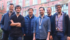
The Fraser Lab team at the University of Sheffield circa January 2017.
This is the latest dispatch from a recipient of a Company of Biologists Travelling Fellowship.
Learn more about the scheme, including how to apply, here, and read more stories from the Fellows here.
Hanna Hakkinen
I am originally a Finnish evolutionary biology student who got fascinated about developmental biology during my exchange programme couple of years ago in Southern Chile (Universidad Austral de Chile). More than a year ago I travelled back to Chile and ever since I have been working at the Developmental Biology laboratory of Dr Claudio Araya to study early vertebrate brain morphogenesis using zebrafish (Danio rerio) as a model system. In contrast to amniote embryos, teleost animals form a neural tube after the generation of a solid neural keel and a solid neural rod at the middle of the tissue. In our current project we have been looking at the early cellular dynamic of neural plate progenitor cells in organizing tissue displacement and internalization to achieve this midline structure.
Thanks for the Travelling Fellowship program of The Company of Biologists last October we had the change to take some critical steps in our original research and begin to tackle the question of how a non-polarized multicellular structure is internalized at the tissue midline in such coherent way. To achieve that we contact Dr Jon Clarke’s lab at the Department of Developmental Neurobiology at King’s College London. During the 6 weeks stay in his lab I had the chance to have access to powerful confocal imaging facility together with his great experience and methodologies in looking at early vertebrate brain formation.
In spite of the increasing knowledge about the molecular signatures during brain development, the underlying cell and tissue morphogenesis are still poorly understood. During my research stay I focused on cellular and sub-cellular dynamics of midline internalization. By combining novel high resolution imaging techniques together with the use of transgenic reporters for subcellular components we have began to characterize contribution of essential cellular dynamics that appear to be critical for tissue internalization and neural formation. We were able to record a series of very unexpected and interesting cell behaviours during this morphogenetic process. In addition, we found how this fundamental sub-cellular and cellular dynamics can also be present on other type of tissue architecture that undergo collective tissue internalization.
On behalf of myself and Dr Claudio Araya we are very grateful for the Company of Biologist for this great opportunity. We really feel that after this stay our project has definitely been taken to a different level, and we will be able to unravel questions which we were unable to pursue due to technological limitations we face here at the end of the world. In addition to achieve some remarkable results for our future publication, for me personally this period in London was also an amazing opportunity to visit a foreign high standard laboratory and learn new skills and make important and delightful contacts. King’s College enormous fish facility with multiple interesting and useful transgenic lines, top-level imaging facilities and especially high class colleagues changed my research stay to a life changing experience. Many thanks for The Company of Biologists!
This is the latest dispatch from a recipient of a Company of Biologists Travelling Fellowship.
Learn more about the scheme, including how to apply, here, and read more stories from the Fellows here.
Tetsuto Miyashita
Pasadena.
Everyone gets different images for the place. JPL. Richard Feynman. The Big Bang Theory. I can’t dissociate the image of Pasadena from lampreys. Lampreys? That blood-sucking, eel-like, slimy fish?
The Company of Biologists Travelling Fellowship allowed me to work on lamprey embryology in Marianne Bronner’s lab at Caltech in summer 2016. The experience was fantastic, although my colleagues in the lab might have felt at times as if I was the blood-sucking lamprey — following them around, asking a million questions, and trying to absorb all I could.
There already is an excellent post about lamprey lab in the Day in the Life in the Lab series by Daniel Meulemans Medeiro’s lab. So I am not going to repeat it here. Also, there is an excellent resource in David McCauley’s website that you should check out. In this post, I will focus on Team Lamprey in the Bronner lab and report about the fabulous people behind the neural crest research in lampreys. Take a look at our group photo.
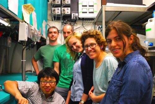
Lamprey researchers at the Bronner lab for the spawning season 2016. L-R: Tetsuto Miyashita (author), Stephen Green, Hugo Parker, Megan Martik, Dorit Hockman, and Aya Jishi.
Stephen Green is the hub of the Team Lamprey. Readers of the Node may recognize his name from the paper on mesodermal development in hemichordates that comes from his PhD dissertation work with Chris Lowe (Stanford University). Now at the Bronner lab, his meticulous and precise work that is showcased in the hemichordate paper has been teasing apart the developmental intricacy of neural and skeletal derivatives of neural crest in lampreys. While running his own research, Stephen is the master of the lamprey facility. He takes such a care of the animals that he was even witnessed walking in the lab past midnight, just to check that everything is running properly for the holding tanks that were set up earlier in the week. Take it as the sign of dedication, diligence, or even something else as you may, he does it matter-of-factly. Team Lamprey owes to him all these years of smooth operation, without which all of our work is unthinkable.
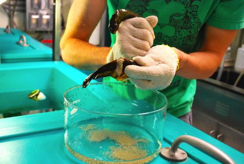
Collecting eggs from a ripe female.
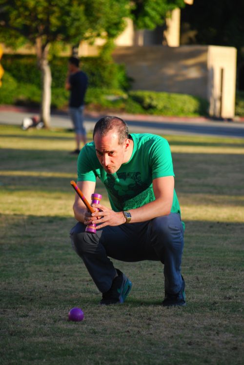
Hugo Parker, a postdoc with Robb Krumlauf at Stowers Medical Institute, is a long-time visitor to the lab. You can read his featured work on the Hox regulation in lamprey hindbrain development here in Nature or here in Bioessays. Hugo pioneered a reporter expression assay for this work when he was a PhD student with Greg Elgar, and he has been using it to study the evolution of regulatory mechanisms in hindbrain rhombomeres. His reporter assay gave lamprey researchers a tool to test functional evolution at the level of enhancers — adding a layer of comparison above gene expression profiles. Aside from serious research, Hugo hosts lab parties in the form of croquet competition in the campus front yard. Despite his competitive edge, and despite his love of this rather mundane sport, he has yet to win the prize, Lamprey Cup.
Hugo Parker, right before hitting the “Submit Manuscript” button.
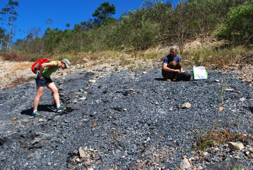
Splitting her time between Oxford and Cape Town, Dorit Hockman travels halfway around the world to work in the Bronner lab. She began her career with limb development in bats (see this paper in PNAS), went through the MBL Embryology course, trained under Clare Baker at the University of Cambridge, and finally arrived at lampreys for her postdoctoral work. Dorit brings computational skills and extensive training in the cutting-edge methods to study non-coding elements (like ATAC-seq). She works on a variety of projects around lamprey development, and one of the early fruits is the fate map of cranial sensory ganglia. Dorit actually joined me on one of my field projects to collect early jawless vertebrate fossils in South Africa. In the photo, she is standing at the locality that produced the world’s oldest fossil lamprey, Priscomyzon.
Dorit Hockman collects a blood sample.
Dorit Hockman (left) visits the fossil locality for Priscomyzon, a stem lamprey from the Late Devonian of South Africa
After completing her PhD with the famed sea urchin GRN guru Dave McClay, Megan Martik has just made a transition to the vertebrate world (see here for her PhD work). She handles two models — zebrafish and lampreys. This summer was her first lamprey spawning season. Megan was practicing with CRISPR/Cas9 and apprenticing in running the lamprey facility. Her research interest ultimately addresses the age-old question: how developmental repertoires differentially evolved between cephalic and trunk neural crest cells. Her recent adventure in China gave her an opportunity to speak with the evolutionary morphologist Shigeru Kuratani, whose lab has been leading the study of lamprey development over the last two decades. A photograph from the conference captures them engaged in what must have been a very lively discussion!
Megan Martik (L) discusses with Shigeru Kuratani (R). Provided by M. Martik.
Aya Jishi, an undergrad at Caltech, is already a seasoned lamprey embryologist. She has volunteered with the lamprey for four years. She began with maintaining embryonic cultures and as an undergrad is now examining lamprey neurogenesis and is often seen working away at a microtome. Although hegraduated with a PhD before this last spawning season, Benji Uy also started in the Bronner lab as a high school student and continued his way through undergrad and PhD working on Sox family in lampreys. These prodigies are making their headways into a promised career in medicine, and the lamprey community will miss their talents
Benji Uy (2nd from left) at the lab croquet party on the Beckman lawn, Caltech.
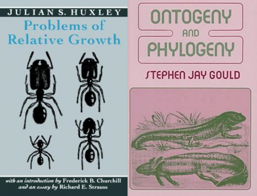
Finally, a bit about myself… I am a paleontologist. More precisely, I belong to a growing population of paleontologists to address evolutionary questions with both fossils and embryos. To me, the interest came naturally when I was a high school student. I worked as an assistant for Philip Currie, a Canadian dinosaur paleontologist who had a long-standing interest in the growth of theropod dinosaurs. His favourite anatomical reference at the time was Edwin Goodrich’s Studies on the Structure and Development of Vertebrates. When I had nothing to do on a cold wintery day in the museum, I would pull out Julian Huxley’s On the Problems of Relative Growth or Stephen J. Gould’s Ontogeny and Phylogeny from his bookshelf and flip through — although the prose of the latter was practically impenetrable for a Japanese high school student.
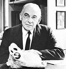
This early education had a profound influence as I developed my research interest beyond dinosaurs, and the Embryology course at the Marine Biological Laboratory reinforced it. I am not the only paleontologist who tried to learn the tricks of developmental biology. My colleagues like Katharine Criswell (the University of Cambridge) and Aidan Couzens (Flinders University) went through the program too. Recently I found in the biographical record that even Alfred Romer, a revered vertebrate paleontologist of the mid-20th century, spent his formative summers (1919-1921) at MBL to study embryology while he was a graduate student (that’s also where he met his future wife, Ruth Hibbard). This explains Romer’s in-depth understanding of development so eloquently displayed in the textbooks and papers he wrote. I feel a bit of connection here — Romer advised Robert Carroll, who advised my advisor.
Alfred Sherwood Romer, perhaps the first vertebrate paleontologist who studied embryology at MBL.
My intellectual meanderings over the last few years led me to lamprey embryology at the Bronner lab. While on the COB Fellowship, I learned CRISPR/Cas9, cloned several genes potentially expressed in the skeletal tissues in the late stage of development, ran multiple rounds of in situ, and basically took an advantage of the environment to interact with the members of the Team Lamprey. As you can see, we all bring in different skill sets and interests that feed into a catalytic reaction over a summer of working together. Certainly, it is no easy work. In the height of the spawning season, you come in to run experiments in the morning, inject in the afternoon, and sort, spread, and collect embryos in the evening. Sometimes most embryos in the batch die. Sometimes you repeat PCRs for weeks just to get a single gene cloned. But even in those hard times when one feels somehow inadequate, it is worth the effort because science is as much about people you work with as about the animals you work on. My gratitude to the COB for awarding a Travelling Fellowship comes down to this point. I brought back from Pasadena more than just data. It gave me an opportunity to interact with this fabulous Team Lamprey and call each one of them my friend. Finally, I thank Marianne Bronner and her lab members for hosting me in such a welcoming community. Check out their work at the lab website here.
A lamprey embryo at around Tahara’s stage 27. Provided by the Bronner lab.
Photo by Argonne National Laboratory (Science Careers in Search of Women 2009) [CC BY-SA 2.0 (http://creativecommons.org/licenses/by-sa/2.0)], via Wikimedia CommonsI want to talk about how you can take your science teaching to the next level, where young people, and especially underrepresented young people (people of color, LGBT, immigrants, girls, etc.), find what you’re teaching engaging, relevant to their lives, and which research shows that if done thoughtfully, enables them to achieve a higher level of learning. I’m not suggesting you change your science content. Instead, I’d like to illustrate the importance of modifying your teaching to be culturally relevant.
Ideally you are already providing hands-on, inquiry-based science experiments, which are known to increase achievement and engagement. [Shameless plug: This is something that a zebrafish program that I work with, BioEYES, does well, so if you need help see our website (www.bioeyes.org), our latest research paper in PLOS Biologythat details our results and how to launch an outreach program in your area, read our Node post from 2010, or contact me directly.] How engaged are your students? Are there some that are struggling, or who aren’t that committed? Do your lessons speak to the diversity of individuals that you are teaching? Are they culturally relevant?
What does this mean, exactly? Culturally relevant teaching was first described in 1994 by Gloria Ladson-Billings, an education researcher, to mean: “a pedagogy that empowers students intellectually, socially, emotionally, and politically by using cultural referents to impart knowledge, skills, and attitudes.” She suggests reframing how we think about and teach students, especially those typically marginalized by the greater society, from a place of need or as a problem to be fixed, to a place that acknowledges the cultural richness and the assets they bring into the classroom and society.
Culturally relevant teaching is based on the following principles: Academic success, cultural competence, and sociopolitical consciousness (Ladson-Billings, 1994). The first principle, academic success, is that we want learners to grow, intellectually. While seemingly obvious, the unconscious biases we educators harbor can sabotage our best intentions. For example, research shows that we call on “poor” performers less often, and spend less time waiting for them to answer. The second principle, cultural competence, is that we introduce students to the global perspectives found in the greater scientific community and the world. Students can then celebrate not only their own cultures, but learn new perspectives as well. And the third and final principal, sociopolitical consciousness, involves helping students to apply their learning to solving some of the world’s pressing problems.
How does a scientist apply these principles in a science classroom? Let’s first look more closely at what a culturally relevant classroom is like.
You get to know your students on a meaningful level. You identify what each student is interested in, what their values are, what their home life is like, their traditions, style of communicating, and how they relate to their community.
You demonstrate caring. By getting to know your students and what they value, you begin to develop meaningful relationships with them and show that you value them, their culture, and their ways of learning, which may be different from traditional ways of learning. This goes beyond merely respecting students and their differences. You build safe spaces for ethnically diverse students to learn and achieve. You provide high expectations for all learners, yet help them succeed in steps, not all at once.
You respond to ethnic diversity while delivering instruction. You actively seek out ways to incorporate your students’ lives and cultures into the science lessons you’re providing.
Now why is this all so important? For one, research shows that academic scores increase when you deliver culturally relevant instruction (Au & Kawakami, 1994; Boykin & Noguera, 2011; Foster, 1995; Gay, 2000, 2010; Hollins, 1996; Ladson-Billings, 1994, 1995; Paris, 2012; Scherff & Spector, 2011). Boutte, Kelly-Jackson, & Johnson (2010) emphasize that: “academic achievement is a central goal of culturally relevant teaching.” When you set high expectations for students, which for starters can be as simple as calling them “scientists” no matter their age, it shows that you believe they can achieve great things. They feel valued. Secondly, many minority students have spent their entire lives having to fit into a dominant culture, while their own culture has been suppressed, oppressed, and devalued. This has a profound affect on an individual’s identity. It forces one to try and maintain one identity at home and in their community, and a separate identity at school. Imagine what that feels like.
Teaching from a culturally relevant framework supports and nurtures student identities and values. It creates a safe space that allows students to excel, and sometimes to fail. But when failing in a supportive environment, you can help them get back up and try again, step by step. It is this scaffolding of learning that helps struggling performers engage with learning and to advance.
So how do you apply culturally relevant teaching to science? For starters, be willing to look at your own beliefs and biases toward others. Take a few implicit bias tests (https://implicit.harvard.edu/implicit/takeatest.html). Understand that we all have unconscious biases. While difficult, seek out ways to challenge your assumptions (https://www.psychologytoday.com/blog/sound-science-sound-policy/201501/overcoming-implicit-bias-and-racial-anxiety). Learn about and interact with other cultures. Put yourself in others’ shoes. Be the curious scientist you know yourself to be. In the process, you might even find ways to mitigate the global stereotype that all scientists are White males that wear glasses and like to blow things up in the laboratory (Finson, 2002).
Next, get to know the population you serve even more. You could start by administering a student inventory (http://www.cultofpedagogy.com/products-for-your-classroom/). Or have the classroom teacher administer a survey. What you learn can help you to build an authentic relationship and can generate ideas for ways to incorporate a student’s culture into your lessons. Don’t make assumptions about who they are. Find out who they are.
Deal directly with controversial subjects such as racism, sexism, homophobia, and poverty. Give them context. You might think about how science has historically been done by and for men, and how this has shaped the field (e.g., medical dosages are prescribed similarly for men and women, yet women sometimes need a lower dose). How might a discussion about this change the way we do/view science? What can you and your students do about it? While genetics education might start with Gregor Mendel, communities like the Native Americans have been doing experiments on corn for thousands of years but didn’t write down their findings because they followed an oral tradition. How might a Native student who brings this prior knowledge into the classroom then respond to a teacher who posits Mendel as the founder of genetics?
Study a wide range of individuals and ethnic groups. Include many perspectives. No one person represents a group. Again, this is true for you as well as each student in your class. What interests you? Where is there commonality with your students?
How might you infuse issues of social justice, for example, into a science activity? One example involves teaching students science vocabulary (http://www.cultofpedagogy.com/culturally-responsive-teaching-strategies/). Author Zanetta Hammond suggests making a game of it, making the activity a social one, or “storifying” it. All three strategies employ the techniques of oral traditions—listening, repetition, memorization and learning—which are common to many cultures. In addition, marginalized populations often share a history of having to pool their resources in order to survive or get ahead, and value their community and group over individual gain. Creating groups where students can socialize and work together is not only inclusive of all cultures, but models the collaborative nature of scientific work.
Help students make connections between the science content, the contributions made by underrepresented scientists, and your students’ lives. This does not mean you change the science content. But you can incorporate data, photos, examples, and information from different cultures into each lesson so multicultural science education is institutionalized in your program and practices, as opposed to being taught in isolation (e.g., during Women’s History Month only). If you are working with African-American learners, you could talk about and show photos of prominent African-American scientists (e.g., biologists Ernest Just and Charles Drew), show data and graphs of diseases that disproportionately affect African Americans, or provide examples of scientific research that has been done to African Americans (e.g., Tuskegee experiment). By utilizing the techniques of science—such as the scientific method, arguing from evidence, and problem solving—you can elucidate and challenge stereotypes and prejudices.
You might frame a genetics unit not around Gregor Mendel, but around researchers of color: Priya Moorjani, a geneticist who has used genomic data to understand the origins of the Indian caste system; Kono Yasui, a biologist who researched the genetics of several plant species; or Rick Kittles who used genetics to trace the ancestry of African Americans. Or you might choose a woman like Barbara McClintock who was not encouraged personally or professionally to study science, but who still went on to win the Nobel Prize for her work in genetics. Here we have several examples that are inclusive of women’s contributions to science (also see http://news.nationalgeographic.com/news/2013/13/130519-women-scientists-overlooked-dna-history-science/). Pay attention to areas of intersectionality. Are there LGBT researchers you know of? Look for these role models, many of which may be “hidden” or ignored, and celebrate them. The key is that you are not teaching specifically to a particular ethnicity or group, but are incorporating different perspectives and creating an inclusive, relevant, and supportive environment for learners from various backgrounds.
While some of these ideas may give you pause —perhaps you feel they require too much effort or take you too far outside your comfort zone—you don’t have to include everything, or even everything all at once (1). Start with small changes, and identify where you and your students share common ground and what you are comfortable with. Feel free to share activities and examples you find in the comments below. Challenge yourself over time to go beyond a casual interpretation of culture in your science classes and in your lab but instead think about how your lessons might allow for opportunities for critical debates on the role and practices of science in society (Ladson-Billings, 2014). I argue that by not expanding upon our current view of science knowledge and oppression’s role in shaping it, we reinforce and privilege Eurocentric hegemonic ideas. By teaching to the whole child, you will gift your students with strong identities, new perspectives, and ultimately will engage them more in science, increase their critical thinking skills, spur greater learning gains, and hopefully help them consider a career in the sciences. And who doesn’t want that?
(1) For a good philosophical framework, see the “typology for multiculturalizing science” at Baptiste, H., & Key, S. (1996). Cultural Inclusion: Where does your program stand? The Science Teacher, 63(2), 32-35. Retrieved from http://www.jstor.org/stable/24149767
References
Au K.H., & Kawakami, A.J. (1994). Cultural congruence in instruction. In E.R. Hollins, J.E. King, & W.C. Hayman (Eds.), Teaching diverse populations: Formulating a knowledge base (pp. 5–23). Albany: State University of New York Press.
Boutte, G., Kelly-Jackson, C., & Johnson, G.L. (2010). Culturally relevant teaching in science classrooms: Addressing academic achievement, cultural competence, and critical consciousness. International Journal of Multicultural Education, 12(2).
Boykin, A.W., & Noguera, P. (2011). Creating the Opportunity to Learn: Moving from Research to Practice to Close the Achievement Gap. Alexandria, VA: the Association for Supervision and Curriculum Development (ASCD). http://www.ascd.org/publications/books/107016.aspx
Finson, K.D. (2002), Drawing a Scientist: What We Do and Do Not Know After Fifty Years of Drawings. School Science and Mathematics, 102: 335–345. doi:10.1111/j.1949-8594.2002.tb18217.x
Foster, M. (1995). African American teachers and culturally relevant pedagogy. In J.A. Banks & C.A.M. Banks (eds.), Handbook of research on multicultural education (pp. 570–581). New York: Macmillan.
Gay, G. (2000). Culturally responsive teaching: Theory, research, and practice. New York: Teachers College Press.
Gay, G. (2010). Culturally responsive teaching, 2nd Ed. New York, New York: Teachers College Press.
Hollins, E.R. (1996). Culture in school learning: Revealing the deep meaning. Mahwah, NJ: Lawrence Erlbaum.
Ladson-Billings, G. (1994). The Dreamkeepers: Successful teaching for African-American students. San Francisco: Jossey-Bass, pp. 17–18.
Ladson-Billings, G. (1995). Toward a theory of culturally relevant pedagogy. American Educational Research Journal, 32(3), 465–491.
Ladson-Billings, G. (2014). Culturally relevant pedagogy 2.0: aka the remix. Harvard Educational Review, 84(1), 74-84.
Paris, D. (2012). Culturally sustaining pedagogy: A needed change in stance, terminology, and practice. Educational Researcher, 41(3), 93-97.
Scherff, L., & Spector, K. (2011). Culturally relevant pedagogy: Clashes and confrontations. Lanham: Rowman & Littlefield Education.
Shuda, J.R., Butler, V.G., Vary, R., & Farber, S.A. (2016) Project BioEYES: Accessible Student-Driven Science for K–12 Students and Teachers. PLoS Biol 14(11): e2000520. doi:10.1371/journal.pbio.2000520
Here are the highlights from the current issue of Development:
ECM: bridging the gap between germ layers
The extracellular matrix (ECM) plays crucial roles during morphogenesis but how it is assembled and patterned in vivo is poorly understood. Here, Yuki Sato, Rusty Lansford and colleagues investigate this by examining the distribution of the ECM component fibronectin (FN) in quail embryos (p. 281). They reveal that FN fibrils form pillars that span the gap between somites and the endoderm. The tissue-specific depletion of FN reveals that both the somites and endoderm provide FN that contributes to these pillars. The authors also observe filopodia-like structures that extend from the basal surface of somatic epithelial cells and are oriented along FN pillars. The formation of these filopodia influences the formation of FN pillars, while the polymerisation of FN is shown to modulate both pillar formation and filopodial elongation. Importantly, both structures are required for proper somite morphogenesis. Finally, the researchers report that blood flow in the nascent dorsal aorta (DA), which is located between the somites and endoderm, controls FN pillar distribution; disruption of DA formation, or occlusion of the DA, leads to a scattered distribution of FN pillars. Together, these findings suggest that pulsations from the DA help establish FN pillars that bridge the somite-endoderm gap and potentially aid communication between these tissues.
Brainy roles for cilia and mTORC1
During vertebrate brain development, neurons and glia arise from a population of self-renewing radial glial cells (RGCs) that contact the cerebral ventricles and bear a primary cilium. Primary cilia are known to play crucial roles in signalling but it is not clear if they are required for morphogenesis. Now, on p. 201, Nathalie Spassky and co-workers show that primary cilia on RGCs are essential for proper ventricular morphogenesis in mice. They first report that ciliary mutant mice exhibit enlarged lateral ventricles (ventriculomegaly) and reduced cortical thickness. The absence of primary cilia also leads to an increase in the size of RGC apical domains. This apical endfoot enlargement, the authors report, is associated with spindle orientation defects and is caused by upregulation of the mTORC1 pathway. Accordingly, treatment with rapamycin – an mTORC1 inhibitor – prevents apical domain enlargement in ciliary mutants and rescues their ventriculomegaly phenotype. Overall, this study reveals a new role for the mTORC1 pathway in regulating ventricle morphogenesis and corticogenesis, suggesting that it constitutes a new potential therapeutic target for the treatment of ventriculomegaly.
Getting to the root of brassinosteroid function
The plant hormone brassinosteroid (BR), which signals through its receptors BR INSENSITIVE 1 (BRI1), BRI1-LIKE 1 (BRL1) and BRL3, is known to regulate hypocotyl elongation but how it functions in the root is less clear. In this issue, Christian Hardtke and colleagues assess the role of BR during cell differentiation in the Arabidopsis root (p. 272). They first show that bri1brl1brl3 triple mutants display protophloem differentiation defects. These defects cannot be rescued by activating BR signalling in adjacent cell files, suggesting that BR acts in a cell-autonomous manner to control protophloem differentiation. Triple mutants also exhibit a small meristem, and the authors show that this can be explained by reduced cell elongation that, together with increased formative divisions in the radial dimension, contributes to the overall reduction in root growth observed in these mutants. Finally, the researchers demonstrate that the protophloem-specific activation of BR signalling can rescue all major aspects of the triple mutant phenotype, thus uncovering a new facet of the non-cell-autonomous effects of BR signalling. Based on these and other findings, the authors propose that BR perception in the protophloem is sufficient to systemically convey BR action within the root meristem.
PLUS…
Primate embryogenesis predicts the hallmarks of human naïve pluripotency
Naïve pluripotent mouse embryonic stem cells (ESCs) resemble the preimplantation epiblast and efficiently contribute to chimaeras. By contrast, primate ESCs correspond to the postimplantation embryo and fail to resume development in chimaeric assays. In their Hypothesis article, Thorsten Boroviak and Jennifer Nichols discuss these differences, highlighting several fundamental differences between rodent and primate early development, and exploit them to predict key hallmarks of naïve pluripotency in primates.
Planar cell polarity in moving cells: think globally, act locally
The planar cell polarity (PCP) pathway is best known for its role in polarizing epithelial cells within the plane of a tissue but it also plays a role in a range of cell migration events during development. In their Review, Crystal Davey andCecilia Moens highlight recent discoveries regarding the localization of PCP proteins in migrating cells and their impact on the cell biology of collective and individual cell migratory behaviors.
To say that biologists have been studying freshwater planarians for a long time is a huge understatement, with one of the earliest known description of planarian regeneration dating back to 1766 (Pallas 1766). Many early naturalists and embryologists marvelled at the planarian’s ability to completely regenerate from any injury, even re-constructing an entirely new body plan from tiny tissue fragments. Even the famed Drosophila geneticist Thomas Hunt Morgan, enjoyed a brief stint experimenting with these flatworms, before moving on to the problem of genetic inheritance (Morgan 1898). Despite this earlier interest, planarians largely fell out of fashion as a model system for developmental biology for roughly an entire century.
I joined Bret’s lab in the Fall of 2010, at a time when planarian research had just enjoyed a dramatic 10-year resurgence. We were now able to specifically visualize the planarian stem cells (also known as “neoblasts”) that granted these flatworms their regenerative powers, and had uncovered some of the genes that regulated their proliferation and differentiation into the various somatic cells of the mature organ systems (Reddien et al., 2005). The genome of the most commonly used lab species, Schmidtea mediterranea, had recently been sequenced (Robb et al., 2008), and many of the fundamental cell signaling pathways, including the Hedgehog pathway, had been assessed for functions during large-scale regeneration events (Gurley et al., 2008; Reddien et al., 2007; Rink et al., 2009). However, significantly less attention had been focused on the planarian nervous system, which must be precisely re-constructed following decapitation, so that the new organ grows to the correct size in proportion with the rest of the body and that all of the mature neurons are regenerated in the proper cellular ratios, thereby bringing about full recovery of brain function.
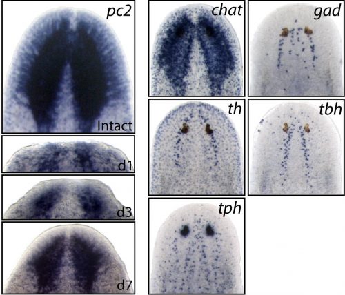
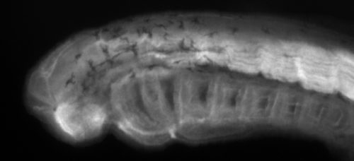
All panels are from whole-mount in situ hybridization experiments. Left panels show brain regeneration for 7 days following decapitation. Right panels show five different mature neuron subpopulations.
Earlier studies demonstrated the surprising molecular complexity of the planarian nervous system, but the specific genes and signaling mechanisms that regulated its construction were still largely unknown. Therefore, we set our sights on some very basic questions; 1) Within the large pool of planarian neoblasts, are there dedicated neural stem cells that are required for brain regeneration? 2) How are specific neural cell types specified/differentiated from upstream stem cells? 3) What extrinsic cell signals control the rate of neurogenesis during regeneration and in homeostatic (uninjured) conditions?
With these broad goals in mind, we did what most people do when you don’t have a concrete starting point, we did a screen. Taking advantage of the fact that planarians are susceptible to RNAi gene knockdown through feeding (a mixture of beef liver paste and E. coli that are forced to express double-stranded RNA for your gene of interest), it was possible to perform a high-throughput RNAi screen. We identified over one hundred planarian homologs of well-established neuronal regulatory genes, including many homeobox transcription factors, and proceeded to knock each one down, testing for possible functions in the planarian nervous system.
Since we were knocking down homologs of canonical neural stem cell regulators such as Pax6 and Sox2, we were really hoping that following decapitation, some of these RNAi-treated flatworms would exhibit major defects in brain regeneration. Alas, as is often the case in science, we never got such striking results. However, what we did notice, was that a fair number of RNAi-treated worms (even without decapitation) exhibited an array of strange/aberrant behaviours (including paralysis, and some very odd involuntary muscular contractions), that we thought could be caused by neuronal dysfunction within the intact nervous system. Therefore, we decided to focus on the transcription factors which yielded behavioural defects after RNAi-knockdown, examining them for neuronal specification defects.
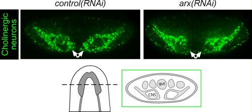
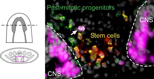
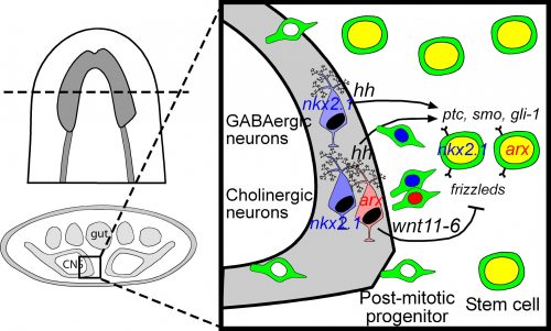
Among these genes causing strange animal behaviours were two homologs of homeobox transcription factors, nkx2.1 and arx. In particular, knockdown of nkx2.1, caused a very striking muscular contraction defect seen above (colloquially called the “cobra” phenotype in our lab), resulting in the animals constantly rearing back their heads so that they swam with their head perpendicular with the rest of the body, similar to a cobra snake. When we examined the brains of planarians after RNAi against nkx2.1 and arx, we found that these worms were missing a significant number of neurons belonging to the cholinergic, GABAergic and octopaminergic neural subtypes, specifically in the medial-most region of the brain (see image below), implicating these two transcription factors in the specification of these neuronal populations. Interestingly, we also found planarian stem cells immediately adjacent to this brain region that expressed nkx2.1 and arx, leading us to speculate that within this microenvironment (see image below), there may be communication between the mature neurons and their upstream stem cells to regulate their own production.
After RNAi knockdown of the arx transcription factor, we observed a significant ablation of medially-located cholinergic neurons in the planarian brain.
By imaging the area in between the brain lobes, we observed planarian stem cells and progenitors that expressed neural transcription factors, such as nkx2.1 and arx. These relatively rare cells may be committed neural stem cells.
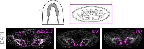
To investigate what this signal might be, we decided to look at the Hedgehog signaling pathway. It was already known that the planarian hedgehog (hh) ligand was expressed in the medial regions of the CNS (Rink et al., 2009). But we showed more specifically that the hh signaling molecule was actually expressed in the very same medially-located neuron subtypes as nkx2.1 and arx. In addition, we found that stem cells located right next to the brain expressed the Hedgehog receptor patched, as well as the downstream effector genes, smoothened and gli-1, demonstrating that planarian stem cells were likely capable of receiving and responding to hh signals coming from the mature brain.
The hh signaling molecule is expressed by mature neurons in the medial brain region, in the very same neurons that are specified by the nkx2.1 and arx transcription factors.
Next, we had to actually test whether hh signaling had any functional consequence on neurogenesis levels. For all of the interesting biology and techniques that are now available within the planarian research community, we still don’t have true genetic tools such as transgenics to perform gene knockout or overexpression experiments in a targeted cell-specific manner. Instead, we had to rely on organism-wide gene knockdown experiments to globally decrease or increase hh signaling, which admittedly I was a little nervous about, due to the wide range of roles that the hh pathway plays in animal development. Luckily, we found that when we knocked down the planarian hh ligand (decreased signaling activity), this resulted in the production of fewer neural progenitor cells as well as new mature cholinergic neurons, while having little to no effect on other tissue lineages (skin, gut & pharynx).
Cartoon model showing how signaling molecules produced by mature neurons (hh and wnt11-6) can communicate with nearby stem cells. Modified from Currie et al., eLife (2016)
We were initially a little confused by the result, since one would expect that in an intact “steady-state” brain, any signals coming from these mature neurons would actively repress the production of new neurons, whereas brain injury or the removal of nervous tissue would promote stem cells to increase their neuronal output. Instead, our results indicate the opposite, where hh signals coming from medial neurons are actually required to promote adult neurogenesis. It is possible that hh acts to counter and balance out the effects of local Wnt signaling, which has been shown to repress brain expansion by limiting neuronal progenitor production (Hill & Petersen 2015). However, it should also be noted that in the mammalian CNS, Sonic Hedgehog has known mitogenic roles, where its local production by differentiating or mature neurons signals back onto neural progenitors and adult neural stem cells to promote cell proliferation levels (Alvarez-Buylla & Ihrie 2014; Ihrie et al., 2011). Interestingly, this wasn’t the only parallel with the mammalian CNS, as Nkx2.1 and Arx are part of a transcription factor cascade within the early ventral telencephalon that specifies GABAergic and cholinergic interneurons (Butt et al., 2008; Vogt et al., 2014). I find it extremely interesting that across this huge evolutionary gap, these same transcription factors and this hedgehog signaling axis between mature neurons and stem cells have retained the same developmental functions in vastly different organisms. It also gives me reassurance about the importance of studying non-traditional model organisms, as the underlying basic biology is often conserved, so our findings from planarians, Hydra, Nematostella, Parhyale and other emerging lab models can be hugely informative for future studies on mammals and even humans.
Butt SJ, Sousa VH, Fuccillo MV, Hjerling-Leffler J, Miyoshi G, Kimura S & Fischell G. (2008). The requirement of Nkx2-1 in the temporal specification of cortical interneuron subtypes. Neuron 59: 722-732. http://www.sciencedirect.com/science/article/pii/S0896627308006302
Gurley KA, Rink JC & Sanchez Alvarado A. (2008). β-catenin defines head versus tail identity during planarian regeneration and homeostasis. Science 319: 323–327. http://science.sciencemag.org/content/319/5861/323
Hill EM & Petersen CP. (2015). Wnt/Notum spatial feedback inhibition controls neoblast differentiation to regulate reversible growth of the planarian brain. Development 142(24): 4217-29. http://dev.biologists.org/content/142/24/4217
Ihrie RA, Shah JK, Harwell CC, Levine JH, Guinto CD, Lezameta M, Kreigstein AR & Alvarez-Buylla A. (2011). Persistent sonic hedgehog signaling in adult brain determines neural stem cell positional identity. Neuron 71: 250-262. http://www.sciencedirect.com/science/article/pii/S0896627311004041
Morgan, T. H. (1898). Experimental studies of the regeneration of Planaria maculata. Arch. Entwm. Org. 7: 364–397. *This paper was subject of one of the Node’s Forgotten Classics posts*
Pallas, P. S. (1766). Miscellanea zoologica, quibus novae imprimis atque obscurae animalium species.Hagae Comitum, apud Pterum van Cleef, Holland.
Reddien PW, Bermange AL, Kicza AM & Sanchez Alvarado A. (2007). BMP signaling regulates the dorsal planarian midline and is needed for asymmetric regeneration. Development 134: 4043–4051. http://dev.biologists.org/content/134/22/4043
Reddien PW, Bermange AL, Murfitt KJ, Jennings JR & Sanchez Alvarado A. (2005). Identification of genes needed for regeneration, stem cell function, and tissue homeostasis by systematic gene perturbation in planaria. Developmental Cell 8: 635–649. http://www.sciencedirect.com/science/article/pii/S1534580705000924
Rink JC, Gurley KA, Elliott SA & Sanchez Alvarado A. (2009). Planarian Hh signaling regulates regeneration polarity and links Hh pathway evolution to cilia. Science 326: 1406–1410. http://science.sciencemag.org/content/326/5958/1406.long
Robb SM, Ross E & Sanchez Alvarado A. (2008). SmedGD: the Schmidtea mediterranea Genome Database. Nucleic Acids Res 36: D599–D606.
Vogt D, Hunt RF, Mandal S, Sandberg M, Silberberg SN, Nagasawa T, Yang Z, Baraban SC & Rubenstein JL. (2014). Lhx6 directly regulates Arx and Cxcr7 to determine cortical interneuron fate and laminar position. Neuron 82: 350-364. http://www.cell.com/neuron/abstract/S0896-6273(14)00161-5
The New Year’s honours list recognises citizens who have made achievements in public life and committed themselves to serving and helping Britain.
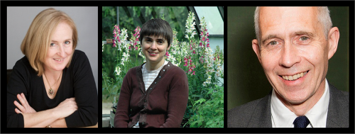
This year three prominent developmental and stem cell biologists were honoured, putting our field in the national news.The Development team were especially happy that two of the newly honoured researchers have connections to the journal: Sir Jim Smith was Development’s Editor in Chief from 2003-2009, and Dame Ottoline Leyser is currently one our editors. Dame Amanda Fisher has also been recognised for her contributions to stem cell and HIV research. The recognition of all three also includes their promotion of women in science.
Dame Amanda Fisher, Dame Ottline Leyser and Sir Jim Smith. Image source: AF, OL, JS
Professor (Henrietta Miriam) Ottoline Leyser CBE Professor Leyser, Director of the Sainsbury Laboratory at the University of Cambridge, is an inspirational scientist who has made seminal contributions to plant biology with direct implications for agricultural crops. Among many other awards, she was given the 2016 Genetics Society Medal in recognition of her work which has maintained the UK’s scientific leadership in this field. She was President of International Plant Molecular Biology and is a Foreign Associate of the US National Academy of Sciences. She has been a passionate advocate of career development for young researchers, especially women, and won the Royal Society’s Rosalind Franklin Award in 2007 for her proposal on combining a research career and a family.
Jim Smith’s bio
Dr. Smith, Director of Science at the Wellcome Trust and Senior Group Leader at the Francis Crick Institute, is a scientific leader who has transformed our understanding of embryonic development, giving insights into genetic defects in children and how stem cells develop into different tissues. He played a leading role in the development of the Francis Crick Institute from its early beginnings to its establishment in 2015. Previously, as Director of the Gurdon Institute and then of the MRC National Institute for Medical Research, he provided leadership in UK science across a breadth of disciplines and helped nurture the next generation of outstanding scientists, taking particular care to promote the careers of women.
Amanda Fisher’s bio
Professor Fisher, Director of the MRC Clinical Sciences Centre at Imperial College, London, has made fundamental discoveries in the molecular biology of HIV, the genomic characterisation of stem cells and the study of epigenetic gene regulation. Her observations of the HIV-1 virus underpinned the complete molecular understanding of the HIV genome and were a basis for the subsequent development of antiretroviral drugs. She is a strong advocate and role model for women in science and has made a significant contribution to the public understanding of science and training and mentoring researchers.
Many developmental biologists have also benefited from the technological advances made by another newly recognised Knight, Shankar Balasubramanian
Professor Shankar Balasubramanian Professor Balasubramanian, Herchel Smith Professor of Medicinal Chemistry at Cambridge University, was co-inventor of Next Generation DNA sequencing, the most transformational advance in biology and medicine for decades. Solexa sequencing, as it is now known, allows an individual genome to be sequenced in a day or two at a cost of less than £1000; previously, sequencing the human genome took years of work and cost billions. His work has spawned an entirely new discipline of Bioinformatics. More recently, he has made major contributions to understanding the role of DNA-quadruplexes in cancer and invented a method for the sequencing of epigenetic modifications.
The Development team also did some filming, asking participants about their views on what made the meeting special, as well as the state of the human development field. You can watch the video, and see a gallery of images from the meeting, below.
The Development team, Seema Grewal (Reviews Editor), Katherine Brown (Executive Editor), Caroline Hendry (Reviews Editor) and Aidan Maartens (Community Manager, the Node)
Nicky Le Blond, The Company of Biologists’ Meeting Organiser
Development posters
Beer and posters
Coffee and posters
Coffee and posters
Coffee and posters
Beer and posters
Beer and posters
Yann Barrandon, Guy Sauvageau and Sally Temple in the panel session on clinical applications
The social event was a trip to the American Optical Museum
Old lenses
Dick, the museum’s curator, shows us around
The American Optical Museum
The American Optical Museum
The American Optical Museum
The American Optical Museum
Coffee and beer in the tavern below the museum
Coffee and beer in the tavern below the museum
Coffee and beer in the tavern below the museum
Coffee and beer in the tavern below the museum
The Development team with editors Austin Smith, Benoit Bruneau and Gordon Keller
 (1 votes)
 (No Ratings Yet)
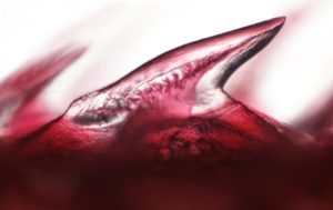

 I am originally a Finnish evolutionary biology student who got fascinated about developmental biology during my exchange programme couple of years ago in Southern Chile (
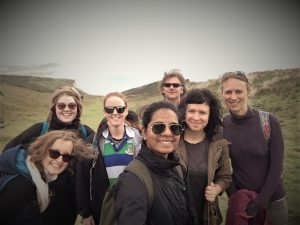

 (7 votes)


 Naïve pluripotent mouse embryonic stem cells (ESCs) resemble the preimplantation epiblast and efficiently contribute to chimaeras. By contrast, primate ESCs correspond to the postimplantation embryo and fail to resume development in chimaeric assays. In their
  The planar cell polarity (PCP) pathway is best known for its role in polarizing epithelial cells within the plane of a tissue but it also plays a role in a range of cell migration events during development. In their