We are currently seeking applications for the role of Reviews Editor at Development. This is a temporary position (maternity cover) anticipated to last up to 12 months, starting June 2017.
Joining an experienced and successful team, this is an exciting opportunity to make a significant contribution to one of the major journals in the field of developmental biology. Development publishes primary research articles, reviews and other front section content across the breadth of the developmental biology and stem cell fields.
Applicants will hold a PhD in developmental or stem cell biology. Post-doctoral and/or previous editorial experience is desirable, although we will provide full training.
Core responsibilities:
Commissioning, handling peer review and developmental editing of material for the front section of the journal
Representing the journal at international conferences and within the wider scientific community
Writing press releases, article highlights and material for Development’s community website ‘the Node’
Creative involvement in the journal’s development
The successful candidate will have a broad interest in science, the scientific community and publishing. Excellent interpersonal and literary skills, enthusiasm and commitment are also essential requirements for the position.
The Reviews Editor will work alongside an experienced in-house team, including the Executive Editor and current Reviews Editor, as well as with our international team of academic editors.
This maternity cover position provides an excellent opportunity to gain experience on a highly successful life-science journal, and offers an attractive salary and benefits. The position is full-time and will be based in The Company of Biologists’ attractive modern office on the outskirts of Cambridge, UK.
The Company of Biologists (biologists.com) exists to support biologists and inspire advances in biology. At the heart of what we do are our five specialist journals – Development, Journal of Cell Science, Journal of Experimental Biology, Disease Models & Mechanisms and Biology Open – two of them fully open access. All are edited by expert researchers in the field, and all articles are subjected to rigorous peer review. We take great pride in the experience of our editorial team and the quality of the work we publish. We believe that the profits from publishing the hard work of biologists should support scientific discovery and help develop future scientists. Our grants help support societies, meetings and individuals. Our workshops and meetings give the opportunity to network and collaborate.
Applicants should send a CV to recruitment@biologists.com, along with a covering letter that summarises their relevant experience, why they are enthusiastic about this opportunity, and their current salary level. Applicants should be eligible to work in the UK and should be able to travel internationally.
Applications should be received by February 28th 2017, though we may be able to consider later applications.
The brown alga Ectocarpus has emerged as a model system for the evolution of muticellularity. Today’s paper, from the current issue of Development, investigates the role and evolutionary history of a gene implicated in Ectocarpus development. We caught up with first author Nicolas Macaisne and supervisor J. Mark Cock of the Station Biologique de Roscoff in Brittany.
Mark, can you tell us your scientific biography and the general focus of your lab?
JMC I am a molecular biologist that worked for many years on various aspects of land plant biology, including nitrogen fixation and self-incompatibility. In 2002 I moved to the Station Biologique de Roscoff, a marine laboratory on the north-west coast of France, with the aim of setting up a laboratory to work on brown algal developmental biology. Working with Akira Peters, we evaluated several brown algae for their potential as model systems and eventually selected the filamentous brown alga Ectocarpus (Peters et al., 2004). The choice of Ectocarpus was based on its amenability to genetic analyses and on earlier work, particularly by Dieter Müller in Constance, that had demonstrated the merits of this species as a laboratory system. The main objective of the laboratory has been to understand the molecular mechanisms that regulate life cycle progression in this brown alga. Ectocarpus has a haploid-diploid life cycle involving an alternation between two multicellular organisms, the sporophyte and the gametophyte. We have been using genetic approaches to identify both key life cycle regulators and regulators of downstream processes during the development of the two generations (Coelho et al., 2011; Peters et al., 2008). In addition, a great deal of effort has been put into generating tools for Ectocarpus, including the sequencing and analysis of its genome in collaboration with the Genoscope in Paris and the VIB in Ghent (Cock et al., 2010). More recently, the laboratory has also become interested in sex determination in the brown algae, a project led by my co-PI Susana Coelho (Ahmed et al., 2014; Lipinska et al., 2015; Luthringer et al., 2015).
And Nicolas, how did you come to work with Mark in Roscoff?
NM I joined Mark’s laboratory in 2011, just after obtaining my Ph.D. under the mentorship of Raphaël Mercier in the National Institute for Agronomic Research of Versailles, France. During my thesis, I studied the genetic regulation of meiotic DNA repair in Arabidopsis, a land plant which is a very well established model for genetics. Thus, my previous research and the model I was employing were quite far removed from Mark’s research on the biology of algae. There are very few labs that study algae, but the unusual life cycle of Ectocarpus, especially the fact that the sporophyte and the gametophyte generations are independent from one another, intrigued me. I realized that this system offered a unique opportunity to study these developmental stages independently. I didn’t appreciate how much I would learn about basic plant biology from this beautiful organism.
I also recognized Mark’s lab as a place where I could challenge myself to learn new techniques and approaches. Working with a model system for which the genome was still not completely assembled and only has a partial genetic map was daunting, but I now feel that I have gained an appreciation for how to set up a new system and look beyond our traditional models for unique insights. This experience was also incredibly valuable for allowing me to learn genome and transcriptome sequencing and analysis. These techniques are absolutely invaluable for the analysis of genes involved in all aspects of developmental biology. Mark’s expertise and the great scientific environment at the Station Biologique de Roscoff are the best assets I could have hoped for in order to achieve my goals.
What makes brown algae interesting models for developmental and evolutionary biologists?
JCM & NM Although many eukaryote groups have evolved simple multicellular forms, we generally consider that only five major groups have evolved complex multicellularity, i.e. possess macroscopic bodyplans with multiple cell types orchestrated by developmental programs (Cock and Collén, 2015). These groups are animals, land plants, brown and red algae and fungi. Complex multicellularity evolved independently in each of these five lineages and comparisons between lineages could therefore provide us with important information about the molecular principals that underlie this key evolutionary transition. Unfortunately, whilst we know a great deal about developmental mechanisms in animals and land plants, the three remaining groups remain almost completely uncharacterised. Of these latter groups, the brown algae are particularly interesting because they include species that rival land plants in terms of their developmentally complexity. We believe that it is important to extend the scope of developmental biology beyond the animal and land plant lineages to effectively address questions about the evolutionary origins of developmental processes associated with complex multicellularity.
“It is important to extend the scope of developmental biology beyond the animal and land plant lineages to effectively address questions about the evolutionary origins of developmental processes associated with complex multicellularity”
The brown algae are also interesting models to address a wide range of more specific developmental questions. Haploid-diploid life cycles, such as that of Ectocarpus, raise the question of how a single genome directs the implementation of two different developmental programs to produce either a sporophyte or a gametophyte at the appropriate stage of the life cycle. Clearly, epigenetic processes must play an important role in this alternation but at present we have no information about how this might be mediated and the epigenetic aspect will be an important avenue for future research. Historically, brown algae have made important contributions to our understanding of early embryogenesis, particularly the establishment of polarity and the asymmetric division of the initial cell (Bouget et al., 1998; Brownlee and Bouget, 1998; Goodner and Quatrano, 1993). In this context, one of the most surprising features of the Ectocarpus life cycle is the alternation between symmetrical and asymmetrical initial cell divisions during the sporophyte and gametophyte generations, respectively. If we look more broadly across the brown algae, these organisms exhibit a bewildering variety of different types of life cycle (Cock et al., 2014) and sexual systems (Luthringer et al., 2014). For example, Fucus species have diploid life cycles and the diecious members of this genus are thought to have diploid phase sex-determination systems (presumably XY or ZW sex chromosome systems) associated with oogamy, whereas the haploid-diploid life cycle of Ectocarpus is associated with a haploid-phase sex-determination system (UV chromosomes) and near-isogamy. Moreover, life cycle and sex-determination systems appear to have varied considerably during the evolution of this group providing an excellent context to link the different types of system with developmental (size, growth habit, reproductive strategy, etc.) and ecological (habitat, biotic interactions, etc.) parameters.
Brown algae also have many interesting features associated with adaptation to the extreme conditions of the coastal environment. These include their unusual cell walls that provide both strength and flexibility, and mechanisms providing resistance to both abiotic (variations in light, heat, desiccation, salinity, etc.) and biotic stresses. Their evolutionary past, which included a secondary endosymbiosis event that gave rise to the stramenopile plastid, provided brown algae with a mosaic of genetic information from various sources, including that of the red algal endosymbiont. Developmental biologists can therefore expect to discover unusual features at the molecular level and the identification of the IMM gene is probably just a foretaste of the novelties to come.
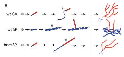
Schematic representation of the early development of the wild-type gametophyte (wt GA), the wild-type sporophyte (wt SP) and the imm mutant sporophyte (imm SP), from Figure 2, Macaisne, et al. 2017. Development.
And you appear to have quite an extensive toolkit for Ectocarpus in particular?
JMC & NMEctocarpus has a long history as a research organism stretching back to the 19th century (Charrier et al., 2008; Coelho et al., 2012). One of our priorities after selecting Ectocarpus as a model organism was to generate a genome sequence and associated genomic and genetic tools including a transcriptomic database, genetic maps and methodologies for investigating gene function. Tool generation is an ongoing process, for example we have recently re-annotated the genome sequence, adding extensive new information and significantly improving the quality of the existing information (Cormier et al., 2016). The Ectocarpus genome is currently one of the highest quality reference sequences available for the stramenopiles. The generation of tools to analyse gene function has been a more complicated process and we still lack one key tool: a robust protocol for genetic transformation. Importantly, the recent development of an RNAi approach, initially for Fucus (Farnham et al., 2013) and subsequently for Ectocarpus (Macaisne et al., 2016), has provided an alternative for reverse genetics and the recent demonstration that forward genetic approaches can be applied in Ectocarpus also represents a powerful tool to analyse gene function. We hope that our demonstration that forward genetic approaches can be applied to brown algal systems will encourage other groups to investigate developmental mechanisms in this group, leading to the emergence of macroalgal developmental biology as a field in its own right.
Can you give us the key results of your paper in a paragraph?
JMC & NM In most brown algae, the initial cell divides to produce two cells with basal and apical identity respectively. This is also the case for the Ectocarpus gametophyte but the sporophyte is unusual because it first establishes an extensive, filamentous basal system before deploying the apical tissues (upright filaments). The first cell division of the sporophyte is therefore developmentally symmetrical, producing the two germ tubes of a symmetrical basal filament (Peters et al., 2008). Sporophytes carrying a mutation in the IMM gene fail to produce the extensive basal system. Instead the initial cell divides asymmetrically to produce a basal rhizoid and an apical upright filament. The mutant sporophyte therefore resembles a gametophyte and the mutation was initially interpreted as causing a partial conversion of the sporophyte into a gametophyte (Peters et al., 2008). In this study, however, analysis of transcriptomic data indicated that the imm sporophyte retains sporophyte identity and we propose an alternative interpretation: that the phenotype of the imm sporophyte represents a more primitive state of the sporophyte, before it evolved the capacity to deploy the extensive basal system. Positional cloning of the IMM locus showed that it encodes a novel protein of unknown function with a repeated cysteine-rich motif that is also found in a gene from the Ectocarpus virus EsV-1. Interestingly, IMM is a member of a large family of 91 genes in Ectocarpus, which we have called EsV-1-7 domain genes after the viral gene that contains this repeated domain. Other stramenopiles either lack EsV-1-7 domain genes or possess only a single EsV-1-7 domain gene suggesting that there has been a massive expansion of this gene family in the brown lineage, perhaps associated with the transition to complex multicellularity (brown algae are the only stramenopile group that exhibit complex multicellularity). A broader search for EsV-1-7 domain genes across the eukaryotic tree of life found a very unusual distribution. In addition to the stramenopile EsV-1-7 domain genes, members of this family were only found in one cryptophyte (Guillardia theta), two chlorophyte green algae (Cocomyxa subellipsoidea and Monorapidium neglectum) and perhaps in one fungal genome (although this latter gene may be a contaminant). It is difficult to reconcile this distribution with vertical inheritance of the gene family. Interestingly, EsV-1-7 domain genes were also found in the genomes of three diverse viruses, suggesting a possible alternative explanation: viral-mediated horizontal gene transfer. This latter hypothesis may be difficult to confirm because horizontal transfer events may have been quite ancient (this appears to have been the case in the stramenopiles for example) but more complete genome sampling for the lineages in question will undoubtedly shed some light on the evolutionary history of this family.
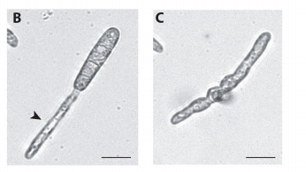
Partheno-sporophyte germlings after introduction with siRNAs targeting imm (B) and controls (C), from Figure 2, Macaisne, et al. 2017. Development
Although IMM seems functionally restricted to the generation of the sporophyte, it is also expressed in the gametophyte: do you think it is doing anything there?
JMC & NM Gametophytes carrying the imm mutation appear to be completely normal indicating that the gene does not have any function during that generation. Of course, we cannot rule out the possibility that there is a subtle or conditional gametophyte phenotype that we have not detected, but the current data indicates that IMM has a sporophyte-specific function. Therefore, in Ectocarpus at least, IMM appears to be specifically associated with the pathway that directs the development of an extensive basal system and delays deployment of the apical system. Nonetheless, the presence of IMM transcripts during the gametophyte generation remains something of a mystery but presumably, if transcription of IMM during the gametophyte generation does not result in any observable phenotype, then there may not have been any selective pressure to prevent IMM transcription during this part of the life cycle.
The IMM gene is conserved in other brown algae, including species that do not exhibit delayed deployment of the apical system. It will be interesting in the future to investigate the role of these IMM orthologues, during both the sporophyte and the gametophyte generations of these other species.
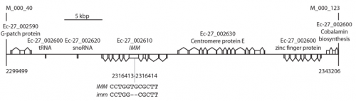
Schematic tree of the eukaryotes showing the phylogenetic position of species that possess EsV-1-7 domain genes, from Figure 5, Macaisne, et al. 2017. Development
Do you want to hazard a guess at what EsV-1-7 domain proteins are doing?
JMC & NM At present we only have functional information of one member of this family: IMM. The phenotype of the imm mutant suggests this protein has a regulatory function because loss of function leads to changes in the expression levels of a large number of genes. The protein sequence doesn’t tell us very much more about its function except that the repeated cysteine-rich motif in the C-terminal part of the protein (the EsV-1-7 repeat) is reminiscent of a zinc finger motif. Our current guess would therefore be that these proteins contain a novel version of the zinc finger motif that allows them to act as regulatory molecules, perhaps as transcription factors for example. We are currently trying to test this hypothesis by assaying for DNA-binding activity and by attempting to obtain three-dimensional structural information for the protein.
If EsV-1-7 domain proteins have a regulatory role, the next question is what are they regulating? The only information we have at present is their expression patterns in transcriptomic data corresponding to various life cycle stages and conditions. These data, which indicate that the members of the gene family have very diverse expression patterns, suggest that the different members of the family may be involved in diverse processes but additional functional analyses will be necessary to confirm this and to explore the functions of each member of the family.
IMM mutants appear to revert to a more ancestral mode of development, and EsV-1-7 repeat genes may have got into Ectocarpus through horizontal gene transfer. So do you think viral transfer facilitated a new mode of development? It’s a very striking idea!
This would seem to be a reasonable explanation based on the unusual phylogenetic distribution of EsV-1-7 domain genes across the eukaryotes, but it is important to underline that the process would have occurred over a very long time-scale. The presence of EsV-1-7 domain genes in stramenopiles, a cryptophyte, two chlorophytes and perhaps one fungal species, together with the presence of related genes in three viral genomes does strongly suggest horizontal transfer, perhaps mediated by viruses. However, the acquisition of EsV-1-7 domain genes by the stramenopile group appears to be ancient because gene family members were also found in oomycete and eustigmatophyte genomes. Several hundred million years would therefore have separated this ancient horizontal transfer event from the evolution of the IMM gene and the associated novel mode of development in the brown algae.
Nonetheless, we agree that the idea that an important brown algal developmental regulator may have originally been acquired from a viral genome is very interesting. We also believe that the identification of IMM underlines the importance of forward genetic screens and the use of diverse model organisms in developmental biology. None of the classical animal, plant or fungal model organisms possesses EsV-1-7 domain genes, so it would not have been possible to identify this family using these systems.
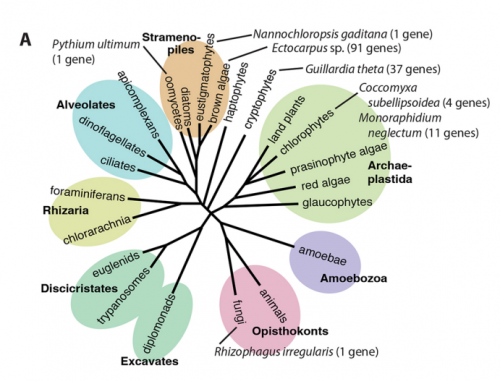
Schematic representation of the 43,708 bp interval on chromosome 27 between the closest recombining markers to the IMM locus, from Figure 5, Macaisne, et al. 2017. Development
When doing the research, did you have any particular result or eureka moment that has stuck with you?
Not really a “eureka” moment, but with the hindsight, I realize how lucky I may have been with some experiments I have undertaken. For example, when I was mapping the mutation causing the imm phenotype, after having scanned the genome of 1699 individuals with more than a hundred molecular markers, I was stuck with a relatively large genomic region containing several annotated genes (and possibly more which were not annotated yet) and no easy way to know which of these genes IMM was. So I decided to adapt a whole-genome sequencing approach to sequence specifically this region of the genome of Ectocarpus. Although this approach was successful in identifying the causal mutation, to my knowledge it had never been performed previously, and I was doing that on an alga. So with hindsight, I think I was a bit overly confident, or maybe a bit naive, in thinking that my approach would work. Thankfully it did, and now I am glad I dared to use it.
“With hindsight, I think I was a bit overly confident, or maybe a bit naive, in thinking that my approach would work. Thankfully it did”
And what about the flipside: any moments of frustration or despair?
NM To be honest, I was a bit afraid when I realized the enormity of data that was obtained from the RNA-sequencing, especially considering the fact I had, at that time, no experience in bioinformatics. However, our team and the bioinformaticians of the Station Biologique were extremely supportive; it has really been a team effort.
More generally, my major frustration is that we cannot yet genetically transform Ectocarpus, preventing us from generating another mutant allele of IMM. This would have made the validation of this gene much easier. However, given the successful use of RNAi for this paper, I am confident that transformation tools like CRISPR/Cas9 targeted mutagenesis will be implemented soon for this model organism. Such tools will have a tremendous impact in the field, hastening considerably the understanding of algal biology.
What about your plans for the future following this paper?
NM Working on a relatively young model organism, with a team that is directly involved in the development of genetic and genomic tools to study it, has been very enriching. I have now joined the laboratory of Judith L. Yanowitz at the Magee-Womens Research Institute in Pittsburgh, PA, where I went back to my primary field of expertise, the regulation of DNA repair during meiosis. I now use the nematode Caenorhabditis elegans as a model organism. Now that I have worked on three different organisms, I realized that having a multi-organism approach is highly beneficial for research as we can take advantage of the similarities and the specificities of each organism to better understand a biological process. The next step for me would be to become a principal investigator to continue my research on meiosis using C. elegans as a model and branching out to less well-developed organisms.
“So far we have only scratched the surface and there is still a great deal to be learnt about brown algal developmental biology”
And where next for the lab?
JMC So far we have only scratched the surface and there is still a great deal to be learnt about brown algal developmental biology, including the identification of the major regulators of the diverse processes discussed above. We are still a long way from having any detailed understanding of developmental processes in brown algae. Our laboratory will continue to focus on life cycle regulation and sex determination, using genetic approaches to dissect the underlying pathways but also developing new approaches, for example to investigate the role of epigenetic processes in life cycle control. We are also very interested in the evolutionary perspective and a recently launched project, in collaboration with Genoscope, aimed at obtaining genome sequences for a broad range of brown algal species will provide the genomic data necessary to place brown algal developmental processes in an evolutionary context.
Ramkumar N, Omelchenko T, Silva-Gagliardi NF, McGlade CJ, Wijnholds J, Anderson KV.
Nat Cell Biol. 2016 Dec;18(12):1281-1291
“It is not birth, marriage, or death, but gastrulation, which is truly the most important time in your life.”
Lewis Wolpert (1986)
Aptly stated by Wolpert, gastrulation is the fundamental process that shapes the morphogenesis of the embryo. It involves the formation of the three germ layers, namely the endoderm, mesoderm and ectoderm, from a single layer of epithelial cells called the epiblast. These germ layers then proceed to give rise to the different tissues and organs in the embryo. Gastrulation involves large scale cell movements, rearrangements and changes in gene expression leading to transformation of cell fates. Among them is a process called epithelial-to-mesenchymal transition (the EMT), wherein epithelial cells lose their apical-basal polarity and adopt migratory mesenchymal behavior. In the mouse embryo, this process happens at the posterior pole of the embryo in a structure called the primitive streak, which is also a convergence point for many signaling pathways in the embryo1. The sheer complexity of gastrulation and its efficiency of execution fascinated me and motivated me to work on it during my PhD.
The bulk of our current knowledge and understanding of the signaling pathways involved in gastrulation comes from the analysis of mutants identified from genetic screens mainly in Drosophila and recently in other animals. Likewise, my journey to the world of Crumbs and its role in gastrulation began with a mouse mutant called wsnp(wing shaped neural plate). wsnp mutants have very little mesoderm-derived tissues and their neural plate resembles the Drosophila wing disc.
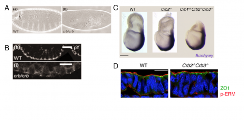
Figure 1: The wing shaped neural plate (wsnp) phenotype. In comparison to their wild-type litter mates, wsnp embryos had a shortened anterior-posterior axis, lacked visible somites and had a completely open neural plate at embryo day 8.5. At this stage, the wsnp embryos resembled a Drosophila wing disc (C), hence the name. Dorsal views, Anterior up (A, B). Scale bar- 150 µm. Image in (C) is from http://www.pbs.org/wgbh/nova/genes/fate-07.html
With the help of deep sequencing and genetic complementation analysis we identified wsnp to be a null allele of Poglut1 (Protein O-glucosyltransferase) which encodes an enzyme that adds O-glucose to the EGF repeats of proteins. At the time we discovered this, Drosophila Notch was shown to be modified by this enzyme2. While I worked on studying the role of Notch glycosylation during mammalian gastrulation, the severity of the wsnp phenotype compared to that of Notch pathway mutants led us to explore the possibility of alternate targets of the enzyme during gastrulation. Among them, the Crumbs family of proteins stood out as likely candidates. In the lab, we had previously isolated a mutant lulu, a null allele of Epb4.1l5, which had a phenotype similar to wsnp at gastrulation, and biochemical experiments had showed a direct interaction of Epb4.1l5 with mammalian Crumbs3,4. With this as our only inkling, we took a leap of faith and decided to explore the role of Crumbs in gastrulation. At that time, there was no precedent for the role of any Crumbs proteins in mouse development, let alone gastrulation. We decided on Crumbs2 because it had a long extracellular domain with EGF repeats that could be glycosylated and the conserved intracellular domain. We waited for about a year to get the conditional Crumbs2 knock out mice and the results made our wait worthwhile. We were really excited that the phenotypes of wsnp and Crb2-/- were identical. We went on to show that sugar modification of Crumbs2 is essential for its membrane localization and that in turn is essential for its function during gastrulation5.
We then focused our attention to our central question – What is the function of Crumbs2 during mammalian gastrulation? We had always known Crumbs as a well-established apical determinant in Drosophila as its role in cell polarity has been well explored there6,7. We were surprised to find that the epiblast of Crumbs2-/- mutants did not have any polarity defects. To rule out the involvement of other mammalian Crumbs proteins in setting up this polarity, we generated triple mutants lacking all known mammalian Crumbs proteins. Any mouse geneticist will tell you – generating triple mutants is not fun! To our surprise, we found that mammalian Crumbs proteins were not required to establish the polarity of the epiblast epithelium. This was in contrast to the case in Drosophila, where Crumbs is essential to set up polarity of the embryonic epithelium. Additionally, in the Crumbs2-/- mouse embryos we found an accumulation of cells expressing E-cadherin and Sox2+at the streak, suggesting that these cells were still epithelial. This was contrary to what we were expecting: as a canonical polarity protein, one would imagine that a loss of Crumbs2 would lead to more cells delaminating i.e. making more mesoderm cells, but we saw the opposite. This suggested to us that the functions of mammalian Crumbs2 could be different from that of Drosophila Crumbs, and got me further engrossed into understanding how mammalian Crumbs2 functions during gastrulation.
Figure 2: Comparing mouse Crumbs null phenotype to Drosophila crb phenotype. (A) Cuticle preparation of wild-type and homozygous crb embryos showing the failure to form a continuous cuticle in crb mutants (anterior is left and ventral down). (B) The ventral epidermis of a stage 12 wild-type embryo and crb mutant embryo stained with an anti-phosphotyrosine (pY) antibody, which labels the site of adherens junctions, shows continuous junctions in wild-type which is completely lost in crb embryos (Apical is down). (C)Wild type, Crumbs2 mutants and Crumbs triple mutants (Crumbs1rd8, Crumbs2-/-, Crumbs3-/-) at E7.5 probed for expression of Brachyury, a marker of the primitive streak. Crumbs triple mutant embryos do not have a more severe gastrulation phenotype than Crumbs2 single mutants (Lateral view, anterior to left, distal down). (D) Transverse section through the epiblast of E7.75 wild type and Crumbs2-/- Crumbs3-/- double mutant embryos immunostained for ZO1 (tight junctions) and pERM (apical domain). The double mutants do not have defects in epiblast polarity (Apical is up). Scale bars in C, 50μm; D, 21μm. The extraembryonic portion of the triple mutant was removed for genotyping. Figures A and B adapted from Ansgar and Knust, 2000.
Our attention was drawn to the cells stuck at the streak. Although we were able to visualize these cells using SEM and X-linked GFP, these were static, 2-dimensional images. Cell delamination from an epithelium is a dynamic process requiring constant neighbor exchange and happens in 3-dimensional epithelium. In order to understand exactly when the cells fail during this process, we had to watch this process live in mouse embryos. While it seemed like the most obvious experiment, it was clearly the most challenging aspect of the project at many levels. At that time, there were hardly any labs that had done live imaging with fluorescent reporters in mouse embryos. While cell delamination had been extensively studied, and imaged in in vitro culture systems, only recently with the advent of better imaging technology and improved reporters scientists have started exploring it in live tissues and embryos. For us, it was a herculean task as we had to work out every aspect of the experiment including the mouse strains to use, then determine the imaging conditions to use while keeping the embryo stable. Additionally, we had to define the temporal dynamics of this process in wild-type embryos before we begin to compare it with our mutant.
As mouse embryos are very sensitive to laser light and the primitive streak is a densely packed epithelium, it is important for the reporter to be very bright to get reasonable data. Initially we tried with reporters expressed ubiquitously in the epithelium, as it would reduce the number of crosses and increase the probability of the desired genotype. However, at that time there weren’t good algorithms to segment these images making their analysis very difficult and unfruitful. To bypass this, we decided to express the fluorescent protein in a random pattern, which helps to create better 3-dimensional reconstructions of individual cells and help in their subsequent analysis. However, this would involve additional crosses and also decrease the probability of getting our desired genotype. After screening many different reporter and CRE lines that we and others at Sloan Kettering had in their labs, we began to lose hope in this experiment, as we weren’t getting the right combination of the two. It was around this time that Kathryn attended a conference where Ann Sutherland presented her lab’s work on imaging with the mT-mG reporter and the EIIA-CRE. They found that this combination gave them a good random pattern and the membrane-GFP (mGFP) was a bright reporter at this stage8. Having seen her beautiful movies with the mGFP, we decided to get these mice and eventually used them for our experiment. Despite our optimization of the imaging, there were many factors in this experiment that were beyond our control. For instance, the probability of getting a mutant with both the reporter and the CRE was low and the percentage of randomness of the CRE was unpredictable. Nevertheless, after many unsuccessful attempts, we managed to get beautiful movies of the cells in wild-type and the mutant streak and were happy to see that the cells in the mutants shrink their apical surface but fail to leave the epithelium. While we focused mainly on shape changes and time to delamination for the purpose of this paper, these movies are a gold mine of data. It is amazing how so many processes are happening simultaneously at the streak. There are cell divisions, tissue level movements, neighbor exchanges to just name a few. With the advent of light sheet microscope, there is undoubtedly lots to learn from just watching these cells live in the epithelium.
GFP positive streak cell translocates from the apical to the basal side of the epiblast in wild type embryo. The epiblast cells in the mouse embryo randomly express mGFP at E7.5 and are imaged from the primitive streak side (posterior). A 3D rendered surface (yellow) was built with Imaris for easy visualization of the individual cell shape changes. Ingressing cells have basal protrusions, translocate their cell body basally and leave the epiblast in less than 2h. In addition to the shape changes we discuss in the delaminating cell, the other cells in this region of the epithelium (streak), which are poised to delaminate are also highly dynamic. Time is h:min. Scale bar,10 µm.
The big debate- Polarity?
Since Crumbs was as a polarity protein, we had to determine whether the defect we saw was specific to gastrulation at the streak or that Crumbs plays a role in the epithelia by virtue of which it affects the streak. My lab meetings would always end on this debate, followed by heated arguments supporting both hypothesis. The only way to test this was how Drosophila geneticists had always done before. We had to prevent gastrulation from happening using Wnt3 mutants (with and without Crumbs2) and see whether the tissue was normal or not. Fortunately for us, we had the right tools out there to do this experiment. Since both outcomes were equally likely and exciting, irrespective of the result, it was one of the most exciting dissections ever. It would answer the question I had since my first lab meeting. I remember discussing the Wnt3 results at my lab meeting: finally, we concluded without any argument that it is the streak, and not the epiblast epithelium, that requires Crumbs2, after all.
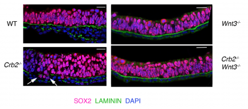
Figure 3: Crumbs2 is required at the streak during gastrulation. Transverse section through the anterior epiblast/presumptive neural epithelium of wild type and Crumbs2 mutants, epithelia of Wnt3-/- and Crumbs2-/- Wnt3-/- double mutant embryos immunostained for SOX2 and LAMININ. Crumbs2-/- mutants have an increase in the thickness of the neural epithelium and SOX2+ve cells are present below the basement membrane (arrows) in the mutants at E8.0. Wnt3-/- and Crumbs2-/- Wnt3-/- double mutant embryos are morphologically identical. The epithelium of Wnt3-/- and Wnt3-/- Crumbs2-/- embryos do not have breaks in laminin expression or SOX2+ nuclei below the basement membrane. Scale bars: 21 μm.
Perspective makes a difference
Traditionally, protein localization in the primitive streak epithelium was observed by immunostaining transverse sections of the epithelium. We too followed suit to look at Crumbs2 expression and were happy to see it localized apically in the cells of the streak. During my trials with live imaging, I began to appreciate the importance of the 3-dimensional nature of the epithelium and thought it was essential to see where Crumbs2 would fit in this 3-D context. The apical surface of the primitive streak epithelium faces the interior of the embryo, making it difficult to access and therefore not many people have looked at polarity proteins en face, in contrast to studies in Drosophila and cell culture. While learning to do en face imaging on neural epithelium, as a fun experiment I decided to look at Crumbs2 localization en face at the primitive streak. Although it took me many trials to get the streak epithelium flat, I was surprised to see that Crumbs2 localization was anisotropic. At first, I thought it could be an artefact of my sample processing. However, after repeated attempts with different approaches to sample processing and appropriate controls I was finally able to convince myself about the anisotropic pattern. Nevertheless, we still didn’t understand what it meant! I had tried co-staining with many other polarity proteins, but none of them had this pattern. I had immunostained for MyosinIIB separately, but it wasn’t until the Roper paper on Drosophila Crumbs in the salivary gland that we decided to probe for MyosinIIB and Crumbs2 together. That’s when it came together. The complementary nature of their pattern was striking and steered us to think of possible mechanisms of Crumbs2 action.
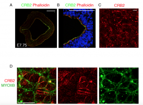
Figure 4: CRUMBS2 localisation at the primitive streak epithelium Transverse section of the primitive streak of wild-type embryo at E7.75 immunostained for CRB2 (green) and Phalloidin (red) showing the expression of CRB2 in both the anterior presumptive neural epithelium and the epiblast and its enrichment in the posterior epiblast. The streak region is magnified in (B). Extended projection of en face view of the apical surface of the epiblast layer of the primitive streak of wild type at E8.5 immunostained for CRB2 (C), showing its anisotropic distribution. The double immunostaining of the streak for CRB2 (red) and MYOSIN IIB (green), showing the reciprocal enrichment of CRB2 and Myosin IIB on different cell edges. Proximal is up. Scale bars: A, B, 100 μm; C,D 10 μm.
We hypothesize that the unequal levels of Crumbs2 in the epithelium must be an important part of the mechanism to determine which cells delaminate first. It’s a very interesting problem which hasn’t received its due. The streak region as defined by the basement membrane breakdown is easily around 4-5 cells wide and runs from the node to the distal tip of the embryo. Despite that, cells delaminate individually and leave the epithelium, suggesting that this process must be highly coordinated and tightly regulated at both cell and tissue levels. We know little about how the cells establish the complementary pattern of Crumbs2 and MyosinIIB, how they define the order in which they delaminate and how they coordinate this with their neighbors. These questions can be addressed by live imaging with appropriate reporters and will offer a wealth of information.
My journey to Crumbs emphasizes the power of genetic screens in identifying new players in gastrulation, and my paper offers a peak into the unexplored territories of mammalian gastrulation. There is a lot to learn by just observing this beautiful process in wild-type and mutant embryos. Hopefully, my work will inspire people to explore the dynamics of this process in greater detail.
References
1 Ramkumar, N. & Anderson, K. V. SnapShot: mouse primitive streak. Cell 146, 488-488 e482, doi:10.1016/j.cell.2011.07.028 (2011).
Here are the highlights from the current issue of Development:
Evolving an atypical developmental programme with IMM
The brown alga Ectocarpus has alternating haploid (gametophyte) and diploid (sporophyte) generations. Morphologically, these are distinguished by a more complex system of basal filaments in the sporophyte, initiated via symmetric divisions before the apical-basal axis is defined and the upright filaments form. This mode of development is unusual – in most brown algae, and in the Ectocarpus gametophyte generation, the first division is asymmetric to establish the apical-basal axis. The immediate upright (imm) mutation of Ectocarpus displays an asymmetric first division, and was initially thought to represent a partial switch from the sporophyte to the gametophyte developmental programme. Here (p. 409), Mark Cock and colleagues identify the gene responsible for this phenotype and provide a detailed analysis of its evolutionary history. The IMM gene is a member of the large, rapidly evolving, EsV-1-7 domain family, which exhibits an unusual distribution across eukaryotic lineages – potentially as a result of horizontal gene transfer. Transcriptional profiling suggests that, rather than imm causing a switch from the sporophyte to the gametophyte programme, the mutation blocks the extensive development of the basal filament system, such that the mutant displays a more canonical mode of sporophyte development. While the molecular and cellular function of IMM has yet to be determined, this gene appears to represent an evolutionary innovation in the Ectocarpus lineage that altered early sporophyte development.
Defining the right MOMent for cell fate decisions
The invariant lineage of C. elegans led to an early assumption that cell fate decisions are largely made cell-autonomously. However, it has subsequently become clear that inductive interactions between cells are essential for fate determination in this system. Moreover, these inductive interactions can be highly complex. The 8-cell stage blastomere, MS, gives rise to a number of body wall muscles. Their appropriate differentiation relies first upon an inhibitory signal from the ABp lineage at early stages, and subsequently upon an activating interaction from the ABa lineage a couple of cell cycles later. It has long been known that the activating interaction depends on Notch activity, but the nature of the signals, and the reason for this complex mechanism of cell fate determination, have remained unclear. Rueyling Lin and colleagues now identify zygotic MOM-2 (a Wnt ligand) as the Notch-dependent signal responsible for both the inhibitory and activating interaction (p. 419). Moreover, they provide evidence that this two-step mechanism is important because early inhibition is required to prevent precocious lineage restriction during this rapid phase of development. These data highlight the complex intercellular interactions, and the robust mechanisms, underlying cell fate determination in even a seemingly simple embryo like that of the worm.
Nan: neomorphic effects in neonatal anaemia
The KLF1 transcriptional regulator is essential for normal red blood cell differentiation, and a particular mutation in this factor is associated with congenital dyserythropoietic anaemia. The Nan mutant mouse, in which the same amino acid is mutated and which displays a semi-dominant phenotype, serves as a valuable model for this disorder. It is known that the mutant protein, Nan-KLF1, can only bind a subset of target sites as compared with the wild type, and this leads to changes in downstream gene expression in heterozygous animals – due at least partly to effects of Nan-KLF1 on the target genes whose sites it can no longer bind. James Bieker and colleagues have recently discovered that the Nan-KLF1 variant can also bind a target sequence not recognised by wild-type KLF1. Now (p. 430), they investigate the phenotypic consequences of this. Importantly, they observe neomorphic expression of a number of genes in Nan/+ heterozygotes, which appears to be due to ectopic binding of Nan-KLF1 to these new target sequences. The downstream genes affected include a number of secreted factors, such as hepcidin – a regulator of cellular iron use – and interferon regulatory factor 7 (IRF7). Thus, Nan heterozygosity in the erythropoietic lineage can confer systemic effects via the inappropriate expression of secreted factors.
PLUS:
Trends in tissue repair and regeneration
The 6th EMBO conference on the Molecular and Cellular Basis of Regeneration and Tissue Repair took place in Paestum (Italy) in September, 2016. As summarised in the Meeting Review by Galliot, Crescenzi, Jacinto, and Tajbakhsh, the scientists who attended discussed the importance of plasticity, biophysical aspects of regeneration, injury-induced immune responses, strategies to reactivate regeneration, links between regeneration and ageing, and the impact of non-mammalian models on regenerative medicine.
Formative pluripotency: the executive phase in a developmental continuum
The regulative capability of single cells to give rise to all primary embryonic lineages is termed pluripotency. Two phases of pluripotency, called naïve and primed, have previously been described. In his Hypothesis article, Austin Smith describes a third phase, called formative pluripotency, that is proposed to exist as part of a developmental continuum between the naïve and primed phases.
Cellular and molecular mechanisms of tooth root development
The tooth root is an integral, functionally important part of our dentition, and understanding how roots develop and how they can be bioengineered is of great interest in the field of regenerative medicine. In their Review article, Yang Chai and colleagues discuss recent advances in understanding the cellular and molecular mechanisms underlying tooth root formation.
“Nothing in biology makes sense except in the light of evolution” (Dobzansky, 1973).
Our knowledge of the evolutionary relationships between all known organisms, the so-called Tree of Life (ToL), is crucial in all fields of biology. Many researchers in evolutionary biology are working on improving the quality and the comprehensiveness of this ToL. This implies better species sampling, more sequencing, improved evolutionary models and new algorithms for phylogenetic tree reconstruction. But displaying this information in a clear and simple way that is also convenient and useful to biologists outside the field of evolutionary biology has been largely underexplored.
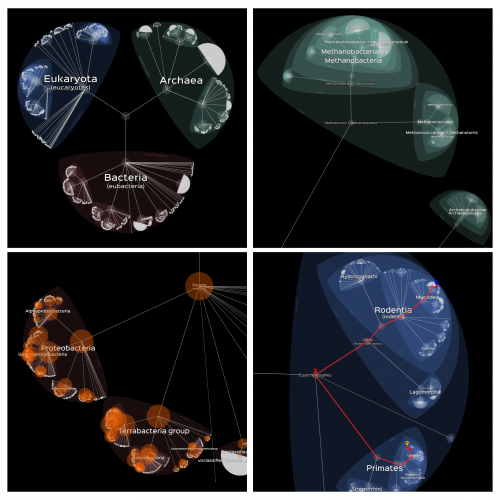
In a paper published last December in PLOS Biology (de Vienne, 2016), I described an online tool, called Lifemap, for exploring the entire Tree of Life (or any large tree) in a zoomable interface (Figure 1). This tool solves many issues that previous methods had, such as the possibility to display nodes with more than two descendants (multifurcations) and the possibility to explore smoothly very large trees of more than 2 million species.
Lifemap relies on tools that were developed for cartography, especially in the context of the OpenStreetMap project, and proposes a new way of representing hierarchical structures. The exploration of the ToL in Lifemap is similar to the exploration of a geographic map: by zooming and panning. It is also possible to click on every node and tip of the tree to have access to additional information (description, picture, link to external resources, etc.), and to search for “routes” in the tree of Life (Figure 1). Finally, an efficient search engine allows for an immediate retrieval of any node or tip by its Latin or its common name.
Figure 1. Different views of Lifemap. Top-left: the three domains of the Tree of Life; Top-right: inside Bacteria; Bottom-left: orange bubbles represent the number of genomes fully sequenced; Bottom-right: a “route” from Homo sapiens to Mus musculus.
Lifemap comes in three versions, depending on the tree that is displayed and the information that is associated to nodes and tips when clicking. A first version displays the tree proposed by the Open Tree of Life (OTOL) project, a second one displays the whole NCBI taxonomy, and a third one displays a simplified version of the taxonomy with pictures and descriptions when clicking on the nodes (this last version is also available as anApp for phones and tablets on the Android market).
In the near future, I would like the NCBI and the OTOL versions of Lifemap to be proper “hubs” giving access to a multitude of information of relevance to researchers in various fields. Because I personally work on comparative genomics, I started adding information on the number of genomes that are fully sequenced according to the NCBI database (Figure 1, bottom-left). This information can be displayed directly on the interactive tree. It is useful for easily identifying species, close to our species of interest, whose genome is available. I also added links on every node and tip to the corresponding web page on the NCBI web site and on other taxonomy web resources. These examples show how flexible Lifemap is at welcoming information from various sources.
I hope that developmental biology will be a field for which information could be displayed on Lifemap as well? I will be happy to consider such options if some interest is declared!
Applications are invited for a Research Assistant/Associate within the Division of Infection & Immunity at University College London in Dr Gillian Tomlinson’s laboratory.
We are seeking a highly motivated individual interested in integrating cutting-edge human and zebrafish models to study the immunopathogenesis of tuberculosis. The post is funded by a Medical Research Council Clinician Scientist Fellowship entitled “Tuning the immune response in tuberculosis”, and combines a human experimental tuberculosis challenge model with studies using Mycobacterium marinum infection of zebrafish to identify and validate host factors that calibrate a favourable immune response in tuberculosis.
The post-holder will be supervised by Dr Gillian Tomlinson based in the Cruciform Building at UCL. Dr Tomlinson works within Dr Mahdad Noursadeghi’s and Professor Benny Chain’s group’s which study host immune responses to infectious diseases at genome‑wide level with a particular focus on tuberculosis (www.innate2adaptive.com). The zebrafish work will be supported by the fully managed world class research aquarium at UCL.
The post is available until 1st March 2020, subject to satisfactory probationary and annual appraisals. There is an established track record for department post-doctoral staff gaining personal fellowships. Independently minded and talented investigators will be encouraged and supported in seeking such fellowship support.
Key requirements
Applicants must have an MSc (or equivalent degree) and/or a PhD (or equivalent degree) in a relevant subject.
Candidates must have experience of working with zebrafish including microinjection and in vivo imaging.
Evidence of significant scientific contribution including publications and presentations at conferences is also essential.
Established by the British Society for Developmental Biology (BSDB) in 2014, The Gurdon/The Company of Biologists Summer Studentship scheme provides financial support to allow highly motivated undergraduate students an opportunity to engage in practical research during their summer vacation. Each year, ten successful applicants spend eight weeks in the research laboratories of their choices, and the feedback we receive is outstanding.
Our final report from the 2016 class comes from Christopher Taylor, who undertook his studentship with Karim Sorefan in Sheffield.
A dual hormone response in Arabidopsis thaliana
The population problem
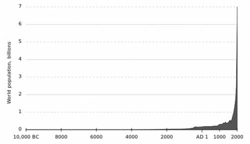
Human population growth is not a new issue. For much of mankind’s history, the species has existed in small, dispersed populations. It is only in the last 100 years where the population has truly boomed with over a fourfold increase in the number of mouths to feed (Fig 1). Demand for food is predicted to increase by 50% by 2050 and already 1 in 7 people are starving, a figure only predicted to increase.
The problems of this unsustainable growth are exacerbated by two issues:
Climate change – in certain areas agricultural productivity is severely reduced by rising temperatures and an increase in extreme events.
Geography – areas with greatest population growth coincide with areas most likely to be impacted by climate change. These include less economically developed countries such as those in the Sub Saharan belt.
Fig 1: The post-industrial ‘population spike’ highlights just how rapidly the human population has boomed in such a short space of time.
Evidently there is an urgent need for a greener revolution.
A solution?
Plants are nature’s biochemical engineers, producing everything they need to grow, survive and reproduce from practically thin air (and a little help from the soil). Plants provide the food we eat, the food our food eats and the food that supports the very ecosystems we depend on. ‘Improving nature’s inefficiencies’ by exploiting plants’ existing biochemical pathways is one way of increasing food production in the finite space we have.
The production, transportation and signalling of plant hormones (phytohormones) allows plants to orchestrate growth and development and importantly for a changing climate, facilitate a response to environmental cues. The classic example of auxin’s role in phototropism may spring to mind. Here auxin exhibits a negatively phototropic response, moving away from the prevailing light direction. This leads to an accumulation of auxin molecules in the side furthest from the light causing acidification of the cell walls and allowing for cell elongation and plant growth. Ultimately this produces a plant (or stem) whose curvature is maximised for intercepting incoming light.
Exploiting these pathways already has agricultural applications. Think of taking a plant cutting, dabbing the base in auxin and growing a whole new plant.
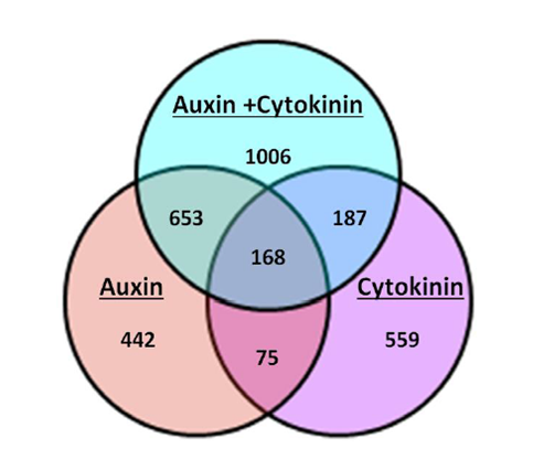
My work in the Sorefan lab in the University of Sheffield’s department of molecular biology and biotechnology (MBB), was to investigate the role of a novel hormonal response produced by auxin and cytokinin. Previous microarray data by the lab revealed an interesting occurrence when Arabidopsis thaliana seedlings were grown under conditions of elevated auxin (IAA) and cytokinin (BAP) concurrently. When grown under a dual hormone combination, the seedlings showed a significant change in the number and types of genes that were either significantly up or downregulated in the microarray. This response was larger than either responses of auxin or cytokinin alone and has been termed the ‘dual hormone response’ (Fig 2).
Fig 2: A Venn diagram illustrating the number of genes regulated by auxin alone, cytokinin alone, a combination of auxin and cytokinin and their overlaps. Nearly twice as many genes have significantly altered expression levels when auxin and cytokinin are combined compared to either hormone alone. (Image credit: James Thackery, Sorefan lab)
Functions of the dual hormone response
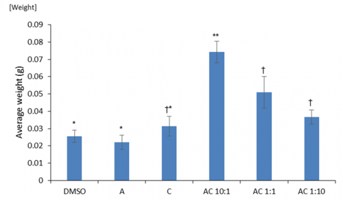
Prior to environmental stresses we tested if there were any general positive growth responses of the dual hormone response (DHR). We investigated the effects of differing ratios of auxin to cytokinin on cotyledon weight. Cotyledons are embryonic leaves that are among the first structures to emerge following germination and their weight is an accurate proxy of seedling health. We found significantly greater cotyledon mass under DHR conditions particularly with ratios showing high concentrations of auxin (Fig 3). Interestingly, this was significantly higher than the effects of just auxin alone, implying that the exogenous application of even a small amount of cytokinin may produce positive benefits.
Fig 3: The effects auxin alone (A), cytokinin alone (C) and combinations of auxin and cytokinin (AC various ratios) on cotyledon weight compared to a DMSO control. Means that share a symbol (*, ** or †) do not statistically differ from one another.
Comparing our lab’s dual hormone microarray data with that of other labs highlighted approximately 11.2% of our identified genes may show some response to heat stress conditions. I produced cDNA from seedlings grown under different combinations of hormone treatments and designed primers for the heat stress responsive genes identified by these microarrays. Using multiple quantitative real-time polymerase chain reactions (qRT-PCR), I confirmed that a number of these heat-responsive genes were also significantly affected by the dual hormone response.
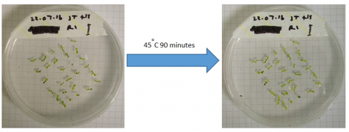
I then conducted an experiment to investigate the phenotypic effects of heat shock on seedlings grown under DHR conditions (Fig 4).
Fig 4: Heat shock experiment before (left image) and after (right image) showing bleaching of the cotyledons as the chlorophyll degrades.
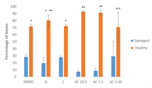
Preliminary data from this experiment suggests that the dual hormone response may convey some resistance to heat stress, as combinations of the two hormones results in significantly fewer damaged / unhealthy leaves than the control or either single hormone alone (Fig 5).
Fig 5: Percentage of leaves that were either damaged or healthy following heat stress under varying hormonal conditions.
Future work aims to quantify chlorophyll more effectively as our own analyses were confounded by the production of purple anthocyanin pigments, which are often produced as a response to stress.
These data may therefore allow us to apply dual hormone responses to agriculture. The dual hormone treated plants were larger and appear to be somewhat de-sensitized to heat stress, which may allow us to manipulate crop plants to convey some heat resistance to them. Knowing the optimal hormone ratios is important for applying this information. Manipulating plants in this way is a promising route for producing ‘climate ready’ crop species and ensuring that we can continue to feed a world of 7 or 10 billion in a vastly deteriorating climate.
Department of Experimental and Health Sciences, Universitat Pompeu Fabra, Barcelona, Spain
Reconstructing the lineage relationships and dynamic event histories of individual cells within their native context is central to understanding how the wide diversity of cell types develops during the construction of an organ. This is a long-standing challenge in biology, because up to now most efforts have been devoted to understand the genetic requirements for cell specification.
The inner ear is an attractive model to take on this challenge as it contains a manageable number of cell types, which develop rapidly on an organized schedule to generate the functional units of the mature organ – the sensory patches, containing supporting cells and hair cells innervated by the sensory neurons (Video 1).
Video 1: Innervated sensory patches in the embryonic inner ear. Animation of an embryo displaying the sensory patches with differentiated cellular neurosensory elements in green at 48hpf. Differentiated hair cells are innervated by sensory neurons of the SAG, which shows the typical segregation into anterior and posterior portion alongside with the segregated projections to the hindbrain.
During the last decades we learnt how neuronal vs. sensory specification was achieved, which signals from the surrounding tissues regionalized the otic vesicle along axes, and that the gradual restriction of cell fates over time was due to a multistep process. However, how sensory and neuronal progenitors behave throughout patterning, proliferation, and morphogenesis remained elusive, and it was difficult to reconcile some of the phenotypes observed in the signaling targeted mutants (Raft and Groves, 2015), due to the limited comprehension of how developmental gene regulatory networks are integrated. We thought that in vivo cellular data could address how patterns are achieved while the cells proliferate and the tissue undergoes morphogenesis, which may affect cell positioning and exposure to signals, and therefore cell specification. We were inspired by the pioneer work of Julien Lewis, who was able to foresee how things would work through in principle simple observations (Haddon and Lewis, 1996).
After a while trying to understand how the different progenitor pools were spatially organized within the otic vesicle using classical technics, it became clear to us that only imaging the whole developing inner ear and going from developed structures backwards in time to founder cells would help us to unveil the answer.
As you can imagine, this turned out to be a bit more complex than expected. It could only be accomplished through high resolution in vivo imaging, and considering that the otic vesicle undergoes extensive morphogenesis at the same time cells specify and proliferate it constituted a major challenge. It required simultaneously tracing the lineages while resolving the kinetics of cell proliferation and fate behavior. Recent developments in 4D-microscopy and cell tracking tools were being developed, but that was something that a small group could not afford to dream about. However, we were lucky enough to learn about the efforts of the lab of a former colleague, Nadine Peyrieras, in developing original methodologies and tools for the in vivo multiscale and multimodal observation of biological processes (Olivier et al., 2010; BioEmergences). After discussing with Nadine, we were excited because we felt we could participate in filling the void between gene regulatory networks and tissue architecture.
Our goal was to address two important questions: i) how neurons and hair cells are specified within similar domains of the otic epithelium; and ii) how from a simple otic epithelium this complex 3D-structure (the embryonic inner ear) is generated with the precise allocation of the neurosensory elements. We undertook this task without being conscious of the enormous effort and all the troubles we had to face, and combined high-resolution imaging with genetic tools using zebrafish embryos. It started our long journey in learning how to image for long periods of time using the best fluorophors, how to use new platforms for image processing, how to write in different programming languages (we became fans of coursera), and most importantly we had to change our prefigured ideas of how things work. Finally, we could record cell lineage information and observe at every time the spatial context of the cells within the whole otic vesicle! This period was very fruitful, and we realized how powerful these tools are and that we can use imaging for discovery.
Video 2:Early organization of neuroblasts within the SAG. Tg[cldnb:lynGFP]Tg[Brn3c:GFP] embryos injected with H2B-mCherry mRNA were imaged, and reconstructed cell centers were color-coded according to their location/identity (see legend). The projection view video (large panel) simultaneously displays the topological organization of cell groups selection and tissue architecture as a projection of the GFP channel (plasma membranes in grey) in x,y,z-axes. The distinct visualization modes displayed on the right hand side allow for a detailed 3D-visualization of data during the analyses. Orthogonal views are used to validate cell tracking, the oblique slice view allows orienting the orthoplane along the embryonic axes, and the rendering view permits to display validated cell centers in the context of the whole image volume.
These experiments led to understand the remodeling of the neuronal progenitor domain upon neuroblast delamination (Figure 1), and revealed that the order and place of neuroblasts’ delamination from the otic epithelium prefigure their position within the statoacoustic ganglion (SAG) (Video 3). Up to date, we knew neuroblasts were exiting the otic epithelium by a process called delamination, and they formed the SAG beneath, but we could not imagine the impact that delamination and morphogenesis had in the spatiotemporal distribution of neurosensory cell progenitor pools. By following the individual progenitors we unveiled that approximately 25% of the cells of the otic vesicle delaminate within few hours, and that the homeostasis of the system was maintained, most probably due to the proliferative capacity of the non-sensory epithelium. The developmental strategy used by distinct progenitor populations differed: neuronal specification is concomitant with proliferation (before/after delamination), while hair cell specification and differentiation lead to postmitotic cells indicating that the final number of sensory cells relies on the control of the progenitor pool.
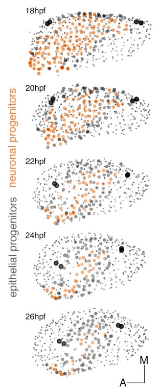
Figure 1:Dynamic map of neuronal progenitors (orange circles) and their epithelial neighboring cells (grey circles) in the context of the whole otic vesicle (grey dots) over time. Color intensity of cell centers depicts the position of cells along the dorsoventral axis of the otic vesicle. The map was built after following the lineages from 18hpf to 26hpf of all encircled cells. Note how neuroblast delamination impacts on the size and position of the progenitor domain (orange circles) over time.
Video 3: The order and place of delamination foreshadows their location within the SAG. A cohort of 144 delaminated neuroblasts was analyzed for time and place of delamination. Reconstructed cell centers were color-coded according to four delamination intervals: 18-20hpf white, 20-22hpf yellow, 22-24hpf orange, 24-30hpf red, or for position along the anteroposterior (AP) axis in the otic epithelium and followed from 18hpf to 30hpf. Note that neuroblasts exit randomly from the delamination domain; those delaminating earlier are located more medially in the SAG than the later delaminating ones prefiguring the gradient of differentiation (middle panel). The relative position of neuroblasts along the AP is maintained from the otic epithelium to the SAG (right hand side panel). Reconstructed cell centers were displayed as colored dots together with the corresponding volume rendering images (plasma membranes in grey).
Moreover, we provide the dynamic map of neurosensory progenitors based on in vivo cell lineage studies (Video 4) supplying a global and temporal perspective to previous otic neurosensory lineages analyses, which were mainly focused on the spatial dynamics of gene expression (Durruthy-Durruthy et al., 2014).
Video 4:Maps of neuroblasts and posterior macula hair cell progenitors in the whole otic vesicle. The neuronal progenitors (orange) and the posterior macula hair cell progenitors (blue) are plotted in the context of the whole otic vesicle (grey dots) at 24hpf. Tether cells are shown as black circles. The animation displays a rotation of otic vesicles around the anteroposterior axis. Note that the two progenitor domains are adjacent, and neuroblasts are located more ventrally while sensory progenitors are more medially.
We are very enthusiastic with this paper because the information about cellular/population dynamics and lineage relationships of neurosensory elements in the inner ear enables us to understand the proportions of the system and provide the cellular data to complement the well-described gene regulatory networks involved in neurosensory specification. We are convinced that for the understanding of how the developmental gene regulatory networks function during tissue degeneration and regeneration, as well as in normal patterning, we need to move forward into 4D-imaging and put previous knowledge in the context of the dynamics of the system.
References
Durruthy-Durruthy, R., Gottlieb, A., Hartman, B. H., Waldhaus, J., Laske, R. D., Altman, R. and Heller, S. (2014). Reconstruction of the mouse otocyst and early neuroblast lineage at single-cell resolution. Cell 157, 964–978.
Dyballa, S., Savy, T., Germann, P., Mikula, K., Remesikova, M., Špir, R., Zecca, A., Peyriéras, N. and Pujades, C. (2017). Distribution of neurosensory progenitor pools during inner ear morphogenesis unveiled by cell lineage reconstruction. Elife 6: e22268.
Haddon, C. and Lewis, J. (1996). Early ear development in the embryo of the zebrafish, Danio rerio. J. Comp. Neurol. 365, 113–128.
Olivier, N., Luengo-Oroz, M. A., Duloquin, L., Faure, E., Savy, T., Veilleux, I., Solinas, X., Debarre, D., Bourgine, P., Santos, A., et al. (2010). Cell Lineage Reconstruction of Early Zebrafish Embryos Using Label-Free Nonlinear Microscopy. Science 329, 967–971.
Raft, S. and Groves, A. K. (2015). Segregating neural and mechanosensory fates in the developing ear: patterning, signaling, and transcriptional control. Cell Tissue Res 359, 315–332.
Established by the British Society for Developmental Biology (BSDB) in 2014, The Gurdon/The Company of Biologists Summer Studentship scheme provides financial support to allow highly motivated undergraduate students an opportunity to engage in practical research during their summer vacation. Each year, ten successful applicants spend eight weeks in the research laboratories of their choices, and the feedback we receive is outstanding.
Our third report from the 2016 class comes from Iona Imrie, who undertook her studentship with Jamie Davies in Edinburgh.
Development of the vascular system in the mouse mesonephros
During the summer of 2016, I was fortunate enough to be awarded a BSDB Gurdon Studentship. The funding enabled me to undertake a research project in the lab of Jamie Davies at the University of Edinburgh. Under the supervision of a PhD student in the lab, David Munro, I studied the vascularization of a primitive and transient murine kidney- the mesonephros.
During mammalian embryogenesis, 3 paired renal organs develop sequentially in a cranio-caudal direction. The most primitive kidney is the pronephros, followed by the mesonephros and then the permanent kidney, the metanephros. Both the pronephros and mesonephros are temporary, with the pronephros being nonfunctional and the mesonephros being functional in some species. The mouse mesonephros comprises 18-26 pairs of tubules. The most cranial tubules connect to the nephric duct, while the caudal tubules do not. The mesonephros first appears at embryonic day 9 (E9), before regressing completely in females. In males, the cranial tubules do not degenerate, instead forming the epididymis. Given that, (a) the fate of the mesonephros is sexually dimorphic and (b) the mesonephric tubules are not uniformly connected to the nephric duct, there could be differences in vascular development that run in parallel with these differences in morphology. During my project, I observed vascular development in order to identify where blood vessels came from and if there were differences in vascular development between sexes and cranial/caudal tubules of the mesonephros.
My project began with dissecting embryos at E11.5. The mesonephroi obtained by dissection were stained with antibodies against CD31, Laminin and Pan-cytokeratin. These antibodies stained endothelia, basement membrane and nephric duct/tubules respectively.
I set out to create a timeline of mesonephric development starting at E10.5 through to E15.5 (Figures 1-4). Dissecting at E10.5 proved difficult due to the small size of the embryo. The gonad develops adjacent to the mesonephros, and I left itattached to the mesonephros in order to observe interactions of vasculature between the two.
Having dissected and stained mesonephroi from various ages, I made the following observations:
E10.5– Aorta branches into mesonephros (Figure 1).
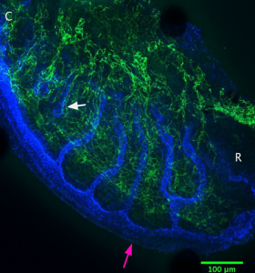
Figure 1: Green=CD31, Red=Laminin. The aorta is shown centrally, between the two mesonephroi. Branches of the aorta going toward the mesonephros can be seen (white arrow). C indicates the caudal end of the mesonephros, and R is the rostral end.
E11.5– Tubules elongate and the mesonephros vascular plexus (mvp) begins to form (Figure 2).
Figure 2: Blue= Pan Cytokeratin, Green= CD31. Compared to E10.5, the tubules look longer. The pink arrow shows the nephric duct, and the white arrow shows the first tubule that is disconnected from the nephric duct. There is a collection of vasculature at the distal end of the tubules where the aorta would be. There are branches into the mesonephros from here.
E13.5– In females, branching of mvp into mesonephros and gonad occurs. In males, branching into the mesonephros occurs but branching into the gonad is not as visible as in females.
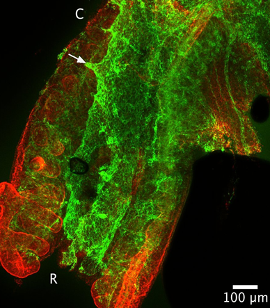
E14.5– Formation of the coelomic vessel on male gonad. Continued branching of mvp into mesonephros/gonad in females (Figure 3).
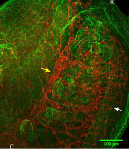
Figure 3: Immunostaining of an E14.5 female mesonephros (right) with gonad attached (left). Green= Laminin, Red= CD31. The white arrow points to the nephric duct and the yellow arrow points to the mvp. Blood vessels seem to branch from the mvp into the mesonephros and into the gonad
E15.5– Branching of coelomic vessel into gonad, and joining of the gonadal artery to this vessel (Figure 4).
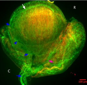
Figure 4: Green=CD31, Red=Laminin. It seems that there is more CD31 staining in this E15.5 male than at earlier ages. The coelomic vessel (white arrow) which first appeared at E14.5 now joins to the gonadal artery (blue arrows). This vessel branches into the mesonephros and bifurcates (pink arrow).
Overall, the vasculature of the mesonephros seems to branch from the mvp and form a network of vessels around the nephric duct. The vessels around the tubules do not look particularly organized. However, confocal slices at 40x objective show vessels wrapping around the tubules. I did not observe glomeruli in the tubules at any age- these structures are rarely found in mouse mesonephroi.
As an adjunct to the main project of vascularization, I tested the functionality of the mesonephric tubules using an assay developed by one of Prof. Davies post doctoral researchers, Dr. Melanie Lawrence. The assay tested the functionality of the tubules to uptake fluorescent anions and cations. The ability of cells in these tubules to transport organic anions and cations would suggest the tubules are functioning as a primitive kidney. It has been assumed that the murine mesonephros has this role, but this has never been proven. Melanie had been using her assay to answer this question, using mesonephroi from different gestational ages. The unpublished data that Melanie has collected shows that these tubules do have the ability to transport organic anions and cations. I was able to help with assaying at some of the gestational time points.
I cultured mesonephroi using the Sebinger culture method, and then assayed the uptake of 6-carboxyfluorescin (anion) through organic anion transporters (OAT) in the basolateral membrane of tubule cells, and uptake of the DAPI (cation) through organic cation transporters (OCT), also in the basolateral membrane. As a control, inhibitors of OAT (Probenecid) and OCT (cimetidine and metformin) were added to mesonephric cultures to show that any fluorescence seen in the tubules was due to uptake via these transporters and not by another process.
The Gurdon Studentship has been an invaluable opportunity. I cannot stress enough how important it is for medical sciences students like myself to spend time in a research lab. As a second year, I didn’t really know what being a scientist would be like. From spending time in the Davies Lab, I have been able to understand and practice techniques that were briefly introduced in lectures, while developing a scientific mind, improving my time management skills and working alongside scientists who are extremely dedicated to their research. I would especially like to thank Jamie Davies for giving me this opportunity, and David Munro and Chris Mills for their guidance throughout my time in the lab.
A position (#121670) is available immediately for a Postdoc Associate to contribute to our studies in neural crest and placodes. The Postdoc Associate will conduct independent research and assist in the training of students in the laboratory of Dr. Lisa Taneyhill at the University of Maryland. Laboratory skills should include the ability to perform various molecular biology and biochemical assays, such as recombinant DNA/cloning; DNA, RNA, and/or protein blotting; immunohistochemistry; and/or in situ hybridization. Experience with microscopy and spectroscopy, chick embryology (including microdissections and electroporation), and tissue culture is desirable. For more information on the lab, please see http://www.ansc.umd.edu/people/lisa-taneyhill. Qualifications: An advanced degree (Ph.D.) in Developmental, Molecular and/or Cell Biology is required. Fluency in spoken and written English is required. Compensation: Salaries are highly competitive, negotiable and commensurate with qualifications. Fringe benefits offered. Applicants must apply through eTerp at https://ejobs.umd.edu. Applications will be accepted until a suitable candidate is identified.
 (1 votes)

 (2 votes)
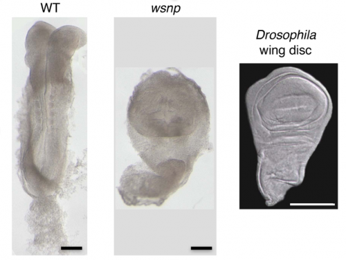
 (6 votes)


 The 6th EMBO conference on the Molecular and Cellular Basis of Regeneration and Tissue Repair took place in Paestum (Italy) in September, 2016. As summarised in the
  The regulative capability of single cells to give rise to all primary embryonic lineages is termed pluripotency. Two phases of pluripotency, called naïve and primed, have previously been described. In his
  The tooth root is an integral, functionally important part of our dentition, and understanding how roots develop and how they can be bioengineered is of great interest in the field of regenerative medicine. In their