The community site for and by
developmental and stem cell biologists

Labome releases Validated Antibody Database (VAD) version 2.2

Posted by , on 22 March 2016

Logo_72dpi_851x315

Developmental biologists use antibodies extensively to study the gene expression during different stages.  However, there is a lack of specific antibodies against many proteins related to development.  In addition, some antibodies yield unspecific and/or irreproducible results.   To help alleviate this antibody quality and specificity problem, Labome sought to organize antibody applications cited in formal publication since 2008 and developed Validated Antibody Database  (VAD).  The most recent version, 2.2,  contains manually curated 143357 entries from 38430 formal articles, covering 35146 antibody products from 110 suppliers.   The suppliers include both commercial entities and non-profit organizations such as Developmental Studies Hybridoma Bank (DSHB) at University of Iowa, and Neuromab from University of California at Davis.   A small number of antibodies from academic researchers are included as well, if these antibodies are validated in knockout models.

One of the side benefits of our curation effort is the identification of cross-reactive species for many antibodies.  Antibodies tend to be developed for human/mouse proteins and tend to be tested by commercial suppliers for their applicability in human or mouse system.   Development models often use more readily manipulatable models such as flies, worms, zebrafish, and frogs.  Labome is able to obtain information about many antibodies having cross-reactivities with the model organisms from the literature.

The database is freely browsable at www.labome.com.    Information about antibody validation using knockout models is also posted at Labome Facebook page www.facebook.com/LabomeNews.

Feedback and suggestions are most welcome.  We hope to work with everybody to develop a useful tool.


Thumbs up (1 votes)
Loading...

Tags: , ,
Categories: Resources

Navigate the archive

Use our Advanced Search tool to search and filter posts by date, category, tags and authors.

Postdoctoral Scientist (m/f)

Posted by , on 22 March 2016

Closing Date: 15 March 2021

Max-Planck-Institut für molekulare Biomedizin


The Max-Planck-Institute for Molecular Biomedicine in Muenster, Germany has an opening for a

Postdoctoral Scientist
(position-code 07-2016)

The position is available in the DFG Emmy Noether junior group of Dr. Ivan Bedzhov that is focused on understanding the self-organization of the pluripotent lineage in mammalian embryos at the time of implantation (I. Bedzhov and M. Zernicka-Goetz, Cell, 2014). The successful candidate will investigate the mechanisms of self-organization of the pluripotent epiblast in the context of the developing embryo and in vitro using embryonic stem cell as a model system.

We are looking for a talented and highly motivated post-doctoral scientist with strong cell and molecular biology background. Previous experience with RNA-seq analysis, genome editing and embryonic stem cells will be an advantage.

The Max Planck Institute for Molecular Biomedicine offers dynamic, multidisciplinary environment with state-of-the-art transgenic, imaging, genomics and proteomics equipment and core facilities. The working language in the institute is English. The institute is located in Muenster, a vibrant city with a highly international academic environment.

The position is initially available for two years with the possibility of extension. Starting date will be as soon as possible. All conditions for the employment will be according to the regulations of the contracts for the civil service (TVöD, Tarifvertrag für den öffentlichen Dienst) level 13 TVöD.

The Max-Planck society is committed to increasing the number of individuals with disabilities in its workforce and therefore encourages applications from such qualified individuals. Furthermore, the Max Planck Society seeks to increase the number of women in those areas where they are underrepresented and therefore explicitly encourages women to apply.

Please send your application (with the position-code 07-2016), letter of motivation, CV including a complete list of publications and the contact information of 2 referees to:
career@mpi-muenster.mpg.de
or
Max Planck Institute for Molecular Biomedicine
Roentgenstrasse 20
48149 Muenster
Germany

Thumbs up (No Ratings Yet)
Loading...

Categories: Jobs

Question of the month- brain organoids

Posted by , on 21 March 2016

Last week, Development announced a special issue on organoids. In vitro organogenesis is a burgeoning new field, with applications in the study of human development, drug testing and ultimately the possibility of producing functional organs in the dish that could be used for transplantation. Every new technological advance brings with it a new set of ethical issues, and this is particularly true with regards to brain organoids. Whilst there have been enormous advances in this area, brain organoids are still far from being functional ‘mini brains’. However, it is not impossible that in the near future we may be able to generate a brain organoid that has a sensory area that is able to functionally connect with a processing centre. At what point could we say that these organoids can ‘think’? And does it matter? This month we are asking:


What are the ethical issues surrounding the generation of brain organoids?


Share your thoughts by leaving a comment below! You can comment anonymously if you prefer. We are also collating answers on social media via this Storify. And if you have any ideas for future questions please drop us an email!


Below is an interview with brain organoid researcher Juergen Knoblich, which may be of interest to you:

Thumbs up (1 votes)
Loading...

Tags: ,
Categories: Discussion

Developing the auxin-inducible degradation (AID) system for versatile conditional protein depletion in C. elegans

Posted by , on 19 March 2016

By Liangyu Zhang and Abby F. Dernburg 


The nematode Caenorhabidis elegans is among the most widely used and powerful model organisms for studying mechanisms underlying cellular and developmental processes. Although a variety of approaches for conditional protein expression have been developed in C. elegans, available tools for conditional protein depletion are far more limited, particularly in the germ line. We were thus motivated to develop a technique to control the abundance of proteins in living animals, which we felt would be a great addition to the toolbox available in this system.

In the Dernburg lab, we are interested in studying the molecular mechanisms underlying meiosis. In many cases we can use genetic mutations to interrogate the roles of individual proteins, but this approach does not work well if the proteins perform essential functions during mitosis, which is required for proliferation of the germ line. We therefore sought to develop a method that would enable inducible, rapid, and quantitative protein depletion in the germ line. After considering a number of possible strategies, we focused on the auxin-inducible degradation (AID) system, which has been applied in cultured cells and single-celled organisms (Nishimura et al., 2009). This approach was originally adapted from the plant auxin perception system. Inducible degradation relies on a small molecule phytohormone produced by all plants, auxin (indole-3-acetic acid). In addition, it requires TIR1, an F-box protein, which forms part of an Skp1–Cullin–F-box (SCF) E3 ubiquitin ligase complex. In the presence of auxin, TIR1 recognizes peptide sequences (degrons) that are present in a large number of target proteins, mostly transcriptional regulators, expressed by plants, and targets these proteins for polyubiquitylation and proteasome-mediated degradation. We thought this system might be transplantable to nematodes, since auxin is a very small, fairly water-soluble molecule, potentially making it easy to deliver to living animals and readily diffusible through tissues.

A rotation student, Ze Cheng, first accepted the challenge of adapting the AID system to C. elegans. He generated a number of constructs to test its feasibility. Because there is no endogenous auxin receptor in C. elegans, he made a strain stably expressing a modified Arabidopsis thaliana TIR1 from a single copy transgene using MosSCI; (Frokjaer-Jensen et al., 2008). He also made a construct to express a degron- and GFP-tagged SMU-2 from extrachromosomal arrays in the germ line. After treating worms expressing both constructs with auxin for a couple of hours, he was excited to observe a disappearance of the green fluorescent SMU-2::GFP signal, indicating the AID system might indeed be useful in C. elegans.

We then systematically expanded and tested the AID system in C. elegans. To maximize the chances of success, we incorporated two amino acid changes in AtTIR1 that were found to increase its binding affinity for degron-tagged substrates and to thereby increase its sensitivity to auxin (Yu et al., 2013). We created strains expressing TIR1 under control of various promoters and 3’ UTR sequences to drive germ line-specific or temporally regulated expression. We used a 44-amino acid minimal degron sequence derived from Arabidopsis thaliana IAA17 (Morawska and Ulrich, 2013) and tagged proteins of interest with this sequence using CRISPR/Cas9-mediated editing (Dickinson et al., 2013). Gratifyingly, we detected efficient inducible-degradation in the germ line when growing the transgenic worms with auxin-containing liquid culture or plates. Excitingly, rapid degradation was consistently achieved within a reasonable range of auxin concentrations (<1 mM), which did not seem to have any effects on wild-type worms. We also found that the position of the degron was quite flexible; it could be placed at either end of a target protein, or even sandwiched between the target and another tag, such as GFP.

Considering the potential usefulness of the AID system for the research community, we further analyzed the inducible degradation of target proteins in the soma of C. elegans in detail. To do this, we made a variety of tissue-specific TIR1 strains with tissue-specific promoters and 3’ UTR sequences. We then combined these TIR1 transgenes with different types of degron-tagged transgenic targets. We found either cytoplasmic or nuclear proteins tagged with the degron can be tissue specifically degraded at various developmental stages in an auxin concentration dependent manner. After trying a wide range of auxin, we also noticed that the degradation is reversible upon auxin removal, with lower auxin doses accelerating recovery. To our surprise, we further detected efficient inducible degradation of targets in early embryos inside the mother and in laid eggs, suggesting promising usefulness of the AID system for studying mechanisms underlying embryo development. Notably, the inducible degradation is also efficient in the absence of food, making it useful for studying starvation-induced processes, such as autophagy and larvae arrest.

When we shared these findings with some colleagues, they were extremely excited about the potential of the system. One of them, Jordan Ward at UCSF, wanted to test the ability of this system to address key questions underlying nuclear hormone receptor-mediated control of developmental gene regulatory networks. He was able to tag two essential nuclear hormone receptors, NHR-23 and NHR-25, with the degron sequence and found that each could be depleted within 40 min, enabling detailed functional dissection of these proteins during development.

Another exciting finding was that the AID system can produce more penetrant phenotypes than depletion by RNAi, not only in the worm soma but also in the germ line. Given the high efficiency of CRISPR/Cas9-mediated genome editing in C. elegans, tagging proteins of interest with the 44-amino acid degron is now quite easy and fast, further augmenting the utility of the AID system for cell and developmental studies in worms.

We have made the AID-related worm strains and plasmids available through the CGC and Addgene, respectively. A large number of worm laboratories have already tried the AID system to deplete proteins of interest in C. elegans. We are getting great feedback from our colleagues, several of whom have told us that the AID system works spectacularly in their hands. In principle, this approach may be applicable to a wide range of other organisms. We thus look forward to seeing the application of this technology in not only worm labs but also other metazoan model organism labs.


Full article at: http://dev.biologists.org/content/142/24/4374.long


References:

Dickinson, D. J., Ward, J. D., Reiner, D. J. and Goldstein, B. (2013). Engineering the Caenorhabditis elegans genome using Cas9-triggered homologous recombination. Nat. Methods 10, 1028-1034.

Frokjaer-Jensen, C., Davis, M. W., Hopkins, C. E., Newman, B. J., Thummel, J. M., Olesen, S. P., Grunnet, M. and Jorgensen, E. M. (2008). Single-copy insertion of transgenes in Caenorhabditis elegans. Nat. Genet. 40, 1375-1383.

Morawska, M. and Ulrich, H. D. (2013). An expanded tool kit for the auxin-inducible degron system in budding yeast. Yeast 30, 341-351.

Nishimura, K., Fukagawa, T., Takisawa, H., Kakimoto, T. and Kanemaki, M. (2009). An auxin-based degron system for the rapid depletion of proteins in nonplant cells. Nat. Methods 6, 917-922.

Yu, H., Moss, B. L., Jang, S. S., Prigge, M., Klavins, E., Nemhauser, J. L. and Estelle, M. (2013). Mutations in the TIR1 auxin receptor that increase affinity for auxin/indole-3-acetic acid proteins result in auxin hypersensitivity. Plant Physiol. 162, 295-303.

Thumbs up (3 votes)
Loading...

Tags: ,
Categories: Research, Resources

From our sister journals- March 2016

Posted by , on 18 March 2016

Here is some developmental biology related content from other journals published by The Company of Biologists.


CoB_DisModMech_AW_RGB


Drosophila as a model to study human disease

Drosophila DMM
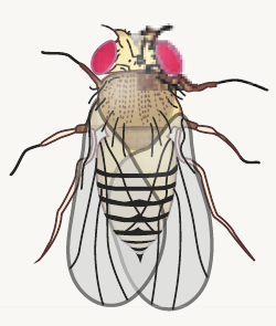
The latest issue of Disease Models & Mechanisms highlights the translational impact of Drosophila research. In this issue, Moulton and Letsou review several Drosophila models of human inborn errors of development (read here), while the poster and review by Bellen and colleagues examine some of the tools and assays available in Drosophila  to study human disease (read here).


Journal typography


Nanog suppresses senescence

JCS Nanog
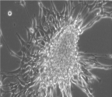
Thummer, Edenhofer and colleagues analysed the outcomes of Nanog gain-of-function in various cell models employing a recently developed cell-permeant version of this protein. They show that  Nanog blocks cellular senescence of fibroblasts through transcriptional regulation of cell cycle inhibitor p27KIP1. Read the paper here [OPEN ACCESS].


skeletalJCS
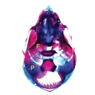
Skeletal development

Panx3 and Cx43 are two important gap junction proteins expressed in osteoblasts. Yamada and colleagues show that Panx3 and Cx43 regulate skeletal formation through their distinct expression patterns and functions. Read the paper here.


Asymmetric cell division in the worm

Phillips and colleagues show that two Dishevelled paralogs have both redundant and non-redundant roles in β-catenin regulation during asymmetric cell division in C. elegans. Read the paper here.


oligodendrocyteJCS
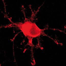
MOBP in oligodendrocyte differentiation

Myelin-associated oligodendrocytic basic protein (MOBP) resembles myelin basic protein (MBP), but the signals initiating its synthesis and function remain elusive. In this paper, White and colleagues show, by several approaches in cultured primary oligodendrocytes, that MOBP synthesis is stimulated by Fyn activity, and reveal a new function for MOBP in oligodendroglial morphological differentiation. Read the paper here.


Journal typography


JEBDaphnia
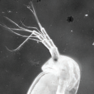
How bacteria can affect development
Mushegian and colleagues examined the effect of temperature and presence of bacteria in diapausing eggs of the water flea Daphnia magna. They show that the presence of bacteria increases successful development of resting eggs at an elevated temperature. Read the paper here.


Water deprivation affects snake development

Dupoué and colleagues examined the effects of water availability on corticosterone secretion in breeding snakes. They show that water deprivation induces an increase in baseline corticosterone level in pregnant aspic vipers, which may subsequently influence offspring growth. Read the paper here.


CoB_BioOpen_AW


Controlling tissue polarity 

BiO panal polarity
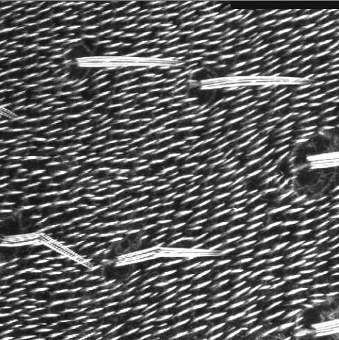
Planar cell polarity sigalling directs the polarization of cells within the plane of many epithelia. Sharp and Axelrod examine the signals that Prickle and Spiny-legs respond to and the mechanisms they use to control the direction of tissue polarity in the distal wing and the posterior abdomen of Drosophila. Read the paper here [OPEN ACCESS].


neural crest BiO
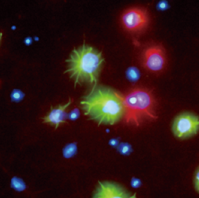
From fibroblasts to neural crest 

Motohashi and colleagues identified the transcription factors specifically expressed in developing mouse neural crest cells, and showed that SOX10 and SOX9 directly converted fibroblasts into neural crest cells. Read the paper here [OPEN ACCESS].


Thumbs up (No Ratings Yet)
Loading...

Tags: , , , , , , ,
Categories: Research

We have PhD position available to start in October 2016 at the University Göttingen

Posted by , on 17 March 2016

Closing Date: 15 March 2021

We have PhD position available to start in October 2016 at the University Göttingen (3 yrs, 75% E 13 TV-L).


The main aim of the project will be to study intra-specific variation in compound eye size in Drosophila melanogaster. The successful PhD candidate will address this question by analyzing the genetic basis of eye size variation in various inbred strains of the Drosophila melanogaster Genetic Reference Panel (DGRP). For all DGRP lines genome sequences are available. For representative strains, developmental transcriptome data using RNAseq will be generated so that data across several scales (genome, transcriptome, phenotype) can be integrated. Similar data will be generated for artificial selection experiments based on a subset of the DGRP fly lines. The successful candidate will work in an interdisciplinary team at the Department of Animal Sciences (Prof. Dr. Henner Simianer) and the Department of Developmental Biology (Dr. Nico Posnien) as well as during an extended research stay in the group of one of the international collaborators overseas.


Please visit the website of the Research Training Group “Scaling Problems in Statistics” (http://www.uni-goettingen.de/en/156579.html) for more details about the general setting of this position.


A detailed description of the project, information about the application procedure and further requirements are available here: http://tinyurl.com/gn3az25


Please forward this job ad to all your motivated future PhD students.


http://www.evolution.uni-goettingen.de/posnienlab/index.html

https://www.uni-goettingen.de/de/92842.html

Thumbs up (No Ratings Yet)
Loading...

Categories: Jobs

A Day in the Life of a Bat Lab

Posted by , on 15 March 2016

I am Aaron Harnsberger, a second year Master’s degree student in the Department of Biological Sciences at Idaho State University in Pocatello, Idaho.  The focus of this lab is on genetic regulatory divergence that results in the diversity of mammalian morphologies.  These morphological differences can be observed at various stages of development.  In this lab we use bats as our model organism.  There are no other members working in this lab besides myself.  There are a growing number of labs throughout the world that also use bats to study evolutionary and developmental biology.  The species of bat that I use is Carollia perspicillata (C. perspicillata) or simply Carollia (Fig. 1).  On average an adult Carollia bat is about 19 grams (Fig. 2).  Although it is possible to maintain Carollia as a laboratory bat (Rasweiler et al., 2009), we do not have a bat colony in the lab so before I can conduct experiments I make trips to the field to collect animals from the wild.


Figure 1: Carollia perspicillata held by John Rasweiler.


At this point, you might be asking, “Why use bats?”.  Bats have some very interesting adaptations that make them a great model for studying evolutionary development.  They are the only mammal to achieve powered flight.  The development of wings where other mammals have paws, fins, hooves or hands is one adaptation that I find fascinating.  Another very interesting adaptation discovered in bats is delayed development.  This is when a pregnant female delays development of the embryo.  In other mammalian species this typically occurs at the blastocyst stage before implantation in the uterus.  However, in Carollia delayed development occurs after implantation at the gastrulation stages (Rasweiler et al., 1997).  This delay may be highly synchronous in colonies, and cues that initiate the delay and end it are still unknown.  Bats can have a long life span when compared to other mammals of similar size.  In addition, bats may respond differently to injury and the healing process may lack inflammation.  There are more features, but as you can see bats are unique models for many biological questions.


Figure 2: C. perspicillata held by Aaron Harnsberger. Carollia is a phyllostomid (leaf-nosed) bat.


Back to the field, which in this case, is the country of Trinidad and Tobago (Fig. 3).  These field collection trips to the island of Trinidad require careful planning and weeks of time to catch bats and collect embryos.  I do not catch the bats on these trips by myself.  The ‘Bat Team’ this year consisted of John Rasweiler, Richard Behringer, Simeon (Patsy) Williams, Joseph Truechen and myself.  Patsy and Joseph are local field assistants who help us find and collect the bats.  The ‘Bat Team’ has consisted of many other biologists that have made this collection trip throughout the years, starting with the initial trip in 2000.


Figure 3: Map of Trinidad taken from Google Maps. We collect Carollia from the northern and central regions of Trinidad.


First and foremost, a collection permit through the Wildlife Section of the Division of Forestry of Trinidad and Tobago is required.  We give them a specific number of bats we would like to collect and plan our trip according to this number.  Next we need a place to process our samples.  This is the Department of Life Sciences at the University of West Indies (UWI) in St. Augustine that has lab space, microscopes and other facilities.  We also need lodging since the trips take several days.  This is arranged through University Housing.  Finally, we rent a car to take us into the deep forest that has enough room for several passengers and our gear.  Once all of this is in order we can begin catching bats.


Figure 4: Joseph, John and Patsy bringing gear up the hillside to catch bats.


To catch bats, we have to travel into the northern or central areas of the island.  We look for certain habitats that can lead us to specific roosting sites.  To achieve this, we conduct scouting trips and spend many hours driving, parking, looking around, taking notes, and if all goes well catching bats.


Figure 5: Field box used to store live bats for safe transport to the lab.


We use lots of different gear to catch bats.  We occasionally need ladders to get into some of the places that bats roost, for example abandoned concrete water tanks (Fig. 6).  On the occasions that we do need a ladder we have to carry it in, sometimes through thick forest (Fig. 4).  We use nets similar to butterfly nets to catch the bats, but much longer to reach high places where they roost (Fig. 6).  After catching the bats, we sort through them, releasing males and checking females for age and stage of pregnancy using external visual appearances.  We keep these female bats in a field box which allows for safe transport of the bats (Fig. 5).  After a morning of collecting, we return to the lab at UWI with the bats to isolate the embryos.  Once embryos are dissected we process them for various types of analyses, including morphological, histological and molecular studies.  To bring the bat embryos back to the lab in Idaho, we obtain an exportation permit, also from the Wildlife Section of the Division of Forestry of Trinidad and Tobago.


Figure 6: C. perspicillata roosting inside an old abandoned concrete water tank. These types of water tanks have a convenient opening on the top that we enter using a ladder. The bats are seen here cohabitating with cockroaches, just visible in the lower edge of picture.


Now that I have the bat embryos to work with I can spend time in the lab back in Idaho conducting experiments.  I use whole-mount in situ hybridization (WISH) to examine the spatial distribution of transcripts for specific genes at different stages of development.  Using the Carollia embryo staging techniques (Cretekos, et al. 2005), I can identify embryos at specific stages of development.  This gives me specific time points to look for gene expression at critical developmental stages (Fig. 7).  Many times these expression patterns are similar yet different from those in the mouse.


Figure 7: Fgf8 expression in Carollia stage 14 embryo after WISH.


I have also used micro computed tomography (uCT) to visualize the tissue anatomy of bat embryos (Fig. 8).  A uCT scan is a high resolution x-ray that is used to make a three-dimensional image.  The differences in contrast allow me to identify different tissue types in the bat embryos.  Again, taking advantage of the Carollia staging system, I have uCT scanned stages of development in C. perspicillata to examine particular tissues of interest for my project.  The data collection was completed in a few weeks’ time.  The image processing and analysis of this data has been an ongoing project in the lab.


Figure 8: Carollia stage 20 uCT image.


As you can see a day in the life of a bat lab is not usually just a day, and some of the work may not be typical in other developmental biology labs (Fig 9).


Figure 9: The 2016 ‘Bat Team’. From left; Richard Behringer, John Rasweiler, Simeon (Patsy) Williams & Aaron Harnsberger.


References


Cretekos, C. J., Weatherbee, S. D., Chen, C., Badwaik, N. K., Niswander, L., Behringer, R. R. & Rasweiler, J. J. IV. (2005). Embryonic staging system for the short-tailed fruit bat, Carollia perspicillata, a model organism for the mammalian order Chiroptera, based upon timed pregnancies in captive-bred animals. Developmental Dynamics, 233: 721-738.


Rasweiler, J. J. IV & Badwaik, N. K. (1997). Delayed development in the short-tailed fruit bat, Carollia perspicillataJournal of Reproductive Fertility, 109(1): 7-20.


Rasweiler, J. J. IV, Cretekos, C. J. & Behringer R. R. (2009). The short-tailed fruit bat Carollia perspicillata: a model for studies in reproduction and development. Cold Spring Harbor Protocols, 2009(3): pdb.emo118.


Tags: Bat, Carollia, WISH, in situ, uCT, microCT, development, ISU, Idaho State University


Node day in the life new doodle squareThis post is part of a series on a day in the life of developmental biology labs working on different model organisms. You can read the introduction to the series here and read other posts in this series here.


Thumbs up (9 votes)
Loading...

Tags: ,
Categories: Lab Life

MSc/PhD Student Position “Using Fruit Flies to Characterize Mechanisms of Nerve Cell Degeneration or Proliferation” University of Alberta, Edmonton, Alberta, Canada

Posted by , on 15 March 2016

Closing Date: 15 March 2021

One MSc or PhD student position is available for a September 2016 start date to study mechanisms determining the degeneration or the proliferation of neurons using the D. melanogaster fruit fly model. The two complementary projects will use common assays to investigate the following questions: i. How does calcium and redox signaling between the Endoplasmic Reticulum (ER) and mitochondria determine neuronal degeneration and inflammation. ii. How does the neurofibromatosis type 2 gene product Merlin determine neuronal proliferation. The project is part of a collaboration between the Simmen and Hughes laboratories that aims to further use the fruit fly model organism for the characterization of the fundamental cell biology behind neurodegeneration and childhood tumors of the nervous system. A publication list and more information about the labs can be found on these two sites

http://www.cellbiology.ualberta.ca/FacultyMembers/ThomasSimmen.aspx

http://www.cellbiology.ualberta.ca/FacultyMembers/SarahHughes.aspx

The location of the research project is at the University of Alberta, the 5th largest University in Canada with world-class biomedical research labs. The University of Alberta Cell Biology graduate program is amongst the best Cell Biology programs worldwide. Edmonton is a culturally vibrant, young city that lies in close proximity to the Rocky Mountains, offering excellent opportunities for sports enthusiasts.

Minimal requirements are competitive grades from an internationally acclaimed University. Applicants need to provide a cover letter that outlines their career objectives and why they would like to enter a PhD program, CV and scanned university grades. Outstanding applicants from Europe are encouraged to apply, but must have excellent English knowledge both spoken and written (high TOEFL/GRE scores or equivalent). Successful applicant will have to enter the University of Alberta Cell Biology graduate program. Detailed instructions for prospective applicants can be found here

http://www.cellbiology.ualberta.ca/~/media/cellbio/Documents/GraduateManualOct2015.pdf

Address further inquiries to Thomas.Simmen@ualberta.ca or Sarah.Hughes@ualberta.ca

Thumbs up (No Ratings Yet)
Loading...

Categories: Jobs

Special Issue on Organoids

Posted by , on 15 March 2016

In vitro organogenesis has exploded onto the stem cell and developmental biology scene. It is now possible to make  miniaturised approximations of many different organs – known as organoids – entirely in vitro, using either pluripotent stem cells or adult tissue stem cells as starting material. Coaxed towards their fate by various signalling molecules and growth factors, these self-organising populations faithfully recapitulate many of the developmental milestones associated with their in vivo counterparts, and can be used to model both developmental and disease processes.


To highlight the terrific progress that is being made in this field, and to draw attention to the enormous potential that organoids hold for understanding developmental and regenerative processes, Development is proud to announce a Special Issue on Organoids.


MelissaProf. Melissa Little – who recently published a spectacular report on growing kidney organoids – will be Guest Editor of the Special Issue, which is scheduled for publication in early 2017. For more information about the Special Issue, including scope, article types and deadlines, click here. You can read an Editorial from Melissa and discover why she’s excited about the emerging organoid field here, or read Catarina Vicente’s “An interview with Melissa Little” here.


Many people share a great enthusiasm for organoid research, especially for how it can be used to study human development. In the video below, Development’s Executive Editor Dr Katherine Brown chats with Dr. Juergen Knoblich, whose report of cerebral organoids (or “minibrains” as they’re known) was heralded as one the major breakthroughs of 2013. In the interview, Juergen talks about why the world was so captivated by his research, whether the minibrains are truly recapitulating development and what the future challenges are for the organoid field.


Thumbs up (No Ratings Yet)
Loading...

Tags: ,
Categories: News

An interview with Melissa Little

Posted by , on 15 March 2016

This interview first appeared in Development.


Melissa Little is a Senior Principal Research Fellow at the Murdoch Childrens Research Institute in Melbourne, Australia. Her lab has studied kidney development and regeneration for over 20 years, recently making notable advances in the generation of kidney organoids from human iPSCs. We chatted with Melissa about her career, her thoughts on the potential of the organoid and stem cell fields, and what she hopes to achieve during her guest editorship with Development.


MelissaHow did you first become interested in biology?

As a child I was fascinated by the world around me. I spent a lot of time camping in the Australian outback and found the plants, animals and insects just fascinating.


You started your research career as a cancer biologist. How did you eventually move on to studying kidney development?

At university I enrolled in science, assuming I was going to study botany or zoology. But I loved my first-year course in physiology, so my primary degree is actually in this area. Molecular biology was just beginning during my PhD, so I worked on the molecular basis of childhood cancer. I worked on Wilms’ tumour (a childhood kidney cancer), before the WT1 gene was isolated, and later worked on WT1 during my postdoc with Nicholas Hastie in Edinburgh. The paradigm at that time was the two-hit hypothesis, i.e. that to get a cancer you needed a hit in both copies of a tumour suppressor gene. This hypothesis was based on retinoblastoma. Whereas the RB1 gene is expressed everywhere, WT1, by contrast, is very confined in its expression during development, being restricted to the urogenital system. So, while mutations in this gene give rise to kidney cancer, they can also give rise to urogenital developmental anomalies. When I returned to Australia I continued to work on WT1. However, I was now based in a research institute with strong developmental biology, such as the work of Peter Koopman and Toshiya Yamada, so I changed direction to study WT1 and other genes in kidney development.


How important were the years that you spent as a postdoc in Edinburgh?

They had an enormous impact. I was a Royal Society Fellow, a relatively new scheme at that time. I had offers in the USA, but I had visited Nicholas in Edinburgh and really wanted to go to the MRC. The unfortunate rule of that fellowship was that after two years I had to return to Australia. But those years in the MRC were incredibly important to me. They shaped how I work as a scientist, how I interact with other scientists and how I approach answering questions in science. Nicholas was an extremely positive mentor and it was great to interact with people such as Wendy Bickmore, Ian Jackson and Veronica Van Heyningen. It was a very formative period of my career.


What are the challenges of establishing your lab in a relatively isolated country like Australia?

One of the surprising things about being in the UK was that amazing scientists wandered through the building on a regular basis. That didn’t happen in Australia. At that time there was no internet. The latest copy of Nature arrived by sea. By the time it appeared in the library it was already three months out of date. The isolation was immense. It was very difficult to keep pace with what was happening in science and to be at the forefront of anything because we were so far behind in our capacity to know what else was happening. I found that acutely oppressive when I came back to Australia in 1992.

The internet completely changed that. It made international collaboration feasible. We have real-time access to journals, we can search for articles (and there is far more published now than anyone could ever consume), we can electronically communicate in real time, and so on. Science has become much more feasible at an international level. Indeed, I have collaborations all around the globe. However, Australia is still a very long way to anywhere. To actually meet someone and talk face to face, which is quite important, you must travel. And I travel extensively. I don’t think people in the USA, for example, understand how taxing that is. I remember doing a talk in Italy where I was on the ground for less time than I was in the air. That is not that unusual, but it is pretty physically brutal. So, Australians travel a lot because they have to.


You initially established your lab in Brisbane but recently moved to Melbourne. What were the reasons for this change?

I was at the University of Queensland for 23 years, and that is a very long time to be in one place. I was in a really excellent institute, but my research evolved to have a regeneration and stem cell aspect to it, and I was relatively alone there in that respect. We were on a large academic campus but quite remote from any hospital. I did my PhD at the Queensland Medical Research Institute, which was located at a hospital, and my thesis supervisor was the head of oncology and haematology, so I had been in an environment quite closely associated with patients. I wanted to move back to an environment that had, first of all, more stem cell biology, and second, access to nephrologists. At the Murdoch Childrens Research Institute I am physically located in a children’s hospital. I now have very close associations with clinical geneticists and nephrologists, and we are setting up a clinic where we derive patient stem cell lines. That has been a really good part of the move.


What scientific questions is your lab working on at the moment?

We are the kidney development, disease, repair and regeneration lab, because we cover quite a wide range of kidney medicine. However, everything we do is underpinned by our understanding of kidney development. That is paramount. My most important message is that stem cell biology on any organ requires you to understand that organ intimately, so understanding development is key. We then use that information to direct stem cells towards a kidney fate in order to understand the relationship between development and disease. We also look at what the postnatal kidney can or can’t do to repair itself. We increasingly do human pluripotent stem cell work, but we still investigate really fundamental developmental biology questions – for example, how cells move, how they communicate with each other, how they self-organise during development, and what genes they express at what time.


Last year your lab published a high-profile paper in Nature, reporting the generation of kidney organoids from human iPSCs. Had this always been a goal of your lab?

It was a very specific and deliberate objective. Around 15 years ago, when stem cell biology was really starting out (around the time that Jamie Thomson derived the first human pluripotent stem cell lines and Perry Bartlett showed evidence that there were postnatal stem cells in the brain) I decided to change our research focus towards regenerative medicine. We started with every option on the table. We didn’t know what the postnatal kidney could do (we now know that it can repair quite well but can’t regenerate), whether there were postnatal stem cells or whether pluripotent stem cells could be differentiated to a kidney fate. In fact, cellular reprogramming was not even discussed at that time. There were a lot of things we wanted to try for kidney regenerative medicine and the differentiation of pluripotent stem cells was one of those. We were very systematic and it took quite a long time to get there.


Your ‘mini-kidneys’ paper was extensively covered in the mainstream media. How was your experience interacting with the media?

It would be lovely to control the media but no one can. Sometimes I cringe when I read what journalists write. They make broad generalisations that might lead a patient to think that a cure is around the corner, which is not true. This is the nature of the media. Some of the interviews I did were, I think, poorly represented, whereas some of them were great. You just roll with it. I don’t worry too much about those events that are less than perfect, because I can’t control them.


The organoid field is a new and exciting area. How much potential do you see in these techniques? Do you think it will be possible to build a full organ in a dish, as the media claims?

I think the media overestimates how far we have come. Organoids are fascinating and a really exciting area, but we are a very long way away from the clinic. ‘Organs in a dish’ is a funny expression, but I actually believe that although we have a long way to go, we will genuinely get there. I think there are some very short-term outcomes from this type of research that fall into the remit of both fundamental and translational biology. This is the first time that we can really start to pull apart human development. The developing human itself is not something we have had any access to, so we do have a circular problem: how can you know that what you are growing from a human cell is actually like a human? From what we can see so far, however, it is a pretty remarkable model. Hence, it really is a door into human development and that is very exciting. From a more practical point of view, I am very interested in what can be achieved with disease modelling and drug screening, even personalised drug screening. However, we need to be vigilant about how we develop these tools. I am already seeing publications claiming that organoids in a dish are accurately modelling disease but there are such challenges with interclonal and experimental variation that I think this still has to be definitively proven.

In the long term what we have is an approach where cells organise themselves based on embryological principles, and this is amazing from a fundamental science point of view. It will actually give us a handle on how cells self-organise. We make a lot of assumptions about how self-organisation works: cells differentially stick to each other or make growth factors that make other cells wander towards them. Now we have models where we can really pull that apart. From a translational point of view, this sort of information will then become an engineering challenge. How do we build these structures with a vasculature? How do we ensure a degree of anatomical correctness that will be helpful for patients? For example, the kidney organoids are currently of no value to a patient that requires a transplant as they have no exiting ureter to remove the urine. The kidney is, I would argue, the hardest thing you could ever try to generate in vitro. It is architecturally completely constrained and its function is totally dependent upon its anatomy. What we have at the moment is too small and indeed dysplastic, so there is a lot that we have to do better. But I think it is achievable with time.


You recently started your guest editorship with Development. What do you hope to achieve in this year with us?

I am really keen to encourage more of the development field to embrace what stem cells can give us, despite some reticence so far. There are enormous opportunities here to look at development in a different way. I want to open the door on investigating human development using directed differentiation, especially using these types of organogenic models (Little, 2016).


How do you see the relationship between the developmental biology and stem cell fields evolving?

I think the nexus between developmental biology, cell biology and stem cell biology is very exciting. There have been amazing advances in imaging in the developmental biology field, particularly by those working on what we would call ‘simple’ organisms, such as the worm or the fly. They have phenomenal expertise in temporal-spatial imaging, right down to the cellular level. Meanwhile, the cell biologists are building tools to look at mechano-biology and real-time reporting of pathway activity. To layer these advances on organoid creation from a pluripotent cell in a dish, which is where stem cell biology is going, is an enormously powerful approach.


What kind of papers would you like to see more of in Development?

I would like to see more papers looking at the fundamental processes governing how cells organise themselves, whether that’s during normal organogenesis in vivo or in a model in vitro. In the past, molecular biology superseded fundamental, anatomical developmental biology. Looking at aspects such as self-organisation was put to one side because it was considered too descriptive. I think it would be valuable to bring these fields back together and ask ‘what is the molecular basis of self-organisation during embryogenesis?’.


You were a member of the Australian Government’s Strategic Review of Health and Medical Research in 2013. Do you think it is important for scientists to play a role in policy?

Yes. I have had the opportunity to play a role in national scientific policy throughout my career. I was not only involved in this review but also in a seminal review of health and medical research in Australia in 1998, when I was still a young scientist. I have played roles within the Research Committee of the National Health Medical Research Council, have advised the federal government on science policy and around the debate on embryonic stem cells, and the state government on biotechnology policy. I simply see this as part of my professional obligations as a scientist. Too many young scientists forget that science is a less tangible product, not like making bricks or building boats. We are still primarily funded by tax payers, in Australia almost exclusively. The tax payers expect health outcomes, particularly in health and medical research. If we do not engage, not just in policy discussions but also in public communication, we only have ourselves to blame if the public loses interest in funding science, or worse, loses faith in scientists. One thing that I learnt very early is that there is a tendency for scientists to think that we just need to educate everyone and then they’ll understand why science is important. It is not about education. People can make decisions without facts and often do. They don’t need to be educated, they need to be engaged with. That is a very different process. It means you need to be in the room, to be having the conversation and discussing what you are doing and why.


What is your advice for young scientists?

Follow your heart. You have to be fundamentally passionate about finding an answer to a question. Have lateral vision and take every opportunity that comes your way. Don’t assume that what you read is right and question everything.


What would people be surprised to find out about you?

First and foremost, I’m a mum with two kids. In high school I was the top student in my year at art and English, not science. Perhaps this is why developmental biology is attractive to me. It is so beautiful. I actually won a prize in creative writing as a teenager. Someone recently asked me whether I still do any creative writing. I write grants and manuscripts. That is a creative process, even if it is describing data. I still paint for leisure, although not often enough.


Reference:

Little, M. H. (2016). Closing the circle: from organoids back to development. Development 143, 905-906

Thumbs up (1 votes)
Loading...

Tags: , ,
Categories: Interview