The community site for and by
developmental and stem cell biologists

Forgotten classics- Regulating the size of the mouse embryo

Posted by , on 6 April 2016


Snow, M. H. L., Tam, P. P. L. (1979) Is compensatory growth a complicating factor in mouse teratology? Nature 279, 555-557

Lewis, N. E., Rossant, J. (1982) Mechanism of size regulation in mouse embryo aggregates J. Embryol. exp. Morph 72, 169-181

Recommended by James Briscoe (Francis Crick Institute)


As our previous forgotten classic demonstrated, much can be learnt from careful observation of embryonic development. Once there is a good basis of what ‘normal’ is though, the next step is to perturb the system. These days we are used to precise genetic manipulations: removing a single gene (or even a single protein domain), affecting only a specific cell type or at a specific stage in development. However, there are certain questions that benefit from more global alterations, such as making the embryo very small or very big. The two papers recommended by James Briscoe use such approaches to address the important question of how the mouse embryo regulates its size.

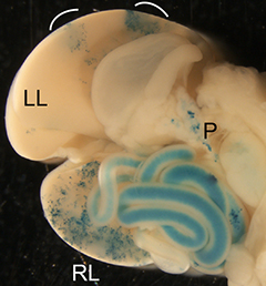
The first study, published in Nature in 1979 by Snow and Tam, did not set out to examine size regulation. Mitomycin C was a known teratogen, i.e. a chemical known to cause embryonic malformations, but its effects on early mouse development were unknown. The authors found that the effects of this compound were indeed very quick: by E7 the compound had caused extensive cell death and the embryos were very small. Yet, while this treatment reduced embryonic viability and the resulting pups showed various defects, they were not noticeably smaller. As the authors stated ‘the mouse embryo can withstand a major disturbance in its early development and recover to such an extent that structural abnormalities rarely emerge’. In short, ‘the mouse embryo can be reduced to around 10% of its normal size at a time when it is about to begin organogenesis, but is nearly normal again before that phase of development is complete.’ What an extraordinary ability to regulate size, and a good example that in science you may find an interesting answer for a question that you weren’t even asking!


teratology embryos

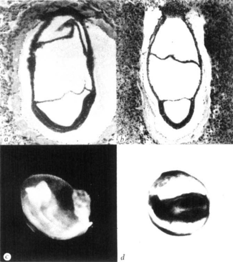
Reprinted by permission from Macmillan Publishers Ltd: Nature (279, 555-557), copyright (1979)


While Snow and Tam inadvertently discovered what happens when a mouse embryo becomes too small, Lewis and Rossant very purposefully tried the opposite in their paper in JEEM (now Development). By 1982, several groups had already shown that if you increase the size of a mouse embryo, e.g. by aggregating several embryos together, the end product is still a viable offspring of normal size. But at which stage of development does this regulation occur, and by what mechanism? To answer this question, the authors compared normal- and double-sized mouse embryos. They showed that while the embryos start off with different sizes, by E6.5-E7 (around the same time as observed by Snow and Tam) they have the same dimensions. They also suggested a mechanism by which this regulation of size may be happening. At E5, normal embryos undergo a period of high mitotic activity while double-sized embryos slow down their cell cycles, giving the normal embryos a chance to ‘catch up’.

As James Briscoe tells us ‘both papers demonstrate the surprising ability of early mouse embryo to regulate its size. Tam and Snow show that ~80% of the cells in an E7 embryo can be killed yet the embryo still recovers (albeit with reduced viability). Conversely Lewis and Rossant show that double sized embryos are twice the size up until ~E6 when they regulate their size back towards normal. Both papers suggest that size regulation happens at around the time of gastrulation, indeed they hint that gastrulation might depend on passing a size checkpoint.’ Despite their important findings, these papers have been cited relatively little, maybe because, as James says ‘the mechanism of size regulation continues to be a mystery today, although it is receiving increasing attention. It is a fascinating example of the self-organising ability of embryogenesis.’


From the authors:

The Lewis and Rossant paper is one of my personal favourites and I still quote it, even if it is often ignored by the field! In fact it is a still unsolved question of how the embryo manages to size regulate so well in such a relatively short period of time. I have been trying to interest new post-docs in the lab with returning to the problem and investigating whether Hippo or other growth control pathways might be involved and/or whether there is a metabolic link to mTor pathways. But so far noone has taken up the challenge!

Janet Rossant, The Hospital for Sick Children (Canada)


The study on the effect of inhibition of DNA synthesis by mitomycin C on embryogenesis was founded on an observation made by Michael Snow: that to account for the increase in cell number during mouse gastrulation, the whole epiblast cell population has to undergo a doubling in about every 6-7 hours and, in particular, a small sub-population of cells would have to proliferate at a much faster rate with a cell cycle time of about 3 hours.  It was not known previously that cell cycling could be that fast in mouse embryos. We hypothesized that this highly proliferative population may be hit more severely by blocking DNA synthesis, and the subsequent deficiency of any cell types in the embryo might reveal the developmental fate of the descendants of this population. As it turned out, the unexpected outcome of the experiment pointed instead to a mechanism for size regulation by compensatory growth. We followed up this study years later where under-sized mouse gastrula were created, not by cell killing, but by removing one to two blastomeres from the 4-cell stage pre-implantation embryo (Power and Tam, 1993). Results of this study further highlighted the attribute of size (or cell number) up-regulation in the control of post-implantation development. The under-sized embryo does not initiate gastrulation until it has attained a threshold cell number and this is accomplished by extending the phase of rapid cell proliferation beyond the normal duration of 24 hours into gastrulation. Recently, there has been heightened interest in the mechanism of size regulation and the control of the timing of morphogenetic events during development and the connection of this process to the mechanosensory activity and the intercellular communication among cells in a community.

Reference: Power, M. A., Tam P. P. L. (1993) Onset of gastrulation, morphogenesis and somitogenesis in mouse embryos displaying compensatory growth. Anatomy and Embryology 187,493-504

Patrick Tam, University of Sydney (Australia)


Further thoughts from the field

These are two really fantastic papers that are indeed often overlooked, and well worth revisiting. Both studies use classic mouse embryology (and teratology – now that’s a word we don’t see too often these days) methods to describe the remarkable regulative capacity of the mammalian embryo. In the intervening (almost 4!?) decades, we’ve come full circle. Armed with sophisticated genetics tools, an increasing ability to accurately measure (and perturb) the behavior of individual cells within a population, and spurned by studies yielding information on (possibly conserved) mechanisms regulating tissue size and scaling in other systems, for example Drosophila, there’s been an upsurge of interest in this phenomenon in the mouse. Interestingly, two recent studies from Miguel Torres’ lab at the CNIC Institute in Madrid, and Tristan Rodriguez’s lab at Imperial College in London, revisited the question in a contemporary setting and pin-pointed the same window of time (approximating to the onset of gastrulation) as noted in the earlier Tam and Rossant studies, when everything seems to have happened, and any outlier embryos have either managed to catch-up or slow-down (Claveria et al., Nature 2013; Sancho et al., Dev Cell 2013). Moving forward, it’ll be interesting to see how our understanding of this phenomenon deepens, especially bearing in mind that achieving the exquisitely invariant size of mammalian embryos (and organs) likely involves the sensing, integration, and reaction to, several inputs.

Kat Hadjantonakis, Sloan Kettering Institute (USA)


Nature has kindly provided free access to the Snow and Tam paper until  June 2016; the Lewis and Rossant paper is freely available.


—————————————–
by Cat Vicente


Forgotten classics doodle squareThis post is part of a series on forgotten classics of developmental biology. You can read the introduction to the series here and read other posts in this series here.


Thumbs up (5 votes)
Loading...

Tags: , , ,
Categories: Education, Research

Navigate the archive

Use our Advanced Search tool to search and filter posts by date, category, tags and authors.

An interview with Abigail Tucker

Posted by , on 5 April 2016

This interview first featured in Development.


Abigail Tucker is a professor at King’s College London, UK and her lab works on various aspects of craniofacial development – from basic, evolutionary and clinical biology perspectives. This year, Abigail will be awarded the first Cheryll Tickle Medal by the British Society for Developmental Biology (BSDB). We chatted with Abigail about her research, her commitment to public engagement and the challenges and rewards of working with emerging model systems.


Abigail TuckerThis year you will be awarded the first Cheryll Tickle Medal by the BSDB. What does it mean to you to receive this prize?

It really is fantastic. Throughout a scientist’s career there aren’t a lot of moments when people come up to you and say ‘that was really excellent science’. There are many awards that you can win when your career is beginning, like PhD or poster prizes and then at the end of your career there are lifetime achievement awards. So it is very nice to have a prize in the middle of my career. To have that recognition, to know that people appreciate the kind of research that I do, is really a great feeling.


This medal is awarded to female scientists. Do you think there is the need for awards that specifically recognise the contributions of women in science?

It would be great if there wasn’t the need for this award, and I think things are changing, but women still face problems that men don’t, particularly with regards to having children and taking that time out. I think we want to encourage women not to worry and to know that taking time out is not going to disadvantage them, but I do hope that at some point the BSDB will find that there isn’t the need for prizes that specifically recognise women. I recently read the Chair’s message in the BSDB newsletter, about how this medal is slightly controversial. It will be interesting to hear other people’s opinions when I give my talk, and find out whether there is support for this initiative.


How did you first become interested in developmental biology?

I did my undergraduate degree in biology at the University of Oxford, and one of the course topics that I studied was developmental biology. I absolutely loved it. It was an extremely interesting subject and it helped that it was taught by really enthusiastic people. I was quite lucky that at that time at Oxford there were lots of wonderful developmental biologists working on all kinds of model organisms. Because of that I decided to stay and do a PhD in developmental biology.


You studied tail bud determination in Xenopus during your PhD, but you now work on craniofacial development. How did your research interests change during your career?

After my PhD I wanted to try a different animal model, specifically the mouse because I was quite interested in genetics. I visited several labs looking for mouse projects and Paul Sharpe had a tooth project that was not at all what I was thinking of. At the time I was more interested in general patterning of the embryo, rather than specific organs. It sounded like a really interesting project though and it turned out extremely well. This project also moved me towards craniofacial development. This is a really interesting area to work on, because there are so many different organs crammed into your head. A kidney is just a kidney, but a head includes the sensory organs, the brain, the skull vault, the jaw, and all the teeth and glands inside it. It is extremely complex and I like that a lot.


What are the scientific questions that interest you at the moment?

We have previously done some work looking at the lineage of different structures in the head and the germ layers that they come from. Now, we are examining whether the origin of a tissue matters in a disease situation. When you tell someone that a specific structure is derived from endoderm or neural crest, their reaction is, ‘and why should I care?’ Actually, it makes a difference. The cells respond differently depending on their origin.

The lab is divided in two halves. There are people who are interested in more evolutionary aspects, asking, ‘why does something develop like this?’ The other side of the lab, mostly clinicians, wants to know what happens when development goes astray. In areas such as the ear or the jaw, we have been able to combine these two aspects together. We now know that things go wrong because of the way the structure evolved in the first place.


Your lab is physically located in a hospital. Does this increase the pressure to introduce a clinical angle into your work?

There is definitely pressure to have a more clinical side to our work, and from a funding point of view it is much easier to get funding for the clinical questions than the evolutionary ones. However, one of the big positives of working in a hospital is that you can actually talk to the clinicians who are working with the patients. You can ask them what the big questions are that they would like to have answered, and that are important to patients. If you are going to be working on a scientific problem you want to make sure that you are asking the right questions, and that your answers will have an impact. We are trying to encourage these interactions between scientists and clinicians by giving honorary contracts to clinicians so that they are members of the department. About half of my graduate students are clinicians who have decided to do a PhD, and they bring that approach to our research.


Your lab works with a wide range of organisms, from more established systems like mouse and chick to curious creatures like snakes, geckos, chameleons and opossums. Why do you use such a variety of systems and what are the advantages and disadvantages?

It is great working with non-model organisms, but everything is a lot slower. When you want to look at the expression of a gene in the snake you have to clone it first, so you always have to add an extra couple of months to any experiment. I hope that other scientists appreciate that experiments are not as quick as they are in other systems. We have had a corn snake colony here at the hospital for over 10 years. They are common pets – so easy to get hold of and nice and friendly. However, they only breed twice a year. So when a reviewer says, ‘could you just repeat that experiment?’, I reply, ‘yes, but we will have to wait until next year when we have eggs again’. That is not ideal. In fact, I have started writing the details of the breeding seasons in our methods sections, so that reviewers appreciate that it is quite difficult to do certain experiments immediately.

There are some questions that you need to go outside the model organisms to answer. The mouse has a really strange derived pattern of tooth development. It only has molars and incisors and it doesn’t replace its teeth. Its incisors are also continuously growing, which is interesting in its own right, but very different from us. If you want to know what is happening in patients missing their second set of teeth, then mouse is not really the model to use. We have to move to something like the mini-pig, that has a full complement of teeth, and we have also recently looked at fruit bats and opossum. There are questions that have not been addressed in the past because scientists were focusing on model organisms. Once you move out into non-model systems you find that the number of unanswered questions suddenly increases dramatically, and that there is enough work to keep you busy for many years.


How do you choose the organisms that you work on? Do you try to find a system that will help you answer your question or does the availability of the organism play a role?

A bit of both. When we wanted to compare marsupial and placental mammal ear development we were very lucky to find the opossum. There are only two or three opossum colonies in the world, and one of them is based in Mill Hill, here in London. That is fantastic. More recently, we wanted a reptile system and we ended up choosing the Madagascar ground gecko. This is not a normal pet gecko that you can easily get hold of, but its eggs have hard shells, which means that you can window an egg as you do in the chick. Being able to do this kind of experiment was worth going through the effort of getting that particular lizard. We actually had a colony here for a while, but because they have hard shells the females very easily get calcium depleted, especially if you are trying to breed them a lot to have access to eggs. After some time, we set up a collaboration with a group in Prague. They have around 2000 Madagascar ground geckos, so it is much easier to collect the eggs from them, rather than trying to raise the animals here.


You are involved in a variety of outreach projects, including science evenings and festivals, and even collaborated with Channel 4 and the BBC. What do you think is the value of such initiatives and would you encourage other scientists to do the same?

I really like talking to the public about my research and I’ve had great feedback. People are really interested when they understand what you are trying to do. It is exciting to share your findings and all the amazing things you can do in science with a different audience that will not read your papers. My lab and I try to participate in three or four activities every year and when I ask for volunteers I am always inundated with replies. I think this is great because it means there is a will on both the side of the scientists as well as the public to come to these events.

Some scientists worry that outreach activities take time away from research, but funding bodies are requesting these initiatives more and more and you can get involved at different levels. I was recently at a local school’s careers day where I talked about working as a scientist. This is a very easy thing to do, whereas if you go to a big festival you have to design a stand, with props and hands-on activities. I really enjoyed our most recent stand about cell death in developmental biology – the idea that cells actually die to shape different organs. There are several good examples throughout the body and it is a concept that people are surprised about – it isn’t something that they thought could happen. We also have an activity about making bioteeth. We discuss what you can learn from animals that replace their teeth all the time, what cells and signals are needed and about the possibility of creating bioteeth from stem cells. This is a lot of fun.


In 2011 you were awarded the King’s College Supervisory Excellence Award. Is mentoring an important part of what you do?

It is. I think it is really important that PhD students have a project that they really like, using techniques that they can do. They should come away from their PhD not only with knowledge of their particular area but also other skills that they have learnt along the way – a whole package. Then they can say, ‘I have a PhD but I can really go out into the world and do many different things’. It is really important to mentor someone through that process.

My PhD supervisor, Jonathan Slack, was supportive but generally had a hands-off approach, but there were lots of people in the lab, like Betsy Pownall and Harv Isaacs, who I could chat to and ask silly questions. I think I am a bit more hands-on. I meet my students every week to discuss what’s been happening and I make sure that everybody knows how to do the techniques. They are shown once or twice what to do and then they carry on at their own pace, without someone looking over their shoulder the whole time. I try to be very encouraging when things are not working out. In science, lots of things can go wrong, so from a PhD project point of view, it is key to have different avenues of ongoing research. Then if one thing doesn’t work, you have another way of looking at the problem. I have also found that PhD students have different interests and you need to tailor a project to the particular student while they progress through their PhD. Flexibility is quite important in mentoring.


What is your advice for young scientists?

I think that old papers are not read as much as they probably should be. When I was doing my PhD, I spent quite a lot of time translating 100-year-old papers from German into English (very badly, I’ve got to say!). It is amazing how many things have already been done and it is really important to know what has been done so that you can start asking new questions. At a recent meeting, someone presented work that made me think ‘I have read this before, but in a paper from the 1970s’. Part of the problem is that the older papers are really long. Normally you quote the paper, or a review that referred to it, but when you read the original you have all the details and you know all the experiments that were carried out. I think it is really important to have as good knowledge of the subject as possible.


What would people be surprised to find out about you?

I was once bitten by the head of a decapitated snake!

Thumbs up (2 votes)
Loading...

Tags: , , ,
Categories: Interview

An interview with Cheryll Tickle

Posted by , on 5 April 2016

This interview first featured in Development.


Cheryll Tickle is an Emeritus Professor at the University of Bath, UK. She dedicated her long research career mainly to the study of limb development in the chick, and has received numerous awards for her contributions to science, including being elected a Fellow of The Royal Society and receiving a CBE from the Queen. This year the British Society for Developmental Biology (BSDB) has created a new award in her honour, the Cheryll Tickle Medal, to be awarded to a mid-career, female scientist for outstanding achievements in the field. We asked Cheryll what this award means to her and how science has changed during her career.


Cheryll TickleHow did you first become interested in developmental biology and why did you decide to study limb development?

As an undergraduate I was most interested in cell biology. I was excited by the idea of understanding how cells behave and how they build tissues in embryos. At the time, developmental biology was usually called embryology, and it was the only subject that seemed totally mysterious and unfathomable: there were no general principles that I could get hold of. So I did a PhD in cell biology in the lab of Adam Curtis, who had just become Professor at Glasgow in the first Department of Cell Biology in the UK. I studied a phenomenon known as ‘cell sorting’, which was very exciting in the 1960s. Malcolm Steinberg, building on earlier work in amphibians, had shown that if you disaggregated cells from the various tissues of the embryonic chick and mixed them together, they would sort themselves out so that cells of the same type preferentially adhered to each other. It was not clear whether a similar process happened in embryos, but it was one possible way by which you could build patterns of differentiated cells.

One of the cell types I used during my PhD in Glasgow was limb bud cells. In the limb you could imagine how cells might get committed at random to become cartilage cells and then ‘sort out’ to the centre of the limb. At that time Donald Ede, who was based in Edinburgh, used to come over to Glasgow with his PhD student Oliver Flint to use Adam’s Couette viscometer to measure cell adhesiveness. Donald was looking at limb bud development in normal chick embryos and in a mutant called talpid3, which was polydactylous and had various defects. His research further reinforced my idea to study limb development and ignited an interest in the mutant. After Donald retired, Dave Burt at the Roslin Institute took on the maintenance of the talpid3 mutant flock. We continued to study it and eventually identified the gene responsible for the phenotype and showed that it is a ciliopathy. Very recently, several groups have identified mutations in the talpid3 gene in patients with Joubert syndrome.

After my PhD I was awarded a NATO fellowship to do a postdoc in America, and I worked with John Trinkaus in Yale for 2 years, on aggregation of cell lines and cell behaviour in early fish embryo development. However, when I went for my fellowship interview I was asked what I would eventually like to work on. When I said ‘limb development’, the person who interviewed me said that he wouldn’t recommend that because everything was already known!


Did any mentors play an important role in your career?

There were three important mentors in my career. I was very fortunate with my PhD supervisor Adam Curtis, my first postdoctoral supervisor John Trinkaus and then of course with Lewis Wolpert, with whom I did a second postdoc funded by the MRC. They all supported me. I was recently asked why it was that a group of successful women embryologists came through from the 1960s and 1970s. I think in my case it was partially the attitude of the men I worked with. They allowed me the freedom to develop and did not put me down.

Another person that was very influential was Bruce Alberts, who I got to work with when he did a sabbatical in the Wolpert lab. He came armed with all these pots of beads and we started using the beads as carriers for applying chemicals to the limb bud. This technique is widely used now, but it was really being developed at the time. We used it to apply retinoic acid to the wing bud. Brigid Hogan was also very supportive during my early career when she was in the UK. Even though I wasn’t in the mouse field, she was supportive, encouraging me to apply for grants and so on.


How do you think that science has changed over the years in which you have been a researcher?

I think people tend to forget that in the 1960s and 1970s there was a lot of emphasis on cell biology, within the confines of what was possible. Quite a lot of the work I did was looking at cells in the limb, their morphology, whether they had gap junctions or not, etc. There were also quite a lot of productive interactions between people in different fields, particularly with mathematicians. During my PhD I collaborated with a statistician, Rob Elton, and we carried out a nearest neighbour analysis to quantitate the degree of sorting out in the aggregates that I had made. Computer modelling was also being used. Donald Ede had made some computer models of limb development, and I believe the computer that he used took up an entire room! These approaches bringing cell biology and/or mathematical modelling to bear on problems in embryonic development are now very much at the forefront of the field. Since my early work on limb development there have been two revolutions: being able to understand the genetic basis of development and the ability to do genome-wide analysis.

When I was carrying out my PhD, there were not so many people doing science, and the funding was easier to obtain. There wasn’t the level of competitiveness that you can see now, although it was very competitive in some areas! We seemed to have a lot more time to think and to ‘undertake scholarship’, such as studying primary papers in detail rather than just reading a review.


What do you think are the exciting scientific questions at the moment?

I think real progress is being made in the area of evo-devo, particularly in this new age of genomics. We are on the brink of learning a lot. There will be a huge amount of data, and for a while it will be very confusing. A little like the Hox gene clusters when they were first discovered. Eventually though it will all become clear. I think in general that developmental biology has not yet reaped the full rewards of the genomic age.


You are now an Emeritus Professor at Bath University. Do you miss doing science?

I really enjoyed doing science. When you discover something there is a real buzz. After a seminar in Switzerland, a member of the audience said that he had enjoyed hearing all about my tedious experiments! But of course I didn’t find them tedious. Doing experiments on chick embryos, cutting bits out, grafting them, observing them develop was always fascinating. So yes, I miss it a lot. No one gives you advice on how to finish your research career! But I am still very busy writing reviews, sitting on committees, and so on.


Over the years you have received many awards, including being elected a fellow of The Royal Society and being the first awardee of the Waddington Medal. What do these awards mean to you?

Receiving these awards is a bit overwhelming, but of course very nice. I remember that when I got the letter about the Waddington Medal I couldn’t believe it! Being a fellow of The Royal Society has been very positive. The Society is very supportive and you have the opportunity to meet people working on different areas of science, which is very interesting. Later on I also became a Royal Society Professor, so the Society directly supported my research, for which of course I am extremely grateful.

I think I received these awards because I was in the right place at the right time. I was lucky in my choice of research field and to work so closely with Lewis Wolpert for many years. The other important thing was the large number of collaborations I established with outstanding scientists in the field. I could combine my expertise with that of people like Denis Duboule and Gail Martin, working with Juan-Carlos Izpisua-Belmonte and Lee Niswander who were doing postdocs in their respective labs at that time. At UCL, I also worked with Anne Warner and Jon Clarke. By pooling our expertise, we were able to do a lot more than I would have been able to do on my own. And of course I had fantastic undergraduates, PhD students and postdocs. I feel that these awards were not just for me but for the whole enterprise.


This year the BSDB created a new award in your honour, the Cheryll Tickle Medal, which will be awarded to Abigail Tucker from King’s College London. What does it mean for you to have a medal named after you?

It is very humbling and a huge honour. I am actually going to present the award to Abigail. I was not involved in the selection of the winner, but I think Abigail is a very worthy recipient. I am very familiar with her work, in particular on how teeth are positioned within the jaw, which very much mirrors some of the work I did on the limb. It is research of fundamental importance, not just for our understanding of tooth development but also more generally.


This medal is awarded to a mid-career, female scientist for her outstanding achievements in the field of developmental biology. Do you think that there is the need for awards that specifically recognise the contributions of women in science?

The Chair of the BSDB, Ottoline Leyser, wrote in a recent society newsletter that all young scientists need encouragement and support, both men and women. However, because there is such a huge attrition in the number of women who go on to embark upon a scientific career, we need to celebrate the success of women and what they achieve. I used to feel that you could sum up the situation of women in science by saying that they are generally undervalued, overlooked and ignored. Fortunately things are changing – although it has been very slow.

There are many types of barriers for women in research. Having children is obviously a big barrier, but not being encouraged, especially if you are in a total male environment, can be a bit demoralising. While I was supported by my mentor, during my PhD in Glasgow I was the only female PhD student in the department. But when I went to Yale there were lots of women students around, and that made a quite a difference. I have tried to encourage other women, but maybe I have not been highly visible. I think that if every woman did something on a personal level we could easily conquer a lot of these difficulties. But of course we need to support our male colleagues as well, particularly at the beginning of their careers.


You said before (in an article in Nature) that current PhD students are not allowed as much freedom or to make as many mistakes as you did during your graduate training. Why do you think this is?

I think this is related to supervisors being too hands-on. I personally think that students should have more chances to make mistakes. In order to learn how to skate you need to fall over, and it is the same with science. You need to make some gaffes and learn from them. However, it is so competitive to get grants that there is increasing pressure to get results and not waste money. There is also more emphasis on training in narrow areas, rather than encouraging creativity. You are constrained to be in a particular way and receive particular kinds of training. It is all a bit too prescribed.


What is your advice for young scientists?

My advice would be to study something that you are really interested in. That is the most important thing. You are going to have to work extremely hard, and there will be quite a lot of lows as well as highs. It doesn’t really matter what the topic is, but it must be personally extremely interesting. I think a key attribute of a successful scientist is doggedness. Also, I would advise you to find people with whom you can work and share expertise. That is the way to make real progress.


You have been a director of The Company of Biologists for several years. How do you see the role of The Company of Biologists in the scientific community?

The Company of Biologists is a charity and not-for-profit publishing company, originally created in 1925 by the zoologist George Parker Bidder III to rescue the British Journal of Experimental Biology, which was failing. It now publishes a portfolio of journals, including Development, and has the aim of supporting research across all branches of biology, including the developmental biology field. I think The Company does not receive enough recognition for what it does. It gives huge amounts of money to the BSDB – both block grants to support its meetings but also travel grants. The Company of Biologists also awards its own travel grants and runs workshops. Sometimes you see a meeting that says ‘supported by The Company of Biologists’ along with many other sponsors, but people don’t really realise how much The Company gives to the field.


What would people be surprised to find out about you?

You could say that I have shared a stage with Bob Dylan. I was awarded an honorary degree from St Andrews University at the same time as Bob Dylan. There was an article in the New Musical Express that had a picture of me standing across the room from him, looking very pleased. One of my ex-students, Sarah Wedden, said that my street cred went up in the eyes of her teenage son!


Thumbs up (2 votes)
Loading...

Tags: , ,
Categories: Interview

In Development this week (Vol. 143, Issue 7)

Posted by , on 5 April 2016

Here are the highlights from the current issue of Development:


TUBB5 analysis yields insights into microcephaly

DEV1126Mutations affecting tubulin genes have been implicated in a range of human neurological disorders but very little is known about the cellular mechanisms that underlie these disorders. Now, on p. 1126, David Keays and co-workers examine how a mutation in the murine homologue of TUBB5 leads to a disease phenotype. Using two new mouse models – a conditional Tubb5 E401K knock-in (which mimics a human mutation) and a conditional knockout – they reveal that Tubb5 perturbation causes a decrease in brain size in mice, mimicking the microcephaly phenotype described in patients with TUBB5 mutations. Although the laminar structure of the cortex is largely maintained in mutants, a loss of upper neuronal layers is seen. The authors further reveal that Tubb5 perturbation causes defects in mitotic progression that lead to massive apoptosis in the brain; in line with this, increased levels of the apoptotic driver p53 are observed. Finally, the researchers note that ectopic progenitors and spindle orientation defects are observed in Tubb5 E401K mutants but not knockout mice, suggesting that the E401K mutation acts via a complex mechanism. Together, these results provide key insights into the pathology underlying tubulin-associated diseases.


A new look for the tumour protein Tctp

Tctp is an evolutionarily conserved protein that has been implicated in cell growth and cancer. Transcripts encoding Tctp are known to be enriched within the axonal compartment of many neurons but how they function in this context is unclear. Here, motivated by the parallels between axon growth and cancer cell invasion, Christine Holt and colleagues investigate the role of Tctp in the Xenopus visual system (p. 1134). They first report that Tctp is expressed throughout the retina, including in retinal ganglion cell (RGC) axons. Using morpholino-mediated knockdown, the researchers reveal that Tctp is required for establishing correct axonal projections in the retina; RGC axons in morphants are shorter and grow in a dispersed fashion as a result of impaired axon extension. Tctp-depleted axons also exhibit a reduction in mitochondrial density and compromised axonal mitochondrial function. Finally, the authors demonstrate that axonal Tctp interacts with Bcl2-related myeloid cell leukaemia 1 (Mcl1) and that its pro-survival activity is required for normal axon development. In summary, these findings highlight a novel function for Tctp, suggesting that it supports axon development in the visual system via regulation of pro-survival signalling and axonal mitochondrial homeostasis.


Growth patterns during liver development

ImpressionThe liver is a vital organ, and understanding how it develops can provide key insights into liver disorders and regeneration. Here, Mary Weiss, Margaret Buckingham and colleagues use a retrospective clonal approach to provide an in-depth analysis of cell behaviour during liver development in mice (p. 1149). Using an Hnf4a/laacZ transgene, which produces β-galactosidase in cells in which a rare recombination event generates a functional lacZ reporter when the liver gene Hnf4a is expressed, two types of clone are identified. The first type, of which there are many, exhibits a limited number of cell divisions and is calculated to arise between E8.5 and E13.5, during a rapid growth phase. The second type, termed a ‘mega-clone’, is larger and is thought to arise from multipotent founder cells at an earlier stage; these generate descendants that also contribute to the pancreas and intestine. The authors note that clonally-related cells form distinct spots or stripes, rather than being dispersed, suggesting that growth is orientated and cohesive. Finally, they reveal that some mega-clones populate just one side of the liver, indicating the existence of a left-right chirality that most likely occurs after liver fate has been established. These observations yield novel insights into the cell behaviours that underlie liver morphogenesis and contribute to liver function.


PLUS…


Lineage specification in the mouse preimplantation embryo

DEV1063Chazaud and Yamanaka discuss recent advances in live imaging, computational modelling and single cell analyses that provide insights into how the first three cell lineages of the mouse embryo are generated.


DEV1075Unlocking the neurogenic potential of astrocytes in different brain regions

Magnusson and Frisén discuss the extent to which astrocytes in different brain regions can behave as neural stem cells, and the molecular and environmental factors that either promote or repress such activity.


Interviews with Cheryll Tickle and Abigail Tucker

This year the British Society for Developmental Biology created a new award, the Cheryll Tickle medal, to recognise the outstanding achievements of a female scientist in the field. In this issue we feature an  interview with Cheryll Tickle, after whom the medal is named, and with its first winner, craniofacial researcher Abigail Tucker.


Cheryll and Abigail


Thumbs up (No Ratings Yet)
Loading...

Categories: Research

This month on the Node- March 2016

Posted by , on 1 April 2016


Focus on organoids

MelissaFollowing Development’s call for papers for their upcoming Special Issue on Organoids, we featured an interview with their guest editor Melissa Little, discussing her career and research on kidney development and organoids. Our question of the month focused specifically on brain organoids and the ethical issues involved. You can share your thoughts by leaving a comment here!


Research

Wolpert cropped – Liangyu discussed how he adapted the auxin-inducible degradation system to C. elegans, providing a new tool for conditional protein depletion in the worm.

– Our latest forgotten classic was a 1967 paper by Tryggve Gustafsson and Lewis Wolpert, where the careful observation of sea urchin development is the starting point to discover the principles governing morphogenesis.


bat sqauredAlso on the Node

– The life of a bat researcher is an exciting one, and includes trips to exotic locations. Find out more in ‘A day in the life of a bat lab‘!

– Ever wanted to influence the way science is portrayed in the movies? Pablo writes about his experience providing scientific advice to a film director.

– A scientific thriller- Katherine reviewed the book ‘Raw data‘, a novel on scientific misconduct.

– You had the chance to vote for you favourite movie from the 2014 Woods Hole Embryology Course, and this amazing video of a Drosophila embryo imaged in 7 channels was the big winner!

– And we remembered theoretical developmental biologist Hans Meinhardt, who recently passed away.

Hans Meinhardt_2011_08c


Thumbs up (2 votes)
Loading...

Categories: Highlights

Woods Hole images 2014 round 2- the winner

Posted by , on 30 March 2016

The results are out! The winner of this year’s movie round from the 2014 Woods Hole Embryology course is… the Drosophila embryo imaged in 7 channels!


Here are the full results:

  • Fly embryo (Dorso-Ventral Split): 29
  • Fly embryo (Sections): 32
  • Fly Eye Disk: 10
  • Fly Embryo (7 channels): 76


Many congratulations to Connie Rich (University of Cambridge, UK), who made this video at the 2014 MBL Embryology Course. Connie’s video will feature on the homepage of Development and this frame will feature in the cover of a coming issue of the journal!

The other great videos that featured in this round were by Shane Jinson and Amber Famiglietti (dorso-ventral split), Carolyn Kaufman (embryo sections) and Jiajie Xu (eye disk).

Thumbs up (1 votes)
Loading...

Tags: , ,
Categories: News

Postdoctoral Scientist – Genetic and Epigenetic Mechanisms of Aging

Posted by , on 29 March 2016

Closing Date: 15 March 2021

Marine Biological Laboratory | Woods Hole, MA | Salary commensurate with experience and qualifications

A postdoctoral research position is available at the Marine Biological Laboratory, in the laboratory of Dr. Kristin Gribble. The MBL is a premier venue for scientific discovery, attracting the world’s leading scientists and students for more than 125 years (http://www.mbl.edu/mbl-facts/). Qualified applicants will have the opportunity to study the molecular genetic and epigenetic mechanisms of aging in a unique and robust experimental invertebrate model system, with a specific focus on the mechanisms of maternal effects influencing offspring health and lifespan (Aging Cell 13:623 2014). This NIH-funded, multidisciplinary research program uses state-of-the-art molecular, genetic, biochemical, and phenotypic approaches to elucidate the fundamental mechanism of transgenerational epigenetic inheritance. The Gribble laboratory is positioned in a rich and diverse research environment amongst a number of vibrant investigators, students and postdoctoral fellows pursuing related genetic questions at the MBL.

Applicants should posses a Ph.D. and/or M.D. in molecular biology, cell biology, biochemistry, genetics, bioinformatics, or a related field. The ideal candidate will have a record of scientific rigor, productivity, and creativity; the ability to work both independently and as part of a team; and a strong publication record. Excellent oral and written communication skills are required. Highly motivated individuals with experience in other model systems and a background in biochemistry, cell/molecular biology, epigenetics, and/or bioinformatics are encouraged to apply.

Qualified applicants must apply for this position via the Marine Biological Laboratory careers website, mbl.simplehire.com/postings/3195. Please submit: (1) A cover letter describing your research goals and motivation for joining the lab; (2) a CV; (3) a 1-2 page research statement; and (4) contact information for three references.

Thumbs up (No Ratings Yet)
Loading...

Categories: Jobs

Obituary: Hans Meinhardt (1938-2016)

Posted by , on 29 March 2016

This obituary first appeared in Development.


Patrick Müller and Christiane Nüsslein-Volhard reflect on the life and career of their colleague Hans Meinhardt.


Hans Meinhardt_2011_08cHans Meinhardt, a pioneer in the field of theoretical biology, died on 11 February 2016 in Tübingen. He made numerous important contributions to developmental biology by spearheading the use of mathematical models to investigate the logic of patterning in complex biological systems. His work expanded our understanding of the mechanisms behind diverse biological processes, from the generation of patterns on sea shells to the evolution of the brain.

Meinhardt grew up in the German Democratic Republic. His family fled to West Germany before the Wall was built. He studied physics at the University of Cologne and received his PhD in 1966. As a postdoc at the European High Energy Laboratory (CERN) in Geneva he gained expertise in computer-based modeling, but over time became more interested in biological processes and decided to move into the emerging field of molecular biology.

In 1969, Meinhardt joined the group of Alfred Gierer at the Max Planck Institute for Virus Research in Tübingen, Germany (which has since become the Max Planck Institute for Developmental Biology). Gierer, a physicist by training, and famous for the demonstration that RNA, not only DNA, could be the genetic material (Gierer and Schramm, 1956), was looking for new challenges in developmental biology. He had started a research group to study the miraculous regenerative capabilities of Hydra, which can self-organize perfect animals even after complete dissociation. Meinhardt’s initial project in Gierer’s department was purely experimental, isolating chromosomal proteins from cow blood cells. Meinhardt was convinced that this could be accomplished without using an ice bucket, arguing as a theoretician that the proteins should be stable at room temperature because they are also stable in living warm-blooded cows. However, discouraged by many wet lab failures, he looked for theoretical rather than experimental challenges.

Stimulated after a seminar by Günter Gerisch (a group leader at the newly founded Friedrich Miescher Laboratory) on oscillations and chemotaxis in the slime mold Dictyostelium, Meinhardt considered applying his expertise in computer modeling to simulate the aggregation of Dictyostelium. Gierer was intrigued by the thought of applying computational methods to developmental biology problems, but suggested that Meinhardt instead use this approach to develop a theory explaining the surprising regenerative capabilities of Hydra. This idea was inspired by two major influences. First, Magoroh Maruyama had demonstrated the importance of positive feedback – selfenhancement – that could dramatically amplify small deviations from initial conditions in diverse processes, from morphogenesis to economy (Maruyama, 1963). Second, neurophysiological work in the neighboring Max Planck Institute for Biological Cybernetics showed that contrast enhancement in the visual system is achieved by a local activation from a stimulus together with an inhibitory effect on surrounding areas of the retina, a mechanism termed lateral inhibition (Kirschfeld and Reichardt, 1964). The synthesis of these two concepts of local self-enhancement and lateral inhibition led Meinhardt and Gierer to formulate a theory explaining the emergence of polarity and pattern from near-uniform states.

Meinhardt and Gierer hypothesized that patterning could be mediated by a short-range activator with strong self-enhancing capabilities, coupled to an inhibitor of longer range that suppressed the expansion of the activator in the surrounding areas. Meinhardt then performed computer simulations to test whether their hypothesis could explain experimental observations. In the 1970s, no biological institute had a computer, so the numerical simulations had to be done using punch cards on a Hollerith machine at the computer center in the University of Tübingen. Much of the theory was based on Meinhardt’s remarkable intuition, which made the tedious computations feasible. Their famous theory of biological pattern formation was published in Kybernetik (Gierer and Meinhardt, 1972), followed by a paper on applications in the Journal of Cell Science (Meinhardt and Gierer, 1974). Although the theory was driven by the experimental work on Hydra, it also provided an important general recipe for self-organization. Strikingly, even in the absence of specific molecular data these models correctly predicted the behavior of several biological systems (Meinhardt, 1982; Meinhardt and Gierer, 2000).

Meinhardt and Gierer’s work is often regarded as equivalent to the earlier work of Alan Turing (Turing, 1952). However, they were not aware of Turing’s paper at the time of submission (Meinhardt, 2006a, 2008; Roth, 2011), and in fact the Meinhardt–Gierer model provided three important advances that were missing in Turing’s work. First, the fundamental concept of local self-enhancement and long-range inhibition, although inherent in Turing’s equations (Gierer, 1981), was not explicitly described in Turing’s paper. Strikingly, Meinhardt and Gierer had also intuitively found the only two possible realizations of self-organizing systems with two components: the activator/inhibitor system and the substratedepletion model (Murray, 2003). Second, Meinhardt and Gierer incorporated realistic pre-patterns that are often found in developing systems – the ‘source density’ – which provides the competence for autocatalysis. Turing’s model instead focused purely on selforganizing mechanisms in a homogenous field of cells. Third, using realistic Michaelis–Menten-based kinetics combined with the source density, the Meinhardt–Gierer models achieved robust and highly reproducible patterns that also scale with tissue size. This was possible because Meinhardt and Gierer introduced saturation kinetics and non-linear terms for the autocatalysis as opposed to Turing, whose models with linear kinetics have a fixed length scale (Roth, 2011). The molecular implementation of the Meinhardt– Gierer models was not strictly specified. Diffusion and degradation of molecules was the simplest way to implement short-range activation and long-range inhibition in the models, but Gierer and Meinhardt also considered other transport and inhibition mechanisms (Gierer, 1981).

In the mid-1980s, Meinhardt proposed models for Drosophila segmentation based on the discovery of mutations affecting segmentation in very specific ways (Nüsslein-Volhard and Wieschaus, 1980). He proposed, using simple logic, that periodic structures such as segments require at least three states to form unambiguous segment borders, not two states as had been previously assumed (Meinhardt, 1984, 1985, 1986). The molecular basis of the genes involved was not yet clear at the time, but the basic idea was to make stripes by assuming cooperation between neighboring cells with mutually exclusive states. Meinhardt’s attempts at modeling embryonic axis formation in Drosophila by self-organizing gradients failed, however, because Drosophila embryogenesis is strongly influenced by pre-patterns of localized maternal determinants and a transmission of this information by complex gene cascades (Akam, 1989; St. Johnston and Nüsslein-Volhard, 1992).

In the 1980s, Meinhardt had key insights into insect and vertebrate appendage development and regeneration (Meinhardt, 1980, 1983a,b). At the time, appendage patterning was explained by the ‘polar coordinate’ model (French et al., 1976). This theory proposed abstract circumferential positional values and complicated rules to explain regeneration experiments, but it was hard to envision how this could be implemented on a molecular level. Meinhardt instead postulated a much simpler and more elegant model, in which the intersection of three compartments could serve as an organizing center for the production of new peaks and subsequent appendage patterning. The theory was initially highly controversial; indeed, three journals rejected his manuscript describing this key insight before it was published in Developmental Biology (Meinhardt, 1983b). However, strong experimental support for this model was subsequently found (Vincent and Lawrence, 1994).

Together with his student, Martin Klingler, Meinhardt developed another breakthrough theory on the patterning of sea shells (Meinhardt, 1984; Meinhardt and Klingler, 1987). The inspiration came when he ordered spaghetti frutti di mare in an Italian restaurant in 1980 and realized that the patterns on the shells on his plate could arise from a self-organizing system. One day, he came into the lab with an excerpt of Doktor Faustus by Thomas Mann. He cited one of the protagonists lamenting on how difficult, if not impossible, it was to ever understand the complicated patterns on cone snails. But Meinhardt had found a general mechanism of how it might work. He published his findings in his classic book The Algorithmic Beauty of Sea Shells, which also supplies the source code that enables readers to reproduce and extend computer simulations of these and other biological patterns (Meinhardt, 1995).

Meinhardt worked on numerous other processes, from bacterial patterning to chemo- and phyllotaxis (Meinhardt et al., 1998, 1999; Meinhardt and de Boer, 2001). He also had a keen interest in comparative aspects of patterning systems, was driven to understand how different organisms solved similar tasks differently throughout evolution, and proposed that an ancestral body pattern evolved into the brain and heart of higher organisms (Meinhardt, 2002).

After his retirement in 2003, Meinhardt continued to work enthusiastically on his projects, publishing more than 20 papers. Unusually for modern times, he published most of his papers as the sole author. His most fruitful collaboration was with Alfred Gierer, with whom he also shared a life-long friendship. Meinhardt’s last publication dealt with the Spemann organizer, developing a unified theory of bone morphogenetic protein signaling and dorsoventral patterning and its relationship with anterior-posterior patterning in different organisms (Meinhardt, 2006b, 2015), but his latest work on planarian regeneration remains unfinished.

Meinhardt was known for riding his bicycle up the large hill to the Max Planck Institute every day, even after he retired. He was an ardent traveler and loved the desert, but was similarly fascinated by the local nature around him, discovering new facets in the old and gaining inspiration for his scientific questions. Much of his work was based on intuition that he then tested with computer simulations. His goal was to find organizing principles, to understand the logic of patterning systems despite apparent complexity, and to develop minimal models with predictive power.

Hans Meinhardt was a happy and dedicated scientist. He spoke softly, and his steel-blue eyes were always full of contagious enthusiasm for his work. Once convinced of a certain strategy, he was stubborn and insisted on his theories, but he could also adjust his models based on new experimental findings. His contributions inspired new generations of biologists to find beauty in algorithms and apply them to the study of life. As Hans was fond of saying: “So wird’s gemacht” – that’s how it’s done.


References

Akam, M. (1989). Drosophila development: making stripes inelegantly. Nature 341, 282-283.

French, V., Bryant, P. J. and Bryant, S. V. (1976). Pattern regulation in epimorphic fields. Science 193, 969-981.

Gierer, A. (1981). Generation of biological patterns and form: some physical, mathematical, and logical aspects. Prog. Biophys. Mol. Biol. 37, 1-47.

Gierer, A. and Meinhardt, H. (1972). A theory of biological pattern formation. Kybernetik 12, 30-39.

Gierer, A. and Schramm, G. (1956). Infectivity of ribonucleic acid from tobacco mosaic virus. Nature 177, 702-703.

Kirschfeld, K. and Reichardt, W. (1964). Die Verarbeitung stationärer optischer Nachrichten im Komplexauge von Limulus. Kybernetik 2, 43-61.

Maruyama, M. (1963). Second cybernetics – deviation-amplifying mutual causal processes. Am. Scientist 51, 164.

Meinhardt, H. (1980). Cooperation of compartments for the generation of positional information. Z. Naturforsch. 35c, 1086-1091.

Meinhardt, H. (1982). Models of Biological Pattern Formation. London: Academic Press.

Meinhardt, H. (1983a). A boundary model for pattern formation in vertebrate limbs. J. Embryol. Exp. Morphol. 76, 115-137.

Meinhardt, H. (1983b). Cell determination boundaries as organizing regions for secondary embryonic fields. Dev. Biol. 96, 375-385.

Meinhardt, H. (1984). Models for positional signalling, the threefold subdivision of segments and the pigmentation pattern of molluscs. J. Embryol. Exp. Morphol. 83 Suppl., 289-311.

Meinhardt, H. (1985). Mechanisms of pattern formation during development of higher organisms: a hierarchical solution of a complex problem. Ber. Bunsenges. Phys. Chem. 89, 691-699.

Meinhardt, H. (1986). Hierarchical inductions of cell states: a model for segmentation in Drosophila. J. Cell Sci. 1986 Suppl. 4, 357-381.

Meinhardt, H. (1995). The Algorithmic Beauty of Sea Shells. Berlin; Heidelberg; New York: Springer-Verlag.

Meinhardt, H. (1999). Orientation of chemotactic cells and growth cones: models and mechanisms. J. Cell Sci. 112, 2867-2874.

Meinhardt, H. (2002). The radial-symmetric hydra and the evolution of the bilateral body plan: an old body became a young brain. Bioessays 24, 185-191.

Meinhardt, H. (2006a). From observations to paradigms; the importance of theories and models. An interview with Hans Meinhardt by Richard Gordon and Lev Beloussov. Int. J. Dev. Biol. 50, 103-111.

Meinhardt, H. (2006b). Primary body axes of vertebrates: generation of a nearCartesian coordinate system and the role of Spemann-type organizer. Dev. Dyn. 235, 2907-2919.

Meinhardt, H. (2008). Hans Meinhardt. Curr. Biol. 18, R401-R402.

Meinhardt, H. (2015). Dorsoventral patterning by the Chordin-BMP pathway: a unified model from a pattern-formation perspective for Drosophila, vertebrates, sea urchins and Nematostella. Dev. Biol. 405, 137-148.

Meinhardt, H. and de Boer, P. A. J. (2001). Pattern formation in Escherichia coli: a model for the pole-to-pole oscillations of Min proteins and the localization of the division site. Proc. Natl. Acad. Sci. USA 98, 14202-14207.

Meinhardt, H. and Gierer, A. (1974). Applications of a theory of biological pattern formation based on lateral inhibition. J. Cell Sci. 15, 321-346.

Meinhardt, H. and Gierer, A. (2000). Pattern formation by local self-activation and lateral inhibition. Bioessays 22, 753-760.

Meinhardt, H. and Klingler, M. (1987). A model for pattern formation on the shells of molluscs. J. Theor. Biol. 126, 63-89.

Meinhardt, H., Koch, A. J. and Bernasconi, G. (1998). Models of Pattern Formation Applied to Plant Development. Singapore: World Scientific Publishing.

Murray, J. D. (2003). Mathematical Biology. Berlin: Springer-Verlag.

Nüsslein-Volhard, C. and Wieschaus, E. (1980). Mutations affecting segment number and polarity in Drosophila. Nature 287, 795-801.

Roth, S. (2011). Mathematics and biology: a Kantian view on the history of pattern formation theory. Dev. Genes Evol. 221, 255-279.

St. Johnston, D. and Nüsslein-Volhard, C. (1992). The origin of pattern and polarity in the Drosophila embryo. Cell 68, 201-219.

Turing, A. M. (1952). The chemical basis of morphogenesis. Philos. Trans. R. Soc. B Biol. Sci. 237, 37-72.

Vincent, J. P. and Lawrence, P. A. (1994). Developmental genetics: it takes three to distalize. Nature 372, 132-133.

Thumbs up (4 votes)
Loading...

Tags: ,
Categories: Research

Database Administrator and 2 Software Developer positions at Xenbase

Posted by , on 24 March 2016

Closing Date: 15 March 2021

Xenbase, the Xenopus model organism database (MOD), is building it’s developer team.

We a looking to hire 2 software developers and a database administrator. These positions are based at the University of Calgary, at the foot of the Rocky Mountains in Calgary, Alberta, Canada.

Ideally, we like to find people who have some biology background, or have worked in other MODs- (e.g., Zfin, Flybase, MGI) or biology databases (e.g., NCBI, UniProt, BeeGee)

Please share this job posting with anyone you think might be interested.

Two Developers: http://careers.ucalgary.ca/jobs/5247311-software-developer-biological-sciences-faculty-of-science

One DBA: http://careers.ucalgary.ca/jobs/5247363-research-associate-biological-sciences-faculty-of-science

Xenbase has received a new grant from the NICHD/NIH to fund its expansion over the next 5 years, but due to a rule about advertising jobs beyond the current grant year, the job ads say “7 months with possibility of renewal”. Don’t let this discourage anyone from applying!

Thumbs up (No Ratings Yet)
Loading...

Categories: Jobs

Lectureships in Biological Sciences, UEA, Norwich

Posted by , on 23 March 2016

Closing Date: 15 March 2021

New Lectureships are available in the School of Biological Sciences, University of East Anglia, Norwich. We are looking for dynamic individuals who complement and extend the activities in BIO in both research and teaching.

BIO’s research portfolio includes: Cell and Developmental Biology, Cell signalling, Musculoskeletal Biology, Cancer, Matrix Biology, RNA Biology and more information can be found on our Research pages.

Details and links to the application forms can be found on our current vacancies page:

ATR1302 (Lecturer in Bioinformatics)
https://www.uea.ac.uk/hr/vacancies/academic/-/asset_publisher/h0n2rDvu3ug8/content/lecturer-in-bioinformatics

ATR1303 (Lecturer in Biomedicine)
https://www.uea.ac.uk/hr/vacancies/academic/-/asset_publisher/h0n2rDvu3ug8/content/lecturer-in-biomedicine

ATR1304 (Lecturer in Evolutionary Biology, Ecology, conservation)
https://www.uea.ac.uk/hr/vacancies/academic/-/asset_publisher/h0n2rDvu3ug8/content/lecturer-in-evolutionary-biology-ecology-conservation

The closing dates for all the above posts are: 12 noon on 27 April 2016.

Thumbs up (No Ratings Yet)
Loading...

Categories: Jobs