Applications are invited for a postdoctoral position at the University of California San Francisco, Broad Center of Regeneration Medicine and Stem Cell Research, in the laboratory of Dr. Katja Brückner.
The laboratory investigates molecular principles of how the peripheral nervous system and its inputs regulate stem cell niches and tissue microenvironments during animal development and homeostasis. We utilize established Drosophila melanogaster models that address the regulation of hematopoiesis by sensory neurons and extrinsic stimuli. The lab also aims to expand the research program into other organ systems in Drosophila, and complementary systems of murine stem cells and self-renewing tissue macrophages. Projects are federally funded by NIH R01 and other NIH and NSF grants.
The ideal candidate is a talented, determined and creative scientist with a solid background in developmental biology and genetics. Prior work with Drosophila genetics, invertebrate or vertebrate neurobiology or hematopoiesis/macrophage biology is preferred. General basic skills in molecular biology, histology and cell-based assays are expected. Candidates must hold a recent PhD, MD or MD/PhD degree (or anticipate such a degree prior to starting the postdoc), have prior publications and be motivated and hardworking.
UCSF and the Broad Center offer a vibrant scientific environment, competitive salary and benefits. Please send a CV, statement of research interests and names and contact information of three references to: katja.brueckner@ucsf.edu
The three-pound lump under our skulls that allows us to speak, run and function in our daily lives is a mass of dozens of types of minuscule cells joined in an intricate web of communication. We pop out of the womb with our brains ready for us to interact with the world, but throughout our lives, new cells are born and others die, connections form and wither. There is a group of cells that makes this possible. They are our brain’s store of stem cells, which can become all of the different types of cells of the brain, including neurons and glial cells.
The brain’s stash of stem cells is important for both maintenance and healing after injury. Some of them become neurons, others glial cells, and some remain as the trusty store of stem cells. When cells change from the stem cell version to a specific type of cell, this is called activation (in scientist lingo). Ana Martin-Villalba and her team of researchers at the German Cancer Research Center wanted to know what makes a stem cell change from the dormant state to this activated one.
They looked at the SVZ (subventricular zone), which is a part of the brain with a hub of stem cells. Using human cells, they looked at the genes expressed in individual cells. Let that one sink in. We can study the genetics of separate cells, and taken together look at the gene expression of whole groups of cells. Based on the gene expression, they distinguished two groups, quiescent and active. The quiescent cells were broken down further into dormant and primed cells, which are ready to become active. The researchers then compared the dormant and active cells, to see how they acted differently. They found that the dormant cells were churning out proteins, the molecules that cells use to function and communicate, whereas the active cells were busy specifying more into specific cell types (a process known as differentiation).
This month’s image looks like a mosaic with each tile representing a single gene in a single stem cell. The cells in this grid were undergoing a simulated stroke, receiving decreased blood flow and oxygen. Simulating this physiological perturbation and examining the response of the stem cells showed that just as there are subsets of cells under normal conditions, subsets of cells respond differently under injury. Not only that, the response showed activation. This study solves a piece of the puzzle of how the cells are being stimulated to become active. A molecule called interferon gamma helps orchestrate the transition.
Understanding the stem cells in our brain can have important implications for helping our brains heal after brain injury or in neurodegenerative disorders.
The use of green fluorescent protein (GFP) has revolutionised the study of dynamic cellular processes in cells, tissues, and whole organisms. Laboratories throughout the world have exploited the simplicity of GFP as an everyday tool to determine protein localisation in cell lines and whole organisms. Fluorescence microscopy now dominates the imaging field although the attainable resolution of these methods has always been a limiting factor. Alternative techniques such as electron microscopy offer higher resolution but have traditionally been viewed as slow and technically difficult.
An electron microscopic method that provides rapid, simple, high resolution, and quantitative detection of GFP-tagged proteins would represent a significant advance in the fields of cell and developmental biology. Ideally this method would be compatible with the new 3D electron microscopic techniques now available, including electron tomography and serial blockface scanning electron microscopy and not require new or highly specialised equipment. We have now developed a new technique that we believe fulfils all these requirements (Ariotti et al, 2015).
We have shown that we can target a recently developed modified peroxidase, called APEX (Martell et al, 2012), to any GFP-tagged protein of interest by co-expression with a GFP-binding peptide directly attached to the APEX-tag (Ariotti et al, 2015). The APEX peroxidase produces an electron dense reaction product at the site of the GFP-fusion protein that allows its visualisation in the electron microscope. With this method whole libraries of GFP-tagged proteins could be localised at electron microscopic resolution in a few days. Moreover, the method was shown to generate remarkably high-resolution localisation data. We can localise proteins to distinct subdomains of microdomains of subcellular organelles such as individual intraluminal vesicles within endosomes and specific protein subdomains of the neck region of caveolae. This method proves to be a very powerful and versatile technique for EM analysis. It is quantitative by linescan analysis, compatible with tomography and serial blockface electron microscopy for 3D analysis of whole cell protein distribution, and offers a simplistic alternative to involved correlative light and electron microscopy techniques that are currently available in the literature.
Perhaps of most interest to developmental biologists is the application of this technique to localisation of GFP-fusion proteins in whole organisms (Ariotti et al, 2015). The effort and cost required to generate entirely new and separate gene edited lines for fluorescence and electron microscopic analyses (by conventional APEX tagging methods) are prohibitive. We bypassed this bottleneck by generating new zebrafish lines that express APEX-GFP binding peptide in all tissues. We designed two different zebrafish lines under the control of two different promoters. The first line exploits a beta-actin2 promoter for ubiquitous and continual expression of GFP-binding peptide-APEX fusion in all cells. The second zebrafish line allows for temporal control of the expression of the EM-marker, as it was engineered under the control of the hsp70I promoter that, after a brief and mild heat shock, induces rapid expression and accumulation of the APEX-GFP binding peptide in the cytoplasm of all cells within in the animal. With a single cross between our gene edited lines and any zebrafish stably expressing a GFP-tagged protein we could show rapid electron microscopic localisation of proteins including those expressed at endogenous levels in vivo. By the exploitation of the hsp70I promoter it will be possible to track subcellular protein distribution changes during development to a resolution of approximately 10 nm rapidly and easily. To further simplify the system, both lines were engineered with a red lens to denote expression of GFP-binding peptide-APEX fusion protein for easy genotyping of progeny.
We envisage that this method will provide a rapid localisation strategy in cells and whole organisms. It does not require new or expensive EM equipment, but rather is adapted from an established protocol that has been standard in the electron microscopy field for decades. While we have only demonstrated the utility of the method in zebrafish embryos the technique will not be limited to this particular animal model and we predict will be of use in other species.
It’s almost the New Year! The Node will be a bit quieter than usual for the next few days and we might take longer than usual in replying to messages.
But not to worry! If you’re craving some developmental biology, you can always have a look at our latest research posts. And if you still need to find a few last minute presents, make sure to check Cat’s science gift ideas, and let us if you’ve found anything that you’d like to add!
The New Year will bring with it new research, so why not share your 2016 research wish list with us? And while you’re at it, have some fun (and find out how far in development you are) with the Node’s DevBio Quiz!
Happy holidays and a happy New Year from the Node!
In October, I attended the latest Company of Biologists Workshop “Transgenerational Epigenetic Inheritance“. Marco Geigges, one of the attendees at the Workshop, has already posted his impressions of the event. While there, I had the opportunity to speak to the organisers and some of the speakers and early career scientists, and we recorded this short movie about the Workshop. We hope this gives you a flavour of the event, and we encourage you to keep an eye out for future Workshops that you might be interested in attending!
A position (#121682) is available immediately for a Research Technician/Faculty Specialist to contribute to our studies in neural crest and placodes. The Technician will conduct research, assist in the training of students, and take part in the management of the laboratory of Dr. Lisa Taneyhill at the University of Maryland.
Laboratory skills should include the ability to perform various molecular biology and biochemical assays, such as recombinant DNA/cloning; immunoprecipitation and immunoblotting; and/or immunohistochemistry. Experience with microscopy, chick embryology, and tissue culture is desirable. For more information on the lab, please see http://www.ansc.umd.edu/people/lisa-taneyhill.
Qualifications: An Bachelor’s degree (B.A. or B.S.) in a related field and prior laboratory research experience is essential. Fluency in spoken and written English is required.
Compensation: Salaries are highly competitive, negotiable and commensurate with qualifications. Fringe benefits offered.
Applicants must apply through eTerp at https://ejobs.umd.edu. Applications will be accepted until a suitable candidate is identified.
A position (#121670) is available for a Postdoctoral Scholar to contribute to our studies in neural crest and placode cells. The postdoc will conduct independent research and assist in the training of students in the laboratory of Dr. Lisa Taneyhill at the University of Maryland. Laboratory skills should include the ability to perform various molecular biology and biochemical assays, such as recombinant DNA/cloning; DNA, RNA, and/or protein blotting; immunohistochemistry; and/or in situ hybridization. Experience with microscopy and spectroscopy, chick embryology (including microdissections and electroporation), and tissue culture is desirable. For more information on the lab, please see http://www.ansc.umd.edu/people/lisa-taneyhill. Qualifications: An advanced degree (Ph.D.) in Developmental, Molecular and/or Cell Biology is required. Degree must be earned no earlier than 2010. Fluency in spoken and written English is required. Compensation: Salaries are highly competitive, negotiable and commensurate with qualifications. Fringe benefits offered. Applications will be accepted until a suitable candidate is identified. If interested, please complete the application process at https://ejobs.umd.edu/postings/search
Sara M. Szczepanski1,2, Amanda M. Baker3, Robert T. Knight1,2
1Department of Psychology, 2Helen Wills Neuroscience Institute, University of California, Berkeley, Berkeley, CA 94720 USA, 3Frontiers, EPFL, Lausanne, Switzerland
About six years ago, Dr. Robert Knight, the founding editor of Frontiers in Human Neuroscience, was attending a meeting regarding the future direction of the review process for a top-tier journal, when he suddenly had the idea of involving kids in the scientific review process. The resulting journal, titled Frontiers for Young Minds, was established by Frontiers in collaboration with Knight in 2013. Frontiers for Young Minds is a non-profit, open-access scientific journal for which young people (8-15 year olds) serve not only as the target audience, but also as critical participants in the review of manuscripts written by established researchers. Scientists either volunteer or are initially invited by an Editorial Board Member or an Editor to write research articles that reframe their own recent discoveries published in peer-reviewed scientific journals, or other core areas within their field, so that their research is put into a broader context to specifically target an 8-15 year old audience. This is meant to give scientists the opportunity to share their work with the younger public, while also ensuring that their research is portrayed as accurately as possible.
Next, a young person or a classroom of individuals may volunteer to serve as a ‘Young Minds Reviewer’ and are then paired with a Science Mentor. Together, Young Minds Reviewers and Science Mentors read and discuss articles chosen by the Associate Editors for review. Anyone between the ages of 8 and 15 is eligible to volunteer as a potential Young Minds Reviewer. Science Mentors include both early career Ph.D. students and post-doctoral fellows, as well as senior scientists and medical doctors, who are willing to serve as a direct connection between a Young Minds Reviewer and the scientific community. A Science Mentor is meant to guide his/her Young Minds Reviewer(s) in providing feedback to the authors of their chosen research article. This is similar to the peer review process that established scientists experience on a routine basis when publishing original research articles. Once the authors revise their article to address the comments and concerns of the Young Minds Reviewer(s) and Science Mentor, their article is ready for publication in Frontiers for Young Minds and our superb illustrator adds a fun and descriptive cartoon to each article (see figure below for an example illustration). The end result is a journal full of freely available scientific articles that are written by leading scientists and shaped for younger audiences by the input of their own peers.
In addition to involving individuals and their Science Mentors, Frontiers for Young Minds is also striving to involve science educators and the students in their classrooms. Science educators provide a crucial role in fostering the scientific interests of students starting at an early age. Frontiers for Young Minds is therefore beginning a program where individual classrooms of students can review a scientific article with the guidance of their teacher and a Science Mentor. For example, a seventh-grade class in Princeton, NJ recently reviewed a paper on the uniqueness of human tool use. One of our current efforts is focused on piloting this program in several inner-city schools in Oakland, California. We aim to involve UC Berkeley Ph.D. and post-doctoral fellows as Science Mentors. Other classroom efforts are planned in Rio de Janeiro and Buenos Aires, again initially targeting schools in less privileged neighborhoods. We hope this burgeoning effort will expose many more young people to cutting-edge scientific findings, to help hone their analytical skills, and to educate them on the scientific process at an earlier age. Our goal is to engage and energize young people, so that they learn science is not only fun, but could also be a viable future career option.
Frontiers for Young Minds aims to include a number of different scientific topics that are of interest to young people. Thus far, these topics include: Understanding Neuroscience, Understanding the Earth and its Resources, and Understanding Astronomy and Space Science. Each section contains articles that focus on subtopics within each of these disciplines. For example, within the Understanding Astronomy and Space Science section, potential article topics based on recent, cutting-edge discoveries may include ‘How our Solar System is Organized’ and ‘How a Black Hole Forms’. In the Understanding Neuroscience section, some interesting previous topics have included ‘How Do We See Color?’, ‘How Our Brains Communicate’, and ‘How Ventriloquism Works’.
We think having a scientific journal focused on content for kids and reviewed by kids is a worthy endeavor for a number of reasons. First, Frontiers for Young Minds enables young audiences to actively engage with the scientific process, connecting them with leaders of the scientific community and challenging them to ask questions and to think critically about scientific problems that at the time may be unsolved. Second, Frontiers for Young Minds is a valuable resource for educating and engaging students in science at an early age. The journal platform enables young people to find out first-hand what it is like to be a scientist. This will hopefully encourage more young people to pursue future careers in science. Finally, Frontiers for Young Minds builds a bridge to more directly connect scientists with the public. All articles are written by the scientists who conducted the initial research, but are written in a format that can be understood by the broadest of audiences. This provides young minds, educators, and the general public with a reliable resource for the latest scientific advances.
Frontiers for Young Minds is and will remain in an open-access format. This means that no one will have to pay for a subscription to access the journal content. Articles are free to anyone with an internet connection. We think this is an important model, since we want young people from all socioeconomic backgrounds to have journal access, not just those whose parents or schools are affluent enough to afford access. This is consistent with the goal of the journal to engage as many young people as possible in the scientific process.
We are implementing plans to expand the number of topics covered within the journal and our current efforts are focused on the addition of Earth and Its Resources, Astronomy and Space Science, and Health as new scientific areas. In the near future we hope to provide a link between each article that appears in Frontiers for Young Minds and the journal article containing the original scientific content from which the Frontiers article is based. This link would assist those who are interested in delving deeper into a particular topic of interest. We also have plans to create Spanish, Chinese, French, and German translations of Frontiers for Young Minds. As mentioned above, one of our key goals is to bring Frontiers for Young Minds into classrooms globally.
If you or someone you know would like to get involved as a Young Minds Reviewer, Science Mentor, Author, or Editor, please see http://kids.frontiersin.org/people for more information.
Funding
Frontiers for Young Minds is run with generous support from the Frontiers Research Foundation and the Jacobs Foundation of Zurich.
Frontiers for Young Minds is a non-profit, open-access scientific journal for which young people (8-15 year olds) serve not only as the target audience, but also as critical participants in the review of manuscripts written by established researchers.
This post is part of a series on science outreach. You can read the introduction to the series here and read other posts in this series here.
Here is some developmental biology-related content from other journals published by The Company of Biologists.
A mouse model for tuberous sclerosis complex
The authors present a mouse model for tuberous sclerosis complex in which the gene Tsc1 is ablated from eye-progenitor cells, leading to the classic hallmarks of the disease. This demonstrates a role for Tsc1 in regulating several aspects of the development of the visual-pathway. Read the paper here. [OPEN ACCESS]
Roles for MAP3K1 in the development and survival of cochlear sensory hair cells
Two papers show that homozygous mutations of the MAP3K1 serine/threonine kinase lead to early-onset profound hearing loss and degeneration of cochlear outer hair cells in mice, demonstrating the role of MAP3K1 in otic development. MAP3K1 is also revealed as a candidate gene for human sensorineural hearing loss. Read the papers here and here. [OPEN ACCESS]
Loss of TRPML1 leads to impaired myelination and reduced brain ferric iron
Mucolipidosis type IV causes impaired motor and cognitive development, progressive vision loss and gastric achlorhydria, and is caused by mutations in MCOL1. Grishchuk and colleages report that Mcoln1−/− mice suffer developmental defects in brain myelination as a result of loss and deficient maturation of oligodendrocytes, possibly through impaired iron handling. Read the paper here. [OPEN ACCESS]
Lessons from yeast for regenerative biology
Dhawan and Laxman examine the key concepts underlying our understanding of stem cell quiescence that can be attributed to studies in yeast, and their implications for regenerative medicine. Read the article here.
Rop is a regulator of dendrite growth
Kim, Parrish and colleagues show that Rop forms a complex with the exocyst, and that this complex predominates in primary over terminal dendrites. Membrane-associated proteins, on the other hand, preferentially diffuse from primary dendrites into terminal ones, suggesting that diffusion supplies membranous material for terminal dendritic growth. Read the paper here.
The function of NRG1, ErB2 and ErB3 in human placental development
ErB2 and ErB3 are two receptor tyrosine kinases expressed in the extravillous trophoblast (EVT) lineage in the placenta. Fock and colleagues demonstrate that neuregulin 1 (NRG1) promotes EVT formation and suppresses trophoblast apoptosis through the activation of ErB2 and ErB3. Read the paper here. [OPEN ACCESS]
Luman is a regulator of osteoclast differentiation
Luman is an ER transmembrane protein that can undergo proteolysis and whose N-terminal fragment can act as a transcription factor. This study shows that Luman can regulate expression, localisation and stability of DC-STAMP, a downstream effector of RANKL, the protein that initiates osteoclastogenesis; making Luman a regulator of osteoclast differentiation. Read the paper here. [OPEN ACCESS]
Dsor1/MEK activates Wg/Wnt signalling
Hall and Verheyen show that Dsor1, a Drosophila homolog of MEK that is activated by Ras, promotes transcription of Wg target genes by interacting with Armadillo and preventing its degradation. Ras-Dsor1 activity seems to be mediated by the insulin-like growth factor receptor. Read the paper here.
Spawning behaviour of the Japanese flying squid
Puneeta and colleagues investigated the spawning behaviour of the Japanese flying squid, comparing eggs spawned in a tank with a temperature gradient with eggs spawned in a tank without a temperature gradient. Only eggs spawned in the tank with the temperature gradient survived. Paralarvae survived for ten days, allowing observation of advanced stage paralarvae. Read the paper here.
The conquest of land by plants over 450 million years ago was one of the most significant events in our planet’s history, and was underpinned by a series of key innovations in plant architecture during evolution (1).
Our group aims to identify the developmental and genetic basis of two such innovations, three dimensional shoot growth and branching (2, 3) in a range of model systems representing different stages of plant evolution.
Our recently published work reports mutants with disrupted branching patterns in a moss (3-6) and ongoing work has identified mutations that disrupt 3D growth.
Your project will build on these advances to identify molecular determinants of body plan in early diverging land plant lineages.
After discussion, applicants should be prepared to supply a 2-page research proposal, a CV and an academic transcript including the names of three referees.
 (No Ratings Yet)
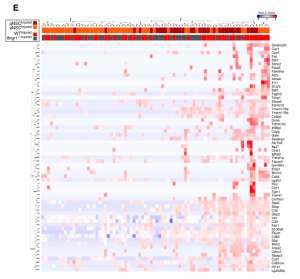
 (2 votes)

 uthors present a mouse model for tuberous sclerosis complex in which the gene Tsc1 is ablated from eye-progenitor cells, leading to the classic hallmarks of the disease. This demonstrates a role for Tsc1 in regulating several aspects of the development of the visual-pathway. Read the paper

 Kim, Parrish and colleagues show that Rop forms a complex with the exocyst, and that this complex predominates in primary over terminal dendrites. Membrane-associated proteins, on the other hand, preferentially diffuse from primary dendrites into terminal ones, suggesting that diffusion supplies membranous material for terminal dendritic growth. Read the paper
  ErB2 and ErB3 are two receptor tyrosine kinases expressed in the extravillous trophoblast (EVT) lineage in the placenta. Fock and colleagues demonstrate that neuregulin 1 (NRG1) promotes EVT formation and suppresses trophoblast apoptosis through the activation of ErB2 and ErB3. Read the paper
  Luman is an ER transmembrane protein that can undergo proteolysis and whose N-terminal fragment can act as a transcription factor. This study shows that Luman can regulate expression, localisation and stability of DC-STAMP, a downstream effector of RANKL, the protein that initiates osteoclastogenesis; making Luman a regulator of osteoclast differentiation. Read the paper
  Hall and Verheyen show that Dsor1, a Drosophila homolog of MEK that is activated by Ras, promotes transcription of Wg target genes by interacting with Armadillo and preventing its degradation. Ras-Dsor1 activity seems to be mediated by the insulin-like growth factor receptor. Read the paper
 a and colleagues investigated the spawning behaviour of the Japanese flying squid, comparing eggs spawned in a tank with a temperature gradient with eggs spawned in a tank without a temperature gradient. Only eggs spawned in the tank with the temperature gradient survived. Paralarvae survived for ten days, allowing observation of advanced stage paralarvae. Read the paper