The community site for and by
developmental and stem cell biologists

Two Neuro-PhD Positions in Manchester: Mathematical Modelling & Neurodegeneration

Posted by , on 21 November 2015

Closing Date: 15 March 2021

indexTwo positions are available as part of two Research Council-funded doctoral training programmes at The University of Manchester, the first one supported by the BBSRC and the second one by the MRC. Both projects involve work on the fruit fly Drosophila as a highly efficient and relevant model organism to study fundamental mechanisms of neuronal ageing and degeneration with unique detail and depth and delivering understanding of high biomedical applicability. Further information about fruit flies as a model organism is available here. Note that Andreas Prokop, who supervises on both projects, drives active programmes of science outreach and public engagement (see here), and students will have unique opportunities to develop transferable skills in science communication, which is of increasing relevance in modern science and an important category on a researcher’s CV. Full details on how to apply for these positions can be found on the UoM BBSRC DTP website.

Normal and mutant primary Drosophila neurons in culture.

Position 1: Advanced imaging and mathematical modelling of ageing and neurodegeneration in the nervous system

Enquiries: Andreas.Prokop@manchester.ac.uk

Project: Do you have mathematics skills, are keen to combine them with biological research and to work on a novel, highly interdisciplinary project investigating processes of ageing and degeneration in the nervous system? To carry out this exciting research you will be trained in and apply advanced live imaging, electron microscopy (EM), tomography, genetics and mathematical/computational modelling.

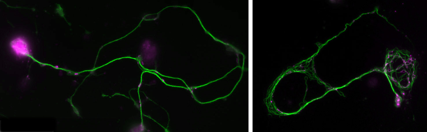
You will study axons, which are the cable-like (~1mm in diameter, metres-long!) extensions of neurons wiring the nervous system. These delicate structures are maintained for many decades in humans. They are key lesion sites in spinal cord injury, trauma and many neurodegenerative diseases (e.g. spastic paraplegia), and we lose 50% of our axons during healthy ageing. The essential structural backbone of axons is formed by continuous bundles of filamentous protein-polymers called microtubules. Disorganisation of these microtubule bundles leads to axon swellings correlating with axon decay, but the underlying mechanisms preventing/causing disorganisation are not at all understood.

To gain this understanding, you will genetically induce MT disorganisation, and use imaging to capture volume and space relationships (EM) as well as the dynamics of MT disorganisation processes (live imaging). Based on your collected data and existing algorithms for MTs, you will develop computational/mathematical models describing MT disorganisation and deduce the underlying rules.

Supervision:

Outstanding supervision and training will be provided through an interdisciplinary consortium of specialists with longstanding expertise in the research areas and technologies/strategies involved in this project:

  • Andreas Prokop (developmental and cellular neuroscience, Drosophila genetics, live imaging, electron microscopy)
  • Simon Pearce (mathematical modelling of biological systems)
  • Matthias Heil (mathematics)
  • Karl Kadler (electron microscopy, tomography, high pressure freezing techniques)

Note, that Andreas Prokop and Matthias Heil are also members of the Wellcome Trust-funded PhD programme “Quantitative & Biophysical Biology” providing further opportunities in this area of research.

Literature:


Position 2: An interdisciplinary approach to unravel mechanistic understanding of Frontotemporal lobar degeneration

Enquiries: SPB@manchester.ac.uk

Project:

Dementia causes enormous personal hardship and costs the UK ~£23 billion every year. The second most common form is Frontotemporal lobar degeneration (FTLD). About 40% of FTLD cases have genetic causes, with >8% involving abnormal intronic GGGGCC hexanucleotide repeat expansions in the C9orf72 gene which can additionally cause motor neuron disease (OMIM #105550). These pathological expansions are actively transcribed and, via bidirectional repeat-associated non-ATG (RAN) translation, generate 5 different aggregate-forming GA, GR, PR, GP and AP dipeptide repeat proteins (DPRs).This project will gain new understanding of this type of FTLD by unravelling neurodegenerative pathomechanisms of DRPs through using interdisciplinary approaches. We will focus on the hypothesis that toxicity is caused by DRP structure, comparable to amyloid plaques in Alzheimer’s disease. The project will capitalise on the complementary expertises of the three supervisors, and a readily available, unique set of 4 GFP-tagged constructs with high, pathologically relevant repeat numbers. The detailed aims and outcomes are:

  1. To generate purified DRPs and perform biochemical and biophysical analyses, in order to understand the reasons for their toxicity and identify useful therapeutic strategies which will benefit patients and their families.
  2. To generate transgenic Drosophila fly stocks to obtain primary neurons expressing the four DRPs. We will use powerful fly genetics and well established cell biological approaches to identify the neuronal death pathway and of the DRP-induced pathomechanisms upstream.
  3. There is substantial proof-of-principle for the use and translational potential of Drosophila To validate identified DRP pathomechanisms in mammalian contexts, we will carry out complementary experiments using well established DRP models in SH-SY5Y cells and inducible neuronal cell lines.

This project is highly interdisciplinary, with the analysis of the dipeptide repeat proteins spanning systems from in vitro solvents to human and fly cell lines. Students will receive a broad training in characterisation of polypeptide structure and aggregation, cell culture, fly and human genetics, and cell biology, all in the context of genetic disease and neurodegeneration.

Supervision:

  • Stuart Pickering-Brown (human geneticist specialised on causes of FTLD)
  • Andreas Prokop (developmental and cellular neurobiologist; pioneered primary neurons of the fruit fly Drosophila as a powerful model to decipher the fundamental cell biology of neuropathologies)
  • Andrew Doig (biochemist specialised on the structural and mechanistic analysis of protein aggregation in the context of neurodegeneration)

Literature

Thumbs up (No Ratings Yet)
Loading...

Categories: Jobs

Navigate the archive

Use our Advanced Search tool to search and filter posts by date, category, tags and authors.

BSDB Gurdon Summer Studentship Report (4)

Posted by , on 20 November 2015

BSDBlogoIn 2014, the British Society of Developmental Biology (BSDB) has initiated the Gurdon Summer Studentship program with the intention to provide highly motivated students with exceptional qualities and a strong interest in Developmental Biology an opportunity to engage in practical research. Each year, 10 successful applicants spend 8 weeks in the research laboratories of their choices, and the feedback we receive is outstanding. Please, read the student reports, kindly sent to us by Oliver Davis from Brighton and Sussex Medical School who was hosted in summer 2015 by Jean-Paul Vincent at the Crick Institute.

Dying for a pattern

Davis-Fig0This summer, I had the glorious opportunity of undertaking a BSDB funded research project in the laboratory of Jean-Paul Vincent at the Francis Crick Institute in Mill Hill, London. My project took place in the lab of Dr Jean-Paul Vincent (www.jpvincentlab.com) under the patient and inspiring tutelage of one of his PhD students, Sam Crossman. Our project investigated the mechanism of apoptosis in the model organism Drosophila melanogaster.Apoptosis is a form of programmed cell death and is an important process in the development of all multi-cellular organisms. Understanding why cells die in certain situations and not in others is of relevance to many areas of health, including embryological disorders and cancer, and my overall research aim was to investigate the role of apoptosis in the developing fly embryo. To do this, I worked with strains carrying mutations in genes required for patterning the anterior-posterior axis. Mutation of these so-called patterning genes can trigger extensive apoptosis in the embryonic epidermis (figure. 1) and therefore provides a useful model to investigate the apoptotic machinery in Drosophila.

Davis-Fig1
Figure 1: Increased apoptosis is observed upon the mutation of many patterning genes. Minimal cell death is observed when wild type embryos are stained with the apoptotic marker cleaved-Dcp1 (a), whilst embryos mutant for the patterning gene fushi tarazu (ftz) have increased apoptosis (b).

The cause of the ectopic apoptosis observed in patterning mutant embryos is not fully understood. One previously suggested explanation is that the cells of the epidermis can sense their ability to adopt the correct fate and undergo apoptosis if they lack the required patterning inputs to do so (Werz et al, 2005). However, if this were the case, there would have to exist an unknown machinery that would allow individuals cells to detect patterning errors and initiate apoptosis as a result.

In order to determine if cells are truly capable of detecting patterning errors, I planned to use a light inducible form of Cre recombinase to clonally remove a lox flanked allele of the ftz gene in a small subset of cells within each segment. If these small clones survive in an otherwise wild type embryo, it would argue against a cell-autonomous system where individual cells monitor their ability to differentiate correctly and would suggest that an alternative mechanism could be in play.


Figure 2. A diagram of a Drosophila embryo with 14 segments subdividing its anterior-posterior axis, which is achieved through the expression of patterning genes.


One difficulty with this plan is that ftz is activated very early on in embryogenesis. As a result, I set out to generate an early acting form of Cre, which could be used to remove ftz before it has carried out its function. To achieve this I spent the first part of my project cloning the Cre enzyme into a plasmid containing the actin promoter. As actin is an important protein in every cell, it is expressed from very early stages of embryogenesis. As a result, we hoped that by using the actin promoter to drive expression of Cre, we could produce the enzyme early enough to remove our lox flanked ftz allele in a timely manner and create mutant cells.

Unfortunately, cloning proved frustrating. Fortunately, it also proved educational. I learnt a lot about the way that experiments work, and how progress in science is more staccato than smooth. One issue I faced was the purification of my final plasmid using an Invitrogen maxiprep column. Each time I purified the plasmid I ended up with a lower yield than required, as I needed enough DNA to send to a company that would use it to generate a transgenic fly. I adjusted a parameter each time, but in the end it may have just been a plasmid with a low copy number as during my last attempt I purified it from a much larger bacterial culture, which finally gave me a sufficient amount of DNA.

The second part of my project was spent optimising a fluorescent in-situ hybridisation (FISH) protocol. I planned to use FISH to label cells expressing the pro-apoptotic gene hid in a series of patterning mutant embryos to characterise the regions where cell death occurs. As a control, I first conducted the protocol with a probe against the segment polarity gene wingless, which is expressed in a row of cells in every segment. This probe was made by someone else in the lab and is known to work, so I used it to learn the steps of the in-situ protocol.

Figure 3: Fluorescent in-situ images of wild and mutant ftz embryos using a wingless probe. a) a wild-type embryo with fourteen stripes; b) a mutant ftz embryo with seven stripes.

My FISH experiments with the wingless probe worked like a treat (figure 3), but difficulties soon followed when I attempted to make a new probe to label cells expressing hid. The stainings I conducted with the hid probe I had made repeatedly failed to work, and every attempt to generate a new probe proved unsuccessful. Disappointingly, I reached the end of my project before I managed to develop a protocol that worked, but I at least learnt plenty of science along the way!

My time in J.P.’s lab has been great for a number of reasons. I’ve come to love the problem solving nature of science and the freedom you get to explore what really interests you. It really is an adventure! However, I’ve also realised how difficult a career in science can be. Whilst this project has inspired me to become a scientist, I wonder if there’s a route into academia that would better suit my background as a medical student and my wider interests in medicine.

Sources

(1) Werz C, Lee TV, Lee PL, Lackey M, Bolduc C, Stein DS, Bergman A. Mis-specified cells die by an active gene-directed process, and inhibition of this death results in cell fate transformation in Drosophila. Development, 2005. 132(24): p. 5343-5352.

Thumbs up (3 votes)
Loading...

Tags: , ,
Categories: Education, Lab Life, Research, Societies

Postdoctoral Fellowship in Neurodevelopment

Posted by , on 19 November 2015

Closing Date: 15 March 2021

Applications are invited from highly motivated and creative individuals who are interested in studying fundamental mechanisms of neuronal migration and axon guidance. The main focus of our research is to understand the molecular and cellular mechanisms underlying the development of neural circuits using the embryonic spinal cord as a model system (http://www.ucmm.umu.se/english/research/sara-wilson/). The fellowship is currently funded for two years and is available immediately. The laboratory is located at the Umeå Centre for Molecular Medicine (UCMM), Umeå University, Sweden. UCMM is an interdisciplinary department, which focuses on questions in basic medical sciences and developmental biology and provides an interactive modern environment with good core facilities.


Requirements: Individuals with a background in developmental biology, neuroscience, molecular and cell biology or related discipline and with a keen interest in developmental neuroscience are encouraged to apply. The successful candidate will have or about to receive a Ph.D. in a relevant discipline and be proficient in written and spoken English.


Technical experience with imaging, vertebrate embryonic model systems – especially chick or mouse electroporation, mouse handling and genetics is a big advantage although training will be given. Experience with molecular, cellular and/or evolutionary biology will be positively considered. The most successful candidate will have a high level of motivation, be creative, organized and rigorous and have the ability to work both independently and within a team.


Please submit your application (reference 2015SW100) by 16th December 2015 to sara.wilson@umu.se by sending the following documents as pdf files:

1) A short cover letter (not more than 1 page) to include a description of your research experience and suitability for the position.

2) Curriculum Vitae including: publication list, technical expertise, names and contact information for three referees.

Informal enquiries may be directed to Dr. S.I. Wilson (sara.wilson@umu.se).

We look forward to your application!

Thumbs up (No Ratings Yet)
Loading...

Categories: Jobs

Organ Design 101: Discovering the rules for building a pancreas

Posted by , on 19 November 2015

diabetes

As we develop from wads of cells to fully formed humans, each of our organs goes through intricate processes to achieve the right combination and number of cells arranged in the proper way.

Research published in PLoS Biology by Yung Hae Kim and her colleagues looks at the development of our hormone-oozing pancreas, which assists digestion. They wanted to know how the cells become different cell types. Cells in developing organs begin without specificity and over time turn into various cell types upon receiving the right signals.

The beauty of this research, a part of the puzzle of how we form, is encompassed in the video below. This moving three-dimensional image is really hundreds of two-dimensional images merged together. The cells shown are a chunk of the pancreas, lit up in white, green, blue, and magenta.  Our cells are constantly churning out proteins that they use to carry out their functions. It is these proteins that the colours are marking. And the colours carry meaning. Blue and magenta indicate two different kinds of cells, distinguished by a specific protein that only that type of cell makes.

From this colorful three-dimensional clip, Kim and her team looked at whether the cells of the pancreas are ones that can produce hormones, or cells that have not specialized to this function. Further down the line, knowledge about when and how the cells become hormone-secreting ones could be important for diabetes research. It is cells of the pancreas that produce the insulin we need to control blood sugar levels. Perhaps we could incite cells that haven’t specialized to make insulin, to secrete it.

For more information on diabetes and stem cells

This research is from the lab of Anne Grapin-Botton, Danstem, part of the HumEn consortiumworking towards making fully functional beta cells for diabetes therapy.

Credit

Kim YH, Larsen HL, Rué P, Lemaire LA, Ferrer J, Grapin-Botton A (2015) Cell Cycle–Dependent Differentiation Dynamics Balances Growth and Endocrine Differentiation in the Pancreas. PLoS Biol 13(3): e1002111. doi:10.1371/journal.pbio.1002111

——

Julia Turan triturated, pipetted, imaged, and analyzed, during her undergrad years studying neurobiology. Since then, she has shifted into the world of science communications, hoping to promote a language of science legible to all. Julia is currently completing an MSc in Science Communication and Public Engagement at the University of Edinburgh.

Follow this link to subscribe to her weekly newsletter of five worthwhile science stories: http://tinyletter.com/juliaturan. Or follow her @JuliaTuran.

Thumbs up (1 votes)
Loading...

Tags: , ,
Categories: Images, Research, Video

Burning down the house II- not-so-bad ideas

Posted by , on 18 November 2015

This Sticky Wicket article first featured in Journal of Cell Science. Read other articles and cartoons of Mole & Friends here.


Embedded Image


“No visible means of support, and you have not seen nothing yet. Everything stuck together. Dum dee dum dum dum dee dum, Baby what do you expect?” Hey, we’re back. And no, I’m not still listening to that song. I’m listening to it again. I do that.

If you’re just joining us, we’ve been talking about bad ideas. Things that keep coming up – observations, conclusions, hypotheses – that we don’t believe but that just don’t go away. Even when they’ve been disproven or replaced by better ideas, here they are again – being cited in reviews, making their way into summary figures, being pointed to as an explanation for something in a paper, being used to make arguments. The field moves past them but they keep coming up for more exposure. And with ‘no visible means of support’ you wondered why I was listening to that song.

So how do we get rid of them? Well, before I tell you my ideas on this, let’s have a look at what is being suggested in the blogosphere (online). No, I don’t pay attention to the blogosphere. But I do listen to the ‘beer-o-sphere’, which I experience at meetings all the time –after the sessions, over a beer, when everyone weighs in on issues like this. The beer-o-sphere is way better than the blogosphere.

So here is the solution from the beer-o-sphere on how to get rid of bad ideas:

Let us publish negative results! Okay, we hear this all the time, not only in the beer-o-sphere. If there were a place to publish negative data, data that say that something doesn’t work, the weight of this will bury the bad idea in a deep grave made of electrons (no, nobody thinks negative data have to be published on paper; it isn’t worth killing trees to kill a bad idea). That should do it. Right?

Well, I agree and I don’t agree. A definitive experiment that proves that an accepted idea cannot be correct, certainly, should be published. But experiments of this type are extremely hard to design, and I suspect that when they are done, they do get published. The sort of negative data that are being discussed in the beer-o-sphere is generally of the sort, “We tried to repeat the experiment and it didn’t work.” People. I’ve said this before and I’ll say it again: Personally, I have trouble reproducing anything – I am not sure I could reproduce an experiment that shows that objects fall down and not up. I do notwant to read about your failures. There are many, many reasons for things not to work and, generally, very few reasons why they do. Yes, having a way for everyone to complain that something doesn’t work, or isn’t reproducible, or gives a different result, is all very cathartic, especially when others have the same problem, but it doesn’t actually prove anything. I don’t mind the idea of publishing negative data but it won’t burn down the house of bad ideas.

Let me give you a counterexample, how we can be wrong about wrong ideas, even when we have compelling negative data. Professor Echidna publishes that after a long search, he has actually found a mammal that is venomous. Quickly the community tries to reproduce this, but no mammals are venomous. So he shows them where he found it. More reports state that all mammals in that area are non-venomous, although many have pouches. Echidna identifies the animal as a duck-billed platypus. Professor Platypus herself publishes that she is definitely not venomous. The community, and the beer-o-sphere, are satisfied that this is a bad idea that needs to go away. But Echidna persists, “I meantmale platypuses.”

Which brings me to a question. Who says a bad idea is wrong? This is one thing to consider. Just because the consensus in the field says something is wrong, doesn’t actually make it wrong. Work can be difficult to reproduce, contain artifacts and look shaky, and still be correct. Male platypuses are, in fact, venomous. I bet you knew that?

But Mole, you say (I’m listening). If something is wrong, and most people in a field know it’s wrong, isn’t it dangerous to give it credibility? At the very least, people will waste their time on it. At the worst, they’ll use the idea to influence policies that could hurt people. Just because something could be correct, despite the experience of other experts in the field, must we give credit to everything that is published? How can we ever get anywhere?

Okay so let’s get something straight. Just because a bad idea seems to persist, it doesn’t mean youhave to use it. In fact, not only shouldn’t you use it but you shouldn’t use anything that isn’t useful for understanding the problem you have set yourself to do (or been set to do, not everyone gets to pick). Be critical, by all means, not only of ideas you question, but all ideas. Examine the evidence. And this goes both ways – if you are told something is wrong, find out why some folks believe it. There are lots of things in your own field you ‘don’t believe.’ But can you make a clear argument for why not? I’m often surprised at how many of us cannot.

So let’s get back to what we can do about this. First of all, be an intellectual. If you doubt an idea, have clear reasons for why. Make an argument. The thing is, while we don’t care (much) to see lots of failed experiments, we are very open to hearing your opinion as to what is wrong and why. And what the correct answer might be. When we write a review, or a commentary, or an opinion piece, be critical and make a case for what is correct and what is not. Be diligent. I promise I’ll be interested in reading that.

My friend Professor Wombat feels it is important to publish contrary data, and he does it regularly. Actually, he does it very well. He also publishes lots of interesting observations that are rigorous and move things forward (it would be a shame if he only set about showing other people are wrong). And he often publishes the contrary positions in places where people will read it (you know, the journals with the nice, soft pages). But here’s the thing – he gets the contrary positions wrong, too. A few years ago, he published a very influential paper that disproved a prominent idea, to great attention. But with time, we came to realize that he had not disproven the idea but, rather, found a counterexample. It did not ‘burn down the house’ of the idea, but refined it. He’s good with that. We’ve talked about it, and I know that he is interested in having the discussion, not proving his own point. This is how we make progress.

So yes, challenge those bad ideas – hold them up in the light and show us why they are bad. And here’s the thing. We have to fight with editors and reviewers, who tell us that challenging the idea is “not new.” As long as it is out there, the arguments against it are just as fresh as ever. And maybe, instead of citing the bad idea, someone will think twice and cite something else. Maybe even you. It’s about having the discussion.

It’s raining and starting to move into evening, and I haven’t had any ‘tea.’ Time to, um, put the kettle on and get back to reading. I’m not too worried about bad ideas when there are so many good ones out there. Many papers are, in the end, wrong on some level, but it is the bits that are right that move us forward. We can burn the house and live in it too – it’s what we do.

Thumbs up (4 votes)
Loading...

Tags: ,
Categories: Discussion

Post-translational regulation of epidermal morphogenesis

Posted by , on 17 November 2015

Closing Date: 15 March 2021

My laboratory is looking for PhD students to study the roles of SUMO proteases in embryogenesis. Our goal is to reveal the main targets of these enzymes during epidermal morphogenesis in C. elegans and their mode of regulation.
Highly motivated candidates with a Master degree in biological sciences and an interest in cell biology, genetics, and/or developmental biology are encouraged to apply. Successful applicants must be fluent in English (both written and spoken) and be able to work independently as well as part of an international team. Preference will be given to candidates familiar with advanced microscopy and molecular biology techniques. Interested candidates should email their CV as well as a brief paragraph describing their research interests and expertise, to Dr. Broday.


Reference

http://www.cell.com/developmental-cell/abstract/S1534-5807(15)00553-5


Limor Broday, Ph.D.

Dep. of Cell and Developmental Biology

Sackler School of Medicine Rm417

Tel Aviv University

Tel Aviv 69978

Israel


Office: 972 3 640-6653

Lab:    972 3 640-8225

Fax:    972 3 640-7432

email: broday [at]post.tau.ac.il


Thumbs up (No Ratings Yet)
Loading...

Categories: Jobs

Developing a new look

Posted by , on 17 November 2015

This editorial first appeared in Development, and it was authored by Olivier Pourquié, Katherine Brown and Claire Moulton.


As you might have noticed, Development has been looking a little different recently, with a new website and a new masthead for the journal (see Box 1 for elements of our new branding). These changes mark the culmination of a series of projects we’ve been working on over the past year at The Company of Biologists, aimed at improving the experience for our readers and at promoting the activities and values of the Company. Our website (and those of our sister journals) has undergone more than just a visual makeover – we have de-cluttered our pages and improved navigability, and the new open source platform will allow us to implement additional functionalities in the future. The consistent design, applied across all our journals, the Company’s own website (http://www.biologists.com/) and that of our community blog the Node (https://thenode.biologists.com) aims to provide clearer brand recognition and a better user experience – we hope you like it!


————————————————————————————————————————————————————

Box 1. A new look for the Company and its journals

The Company of Biologists logo:

Embedded Image

Development’s new masthead:

Embedded Image

————————————————————————————————————————————————————-


Since you are reading this editorial, you are probably at least reasonably familiar with the journal, and many of you will know something about The Company of Biologists, the publisher behind it. But we’d like to take this opportunity to let you know a bit more about who we are, what we do and how we help the scientific community. The Company of Biologists is a not-for-profit organisation dedicated to supporting and inspiring the biological community. At the heart of the Company are our five journals – Development, Journal of Cell Science, Journal of Experimental Biology, Disease Models & Mechanisms and Biology Open – and our primary mission is to publish influential and innovative science, providing a valuable forum for sharing scientific knowledge. Our journals cover a broad range of the life sciences, from comparative physiology to drug discovery, but they all aim to provide an important platform for their respective communities: to disseminate research to the community in an accessible manner and to make the publication process as pain-free as possible (for some of the benefits of publishing in Development, see Box 2). For example, at Development we recently changed the way we ask our reviewers to assess papers (Pourquié and Brown, 2015), with the aim of easing the path to publication while still maintaining our high standards. We hope this is making a difference for authors (we welcome your feedback), and we will continue to review and improve our processes.


————————————————————————————————————————————————————-

Box 2. Key information about the policies, processes and benefits of publishing in Development

  • Free to publish: no page or colour charges.

  • Editorial decisions made by expert Academic Editors who know and understand your field.

  • Fast publication, online and in print.

  • Avoidance of multiple rounds of review wherever possible; strong commitment to publishing invited revisions.

  • Scoop protection: we won’t reject your manuscript on the grounds that a competing paper has come out after yours was submitted.

  • Author-pays Open Access publication option available.

  • All articles freely available online six months after publication; online archive dating back to 1953.

  • Open attitude to sharing research results and data on pre-print servers and data repositories.

  • Informative and provocative Review and Hypothesis-type articles written by leaders in the field.

  • All articles copyedited by experienced scientific editors.

————————————————————————————————————————————————————-


Another way we have tried to help in reducing ‘the pain to publish’ is through our newest journal, Biology Open (BiO; http://bio.biologists.org), which we launched as a response to community feedback. As a fully Open Access journal, BiO supports the rapid publication of scientifically sound research across the biological sciences, without making judgement on the ‘impact’ of the work. Importantly, papers rejected from Development can be transferred to BiO, where their editors (including developmental biologists Anna-Katerina Hadjantonakis, Yishi Jin and Jenny Nichols) can use the Development referee reports to make swift decisions on the potential suitability of these papers for BiO, thus streamlining the process and hopefully allowing authors to get their research results out to the community more quickly (see Box 3 for recent BiO papers of interest to the developmental biology community). While we know that developmental biologists have a wide choice of specialist journals to which they can submit, we believe that BiO provides a valuable alternative – particularly in cases where the prime objective is to get a paper published with minimum hassle.


————————————————————————————————————————————————————-

Although our journals are our most important output, the Company is much more than just a publisher. As a UK-registered charity, we believe that the profits from publishing the hard work of biologists should support scientific discovery and help develop future scientists. Each year, we provide significant financial support to the community through our various charitable programmes, overseen by our dedicated Board of Directors – distinguished practising scientists who give up their time free of charge to help oversee the Company’s activities.

So where does this money go? We run a very active Meeting Grants programme (http://www.biologists.com/grants/), providing support to help defray the significant costs of putting on a conference. We also believe in supporting young scientists to learn new techniques, make new scientific connections and experience different scientific cultures: our Travelling Fellowships provide funding for researchers to make collaborative visits to other labs (http://www.biologists.com/travelling-fellowships/; see also Box 4). We also provide large grants to several societies, including the British Society of Developmental Biology (BSDB; http://bsdb.org/). Part of this money is used by the society to help run its annual meeting, while the rest is dedicated to a travel grants programme, to which BSDB members of any nationality can apply for funding to help pay for attendance at conferences across the globe (see http://bsdb.org/membership/meeting-grants/company-of-biologists/).

————————————————————————————————————————————————————-

Box 4. A Development Travelling Fellowship story: The benefits of global collaboration

Mirana Ramialison has been able set up her own lab at the Australian Regenerative Medicine Institute in Melbourne – thanks in part to Development.

Mirana had been a bioinformatician post-doc in the laboratory of Professor Richard Harvey, at the Victor Chang Cardiac Research Institute in Sydney. Her project consisted of deciphering the regulatory network that controls heart development and how this network might be perturbed in disease conditions, such as congenital heart disease. This involved genome-wide information obtained using a mouse cell line (HL-1 cells). Mirana wanted to further investigate how this cardiac gene regulatory network changed (or not) during evolution. This new project required genome-wide information on cardiac-specific cells from different organisms, including zebrafish. The laboratory of Dr Eileen Furlong at EMBL Heidelberg in Germany had published a new method, BiTS-ChIP-seq (Bonn et al., 2012), that allows researchers to obtain genome-wide information from tissue-specific cell types in vivo. Mirana saw that using this method in the developing zebrafish, in a transgenic line that specifically labels heart nuclei, would help gain insight into the nature of this gene regulatory network in vivo.

The published protocol was detailed and complex and included many techniques that Mirana has never performed before. The quickest way to get these experiments going was to get ‘hands-on’ experience of this protocol – but Australia is far from Heidelberg. A Travelling Fellowship from The Company of Biologists made the visit possible, providing Mirana with the hands-on experience to learn the protocol and its ‘tricks’, saving months of trials to get it right.

The Travelling Fellowship also had repercussions beyond research; Mirana was in the process of looking for an independent position and finding grants to fund it. The collaboration with Dr Furlong’s laboratory on the BiTS-ChIP-seq experiments gave the evidence of collaboration that the Australian Research Council needed. Not only did the Travelling Fellowship help save time on experiments, it strengthened connections around the world and helped Mirana to start her own laboratory.

————————————————————————————————————————————————————-


More recently, we started hosting our own series of Workshops (http://www.biologists.com/workshops/), bringing together small groups of leading experts and young scientists (who can apply to attend with no registration fees) from a diverse range of scientific backgrounds for the cross-fertilisation of interdisciplinary ideas. The unique format of these Workshops has proved a huge success, with overwhelmingly positive feedback from organisers and participants alike, and we welcome proposals for future events. Finally, we are now organising Meetings on topics of particular interest to the journals (http://www.biologists.com/meetings/). The first of these focussed on the emerging field of human development (for a review of the Meeting, seeMedvinsky and Livesey, 2015) and was such a success that we will be running a second Meeting on the same topic in 2016 (see http://www.biologists.com/meetings/from-stem-cells-to-human-development-2016/) – we hope some of you will be able to join us!

We hope this brief overview of the activities of the Company has given you some idea of how we strive to help the scientific community. For further information, we encourage you to browse our website (http://www.biologists.com/) to see how you – whether you’re starting out in your career or you’re an established group leader – can benefit from what we do. Our ethos, ‘Supporting biologists, inspiring biology’, influences all that we do. We hope that by publishing in, reviewing for and reading Development you will continue to support the journal and the Company and, through us, the community at large.

Thumbs up (2 votes)
Loading...

Categories: News

In Development this week (Vol. 142, Issue 22)

Posted by , on 17 November 2015

Here are the highlights from the current issue of Development:


Nodal: sustaining Shh expression

Embedded Image

Underlying the developing vertebrate forebrain is the prechordal mesoderm, which secretes sonic hedgehog (Shh) at a precise developmental time. The tight temporal regulation of this morphogen is crucial for the specification of several ventral cell types in the forebrain. However, little is known about the signals that limit Shh expression temporally. Nodal is expressed in the prechordal mesoderm and had previously been suggested to interact with Shh during ventral forebrain development. Now, using the chick embryo, Marysia Placzek and colleagues (p. 3821) show that Shh expression in the prechordal mesoderm is regulated by proNodal, the precursor of Nodal. Surprisingly, proNodal maintains Shh expression by a non-canonical route: binding to and activating FGFR3. Through this route, proNodal antagonises BMP7 and pSmad1/5/8, which suppresses Shh expression. Together with previous findings, this study suggests that whereas Nodal operates through canonical signalling to induce prechordal mesoderm, it acts via a non-canonical route involving FGFR3 to control the expression of Shh in the prechordal mesoderm.


Hh puts the pressure on boundaries

Embedded Image

The Drosophila wing originates as an imaginal disc, which is divided into anterior and posterior domains separated by a straight antero-posterior (AP) boundary. This barrier is characterised by an actomyosin cable and increased mechanical tension at cell junctions, termed cell bond tension. Engrailed and Invected, expressed in the posterior compartment, maintain the straight morphology of the AP boundary both through the induction of the morphogen Hedgehog (Hh) and via an Hh-independent mechanism. How do such signalling pathways regulate cell bond tension at the AP boundary? In this study (p. 3845), Christian Dahmann and co-workers show that the difference in Hh activity between the two compartments drives the local increase in cell bond tension along the AP boundary and is required to bias cell intercalations to maintain its straight shape. Furthermore, increased mechanical tension is generated autonomously at the boundary and does not depend on the actomyosin cable or the Hh-independent mechanism that contributes to the preservation of the AP boundary shape. By linking the molecular players and mechanical determinants, this study sheds light on the mechanisms governing the physical separation of adjacent cell populations destined to different cell fates.


Deciphering genome imprinting

Embedded Image

Following fertilisation, a genome-wide demethylation wave reprogrammes the genome. However, in mammals, certain loci can remain methylated specifically on the maternal or paternal chromosome, i.e. imprinted, in somatic cells. It was previously shown in transgenic mice carrying a fragment of the H19imprinting control region (ICR) that the paternally inherited H19 ICR does not need to be methylated in the germline to be imprinted, pointing at the existence of an unknown epigenetic mark inducing post-fertilisation methylation of that locus. Now, using the same H19 ICR transgenic line, Keiji Tanimoto and co-workers (p. 3833) show that H19 ICR imprinting is achieved through maternally inherited DNMT3A- and DNMT3L-mediated de novo methylation. This process is also at play at the endogenous H19 locus. Further, the authors identify the sequences responsible for the post-fertilisation methylation of the transgenic H19 ICR and show that their removal from the endogenous locus leads to partial H19 ICR demethylation and delayed embryonic growth in the offspring that inherited the mutation. These results provide a mechanistic understanding of the contribution of de novo methylation to genomic imprinting in the absence of germline methylation, though the nature of the putative epigenetic mark that directs this methylation has yet to be discovered.


A genome-wide view on cell differentiation

Embedded Image

The acquisition of specific cell fates in the early embryo is driven by changes in gene regulatory networks that induce differential expression of effector genes to ultimately instruct a specific cell fate. The activation of such effector genes has been well characterised in time for individual genes, but to a much lesser extent in space. Here (p. 3892), Julius Barsi, Eric Davidson and colleagues performed a quantitative transcriptomic analysis of effector gene activation on a genome-wide scale in six cell populations isolated from different regions of pregastrular and early gastrula sea urchin embryos. With this approach, the authors identify a set of effector gene transcripts shared by the different cell populations. Surprisingly, this shared set of genes is not as large as previously thought. Indeed, the authors show that spatially distinct populations in the early embryo actually display profound differences in effector gene expression long before morphological differences in cell types can be distinguished. This study sheds light on the mechanistic essence of embryonic differentiation and provides a large-scale transcriptomic dataset, a rich resource for the developmental community.


Distilling principles of tubulogenesis

Embedded Image

In the developing Drosophila trachea, the maturation of tracheal terminal cells involves the generation of gas-filled tubular branches. However, the mechanism underlying subcellular lumen formation in these cells remains unknown. By adapting high pressure freezing and freeze substitution techniques toDrosophila larvae and performing transmission electron microscopy, Mark Metzstein and Linda Nikolova (p. 3964) show that, contrary to previous belief, lumen formation is not achieved by the direct fusion of cytoplasmic vesicles. Instead, the authors find that it requires a previously undescribed intermediary membrane-lined multivesicular compartment. In this compartment, vesicles assemble and then fuse into a nascent lumen. By further adapting their ultrastructural imaging technique to preserve the fluorescence of protein reporters and performing correlative light and electron microscopy, the authors show that the resolution of the multivesicular intermediate into a mature lumen requires Rabconnectin-3-mediated acidification of the compartment by the V-ATPase proton pump. The tools developed in this study to analyse tubulogenesis in the trachea and the insights provided on the mechanisms underlying this process are likely to contribute to the understanding of lumen formation in other organs.


Hox6: establishing a dialogue during pancreas development

Embedded Image

The development of the pancreas, a secretory organ that expands from the endoderm into the surrounding mesoderm-derived mesenchyme, requires a dialogue between its endodermal and mesodermal components. How do these two compartments communicate? To understand the molecular basis of this cellular cross-talk, Deneen Wellik and colleagues (p. 3859) have analysed pancreas organogenesis in mouse, finding that Hox6 genes, a group of patterning genes expressed in the pancreas mesoderm but not in the endoderm, play a crucial role in this process. Indeed, the genetic loss of all Hox6 paralogues results in mild defects in branching and in exocrine differentiation, and a drastic loss of mature endocrine cells. Mechanistically, the authors show that Hox6 depletion results in decreased expression of mesenchymal Wnt5a, a morphogen crucial for pancreas development. This then leads to the loss of the expression of two Wnt inhibitors, Sfrp3 and Dkk1, in endocrine progenitors. Hence, as repression of Wnt signalling in developing endocrine cells is crucial for their differentiation, this study highlights that regional mesodermal patterning cues are essential for the establishment of the mesenchymal/endodermal crosstalk necessary for pancreatic development.


PLUS:


Developing a new look

Embedded ImageAs you might have noticed, Development has been looking a little different recently, with a new website and a new masthead for the journal. These changes mark the culmination of a series of projects we’ve been working on over the past year at The Company of Biologists. Read more about these changes in the Editorial on p. 3803


Glia in mammalian development and disease

Figure1The past few decades have witnessed a flood of studies that detail novel functions for glia in nervous system development, plasticity and disease. Here, Bradley Zuchero and Ben Barres review the origins of glia and discuss their diverse roles during development, in the adult nervous system and in the context of disease. See the Development at a Glance article on p. 3805


Next generation limb development and evolution: old questions, new perspectives

Fig. 1.In recent years, systems biology approaches have aided our understanding of the molecular control of limb organogenesis, by incorporating next generation ‘omics’ approaches, analyses of chromatin architecture, enhancer-promoter interactions and gene network simulations based on quantitative datasets into experimental analyses. Here Aimee Zuniga reviews the insights these studies have given into the gene regulatory networks that govern limb development, the fin-to-limb transition and digit reductions during evolution. See the Review on p. 3810


Thumbs up (No Ratings Yet)
Loading...

Categories: Research

PhD and postdoc positions at IST Austria

Posted by , on 16 November 2015

Closing Date: 15 March 2021

PhD and postdoc positions are currently available in the lab of Anna Kicheva at IST Austria, working on vertebrate neural tube development. Candidates with background in developmental, cell or molecular biology or at the interface between biophysics and biology are encouraged to apply.

During development, tissues increase considerably in size at the same time as cell type diversity is generated. How these processes are coordinated to achieve the correct size and morphological proportions of organs is a fundamental question in developmental biology. While much is known about how extrinsic signals, called morphogens, control the specification of cell identities, the mechanisms of tissue growth control are not well understood. Our aim is to gain a quantitative understanding of how neural progenitor cells in the developing spinal cord interpret morphogen signalling to regulate their progression through the cell cycle. We will also investigate how tissue growth affects the formation of morphogen gradients and pattern specification. The work will involve live imaging of mouse and chick embryos in both ex vivo and in vivo assays, as well as collaborating with theorists to analyse and interpret data.

IST Austria is a young international research institute close to Vienna, with a strong focus on interdisciplinary research and access to excellent facilities. PhD student candidates are required to apply through the IST graduate school, deadline January 8, 2016. To apply for a postdoc position, please email me at anna.kicheva@ist.ac.at with your CV, motivation letter and contact information for 2-3 references.

For more information: Kicheva lab IST, anna.kicheva@ist.ac.at


Thumbs up (No Ratings Yet)
Loading...

Categories: Jobs

Post-doctoral Research Associate, UCL, in London

Posted by , on 13 November 2015

Closing Date: 15 March 2021

We are seeking to appoint a Post-doctoral Research Associate to undertake research on the molecular mechanisms of eye induction/development and in vitro eye construction from mammalian stem cells with Prof. Shin-ichi Ohnuma.

The work brings together a range of multi-disciplinary approaches including in vivo analysis of Xenopus eye development, in vitro stem cell biology, and molecular biology, in conjunction with a variety of imaging and data analytical approaches.

This project builds on the recent publication of the group: Luehders, K. et al, Development (2015) 142, 3351-3361.

Applicants should have a PhD and research experience in developmental biology. Previous experience in Xenopus development and/or stem cell biology is preferable. Applicants will be self-motivated, have the ability to plan and interpret experimental studies as well as have excellent communication skills and ability to write well.

The position is available for 36 months in the first instance.

The laboratory is well equipped and is part of UCL Institute of Ophthalmology. The Institute comprises about 40 research groups spanning a research area encompassing visual development, stem cell based study, and therapy of retinal diseases. UCL has active neuroscience, developmental biology, and stem cell research communities and the group has established collaborative links within the institute and UCL, as well as with national and international colleagues.


UCL Reference: 1514567


Applicants should apply online through UCL Job search webpage. Please input the number above, fill in your information and apply.


Informal inquiries may be addressed to s.ohnuma@ucl.ac.uk


Deadline of application: 6 th Dec 2015

Thumbs up (No Ratings Yet)
Loading...

Categories: Jobs