The community site for and by
developmental and stem cell biologists

2015 Gordon Conference – Developmental Biology

Posted by , on 14 July 2015

The Developmental Biology Gordon Conference is held every two years and this year it was held in the picturesque Mount Holyoke, MA, USA. This conference’s mission is to bring together people from research institutions all across the globe who are studying developmental biology which lies at the cross roads of all the Life Science, integrating investigations at molecular, cellular and tissue and organismic levels. The 2015 Gordon Conference on Developmental Biology presented the most recent, cutting-edge research in the field.

It was a great conference with a fantastic line up of speakers which kept us engaged through all days of the conference. It was a superb learning experience with wonderful interactions and discussions. The highlight of the conference were two talks by Dr. Victor Ambros and Dr. Gary Ruvkun both co-recipient of 2015 breakthrough prize for discovery of microRNAs using worm C. elegans as the model system. A predominant number of speakers (>50%) at the conference were using C. elegans as their model system and a large subsection of the research presented dealt with genomic level events many of which directly or indirectly involved microRNAs. It goes to show how much the field of microRNAs has evolved in the past couple decades and many researchers are working to understand the role of microRNAs.

All and all it was a wonderful conference. Given that it was a Developmental Biology conference, I went home still yearning for a broader exposure of different model organisms (in addition to C.elegans) and studies that span the breadth of developmental biology at the cellular, tissue and organismic levels in addition to genomic level.

Thumbs up (1 votes)
Loading...

Tags:
Categories: Events

Navigate the archive

Use our Advanced Search tool to search and filter posts by date, category, tags and authors.

An interview with Brigid Hogan

Posted by , on 11 July 2015

This interview was first published in Development.


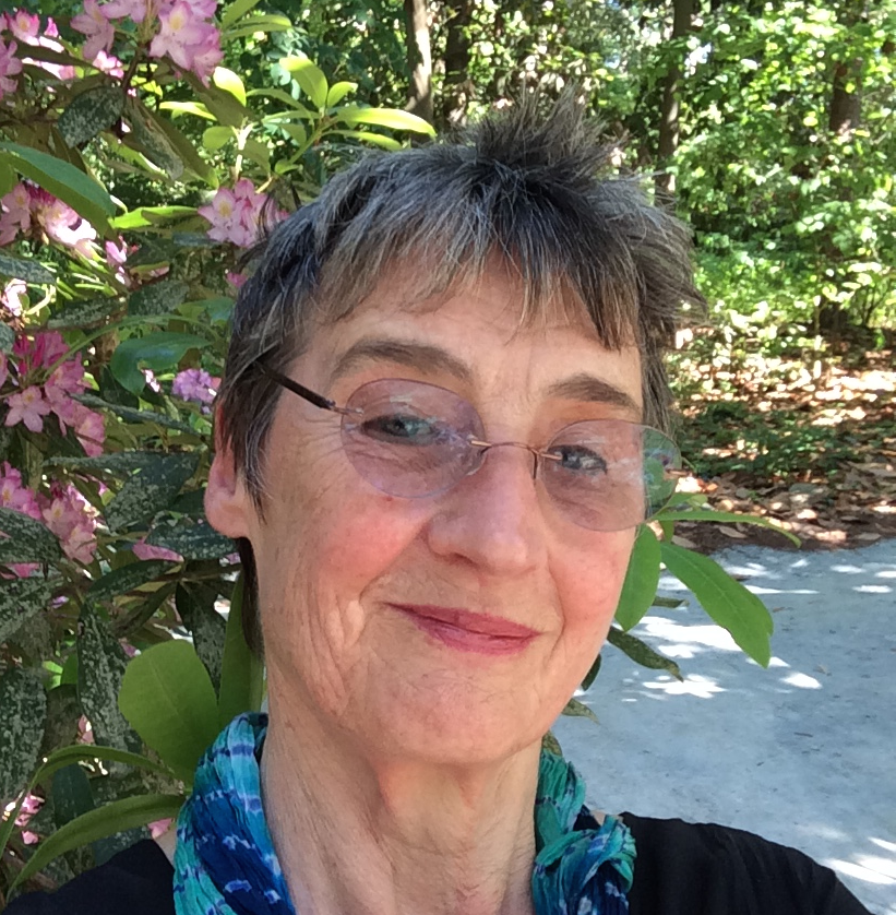
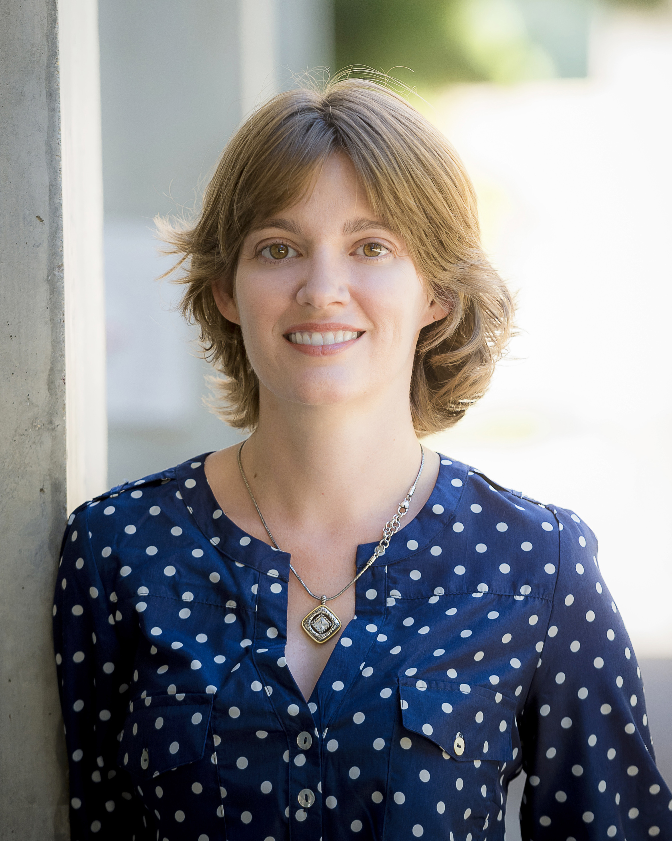
Brigid Hogan is a developmental biologist who has worked extensively on the early stages of mouse development and is now unravelling the mysteries of lung organogenesis. She is the George Barth Geller Professor and Chair of the Department of Cell Biology at Duke University Medical Center. Brigid is also the winner of the 2015 Society for Developmental Biology (SDB) Lifetime Achievement Award.


Brigid HoganHow did you develop an interest in biology and was there someone who inspired you?

The first person who inspired me was my grandmother. She liked gardening, and I distinctly remember her showing me how to plant seeds in a little bed that she said was my garden. I was fascinated by how these seeds would turn into flowers. When I grew older I became interested in bird watching and collecting flowers.

When I was in school I didn’t do very well in subjects like history and Latin, but I always did really well in biology. I attended an all- girls high school, but the year I reached 16 the women teachers who had taught chemistry and biology retired. The school couldn’t find any women replacements, so they hired Mr Jones. He was very interested in DNA, chromosomes and molecular biology in general, so he swept aside all the stuff we had been doing before and said we were going to do experiments, such as dog testis squashes of chromosomes. This was very exciting for me, and a great inspiration. I also joined other activities outside school. On the other side of the hill was the boys high school and Mike Ashburner, the Drosophila geneticist, attended that school. We both belonged to the Middle-Thames Natural History Society, and the society’s weekend meetings took place in areas such as Burnham Beeches woods, where Mike photographed flowers while I was interested in birds and, indeed, any natural history.


Both your parents were artists and you have said before that you “view embryos as a thing of beauty”. Does your artistic sensitivity influence the way that you view scientific problems?

My father died when I was really young, so it was really my mother who was the biggest influence. She had been an artist, and we had lots of books about art that we used to look at. Developmental biology didn’t quite exist while I was at university, but the papers and topics that interested me always had a visual element. I remember hearing about Drosophila genes associated with segmentation from David Ish-Horowicz when I was in Mill Hill. I would listen to his talks but didn’t really get it. It wasn’t until I went to a seminar given by Mike Akam, in which he presented some of the first in situ hybridisation patterns for Ubx, and saw his pictures of the stripes of mRNA distributions that I finally understood what it was all about. The visual input was always tremendously important for me and it still gives me enormous pleasure to look down a microscope at embryos and tissues and wonder how they develop.


You did your degree at the University of Cambridge, where you experienced negative attitudes from male faculty. How have attitudes towards women in science changed during your career?

Fortunately, attitudes have changed, and there is a lot of external pressure for them to change. In fact, yesterday I was talking to some Cambridge students and recounting some of the bad experiences I had as an undergraduate, and they were quite shocked. If this kind of sexual harassment happened now it would be immediately reported. In those days it was all swept under the rug; you just put up with it. I am amazed that I survived and stayed interested. It would have been terribly easy to give up, and it must have been the sheer passion for what I wanted to do that kept me going. It is a real pity because I think I might’ve been much happier if the attitudes of teachers to women students would have been different back then.

The harassment has largely gone, but there is still a long way to go. I have just been to the Wellcome Trust meeting on The Biology of Regenerative Medicines and around 85% of the oral poster presenters were young women. I don’t know what happens to them, but the number of senior women speakers was by no means the same proportion. The real problem is how to combine a career with having a family. Confidence is also an issue, to overcome all the stresses and strains during your career progression. These stresses affect both men and women, but I think women often feel more insecure and take criticism much more personally. In addition, although people are much more aware of women’s issues, there are other problems to solve. If we think it is difficult for women in science, it is even more difficult if you are an African American or Hispanic, at least in the USA where I work. There is a huge diversity problem.


You established yourself as a developmental biologist with work on early mouse development and organogenesis. However, this was not how you started your career. You worked on sea urchins during your postdoc and mouse teratocarcinoma cells in the early days of your lab. How did your interest in mouse embryology develop?

I have always been very interested in embryos, even as an undergraduate, and I would read a lot about developmental biology. However, in those days there weren’t any classes in cell biology or developmental biology, so I chose to do my PhD in a topic that was very exciting in Cambridge then – protein synthesis and RNA. However, when I finished my PhD I said to my advisor: “I really want to work on embryos, do you know anybody with whom I can do a postdoc?”. My advisor suggested that I worked with Paul Gross, who was studying sea urchin embryos at MIT. In the end, I didn’t find sea urchins so exciting, especially because the availability of the material was a little sporadic (during the winter you had to wait for shipments to come from California), but it was a wonderful experience being at MIT. It was so completely different to Cambridge in the UK – I remember being overawed by the size of the biology department!

Mostly for personal reasons I eventually decided to come back to the UK. I got a job as a lecturer at Sussex University but was dissatisfied there, and ended up moving to work with John Cairns at the Imperial Cancer Research Fund (as it was then) Mill Hill laboratory in London. John was hugely influential in my career because he gave me the freedom to look around and find a research topic. Initially, I started working with F9 embryonal carcinoma cells and gene expression changes as they differentiated into extraembryonic endoderm in response to retinoic acid. By this time I had two terrific postdocs, Denise Barlow and Markku Kurkinen, who brought molecular biology skills to the lab. F9 cells start making lots of extracellular matrix proteins when they differentiate, so this led us to beat big groups in Germany and the USA in the first cloning of the genes for laminin and type IV collagen. This work was very exciting, but in the end it wasn’t really developmental biology. I still hankered after the embryo, and so I started trying to isolate pre-implantation embryos. I found this really challenging on my own, so I contacted Anne McLaren at the MRC Unit for Mammalian Embryology at University College, London. Besides John Cairns, Anne was the most influential person in my career and the best possible person I could have found to help me. She was such a brilliant scientist, so encouraging and supportive. She and her colleagues, and people like Janet Rossant, Liz Robertson, Ginny Papaioannou and Allan Bradley – all of whom had grown up knowing how to manipulate embryos – were incredibly kind, supportive and generous with me. So was Gail Martin, who was working on embryonal carcinoma cells in London then. So if there is one take-home message from my career it is that it can take a long time to get to where you want to be!


You have been very involved in the development and teaching of techniques in mouse embryology and transgenesis. Did your interest in this develop during those early days?

In those days I would visit Anne’s lab, and people such as Mike Snow and others would answer my questions: “what medium do you use for this?; how do you do this experiment?; show me precisely how you do the dissections”. They would pull out a drawer and fumble around for a bit of paper and say “Oh, I think this may be the formula”, and I would snatch these pieces of paper and take them back with me. I gradually realised that what beginners like me needed was a handbook like Joe Sambrook’s famous cloning manual. I also thought we needed a course where experts could teach us how to collect embryos and manipulate the early post- implantation stages. I kept on mentioning this to people and everybody said it would just be too difficult, that no one would support or pay for the course. Then, when I was at Cold Spring Harbor, I was at lunch and Jim Watson sat down opposite me and just said: “What’s new?”. I realised that I needed my two-minute elevator speech to say something that would catch his attention. I told him that there were some really exciting developments in mammalian embryology and molecular biology, and that I really wanted to run a course but was being told I couldn’t do it. He just stood up and walked off and I thought “Oh, I’ve annoyed yet another person”. I finished my lunch, left the dining hall and started walking away when he ran down from his office with a piece of paper in his hands saying: “It’s all arranged, it’s all arranged! You’ll do a sabbatical here and we’ll run a course with Frank Costantini and Liz Lacy”. He had been trying to recruit them to Cold Spring Harbor because they had made the first transgenic mice in Oxford and had just started their own labs in New York. So I helped run the course, and wrote the manual, which was eventually published by Cold Spring Harbor. Frank and Liz were co-authors, and of course it included their technologies for making transgenic mice, which is what people were really excited about. Every now and again I would push a little bit of post-implantation embryo at someone and say “Don’t you think this is interesting”, and they would say “Oh yes, but I want to inject my DNA”. It took a while for the course to gradually evolve into what it is now. It moved from transgenic mice to ES cells, making chimeras and now making iPS cells and organdies.


Your lab is currently interested in understanding lung development. Why did you decide to focus on this organ in the last few years?

There was a short period of time when you could become interested in almost any organ system, because you would make a knockout homozygous mutant mouse and you didn’t really know what sort of phenotype you were going to get. My lab went through a stage when we were interested in many different organs and their development, because of the role of the BMPs and Fox genes that we had cloned and for which we had reporters.

But the lung has fascinated me since my early days in London. At Mill Hill we had access to about twenty different strains of mice and you could just ask for mated, timed embryos of these different strains. I looked at all of them and was fascinated by the fact that the lung branching pattern was the same. When we were working with BMP4 and FGF10 we noticed that these proteins are expressed in the developing lung, in the epithelium and mesenchyme of the growing buds. I had a brilliant student, Molly Weaver, who loved doing manipulations, cutting up buds and showing that they grew towards beads soaked in signalling factors. This work was incredibly exciting to me, and it seemed that it was opening up an important area of developmental biology: epithelial/mesenchymal interactions and organogenesis. I also ultimately focused on the lung owing to funding. I had grants from the National Institute of Child Health and Development, but they always cut their grants by 25% after you’ve got one, so it was very difficult to keep going. So I applied to the National Heart, Lung, and Blood Institute and got a grant to look at lung development. They didn’t cut the grant by 25%, so I wrote another… There were lots of interesting questions, but you can’t really focus and ask important questions about many tissues simultaneously. It is difficult to be competitive in many fields.


You have been involved in the past in high-level discussions of the ethics and regulations of embryology. You were the co-chair of the 1994 NIH Human Embryo Research Panel, and a member of the 2001/2 National Academies Panel on Scientific and Medical Aspects of Human Cloning. What do you think are the next big ethical challenges in the field, and what role should scientists play in these discussions?

In the ethics discussions I was involved in at the NIH my role was very much just to tell the committee the basic facts of early embryonic development. I remember explaining that if you separated an embryo into four blastomeres and put them back, you weren’t necessarily going to get four babies. I suppose I was quite good at explaining, perhaps from having taught in courses. It was a hugely interesting experience and I was deeply inspired by Anne McLaren, who had been the pioneer in being involved as a scientist in such ethical discussions.

At the moment the hot topic is undoubtedly the genetic manipulation of the human embryo by CRISPR/Cas9 technology. It is a very powerful technique, but it is far too soon to apply it to humans. A lot more basic research has to be done on possible side effects. If one of the parents carries a mutation, you don’t know which embryos are carrying the mutation. So which ones do you choose to repair? Is this necessarily better than just pre- implantation genetic diagnosis, where you keep the embryos that don’t have the mutation? You could also apply this technique to stem cell populations that could be replaced without having to change the genome of the whole human. However, this strategy has the problem of how you get these cells back into the damaged tissue. The real danger is that the promise is all blown out of proportion based upon preliminary results. The big challenge is going to be to make sure that people don’t move too fast, getting everybody’s consensus and agreeing on a course of action together.


Later this year you will receive the SDB Lifetime Achievement Award. Does this prize have a special significance for you?

I’m very grateful. The SDB is a great organisation and I’ve got many friends there. It gives me enormous pleasure and is a boost to keep going. It is very gratifying to feel that, in spite of all the early struggles one had, I have been very lucky in the colleagues, friends and people who helped me. This is another example of people recognising me and being nice to me.


What is your advice for young scientists?

You have to be passionate. That is what kept me going during the dark days of my undergraduate, PhD and early postdoc. It took quite a long time before I found the mouse embryo, Anne McLaren and the community of mammalian developmental biologists. I kept going because I just felt I wanted to work on embryos and probably because I picked up a tenacious attitude along the line. Maybe that has not necessarily always been good, since I have the reputation of being a little abrasive at times, unlike someone like Anne who was enormously diplomatic. You mustn’t be too aggressive in what you want, but you still have to be very tenacious. It is also important to find a community, a life partner and/or a group of friends who will support you and encourage you.


Thumbs up (7 votes)
Loading...

Tags: , , , , ,
Categories: Interview

Postdoctoral Position Available: Gene Regulation and Druggability of Normal and Tumor Stem Cells

Posted by , on 10 July 2015

Closing Date: 15 March 2021

Description
Postdoctoral positions are available in the Markstein Laboratory at the University of Massachusetts at Amherst. We study how stem cells respond to natural and synthetic chemicals in the environment over the course of normal development and tumor progression. We recently showed that stem cells proliferate into small tumors in response to a subset of FDA approved chemotherapeutics, highlighting the clinical importance of understanding how stem cells respond to their chemical environment. We employ Drosophila genetics, chemical screening, tumor modeling, transgenics, genomics, and confocal microscopy. To learn more about our laboratory visit: http://marksteinlab.org.


Qualifications

The ideal candidate will have recently completed their Ph.D. in molecular biology or a related field. Experience with Drosophila genetics and/or cell biology is highly desirable.


How to Apply

To apply, send a cover letter explaining your past and future research interests, plus your C.V. with contact information for three references. Email your application to Dr. Michele Markstein at mmarkstein [[at]] bio.umass.edu.

Thumbs up (No Ratings Yet)
Loading...

Categories: Jobs

A Day in the Life of an MBL Embryology Student – #embryo2015

Posted by , on 10 July 2015

For the second instalment of our blog from the 2015 Woods Hole Embryology course, we decided to do something a little bit different this time around, and write a “Day in the Life” style blog, to complement the excellent Day in the Life of a Model Organism series which The Node has recently been running.  Our experience here at the Embryology course is hard to put into words, but perhaps this will give you a feel for what goes on here on a day-to-day basis.


A typical day in the life of an MBL Embryology student:

0730: The embryology students begin to stir, as the collective gargle of 24 alarm clocks echoes through the halls of the Brick Dorm where we’re all staying. As the mass brawl to get in the shower begins, at least 4 people will be locked out of their bathroom.

0800: The embryologists slowly begin to trickle out of the Brick Dorm and take the 30 second walk over to Swope for breakfast. There will be a relatively small group of early risers present who were victorious in the battle to get in the shower. We fight the hoards of other students to get one of the big tables, as we always try and sit together – including coursemates, teaching assistants and faculty. The conversation frequently falls to either science or observations about the quality of the food. Some mornings the conversations revolve around reconstructing the events of the previous night and figuring out who stayed up the latest (or who went to bed at all). Before we head out for lecture, we all make sure to fill up our coffee cups – we’re going to need it.

0900: Another 30 second walk and we’ve arrived at the Speck Auditorium for the morning lecture. Richard and Alejandro alternate introducing the speakers. When Richard introduces someone, they are often subjected to an interview that includes asking the names and occupations of their parents, a la James Lipton. The speaker uses a repurposed fishing rod as a pointer. The first talk is usually a general background to an organism, a concept, or a methodological approach.

0910: At least one oversleeper rushes in.

0930: The latecomer can be witnessed engaging in the “head-drop behavior” so famed among MBL students by the fourth week of the course.

1000: More coffee is required immediately. A core group of caffeinistas heads off to Pie in the Sky, the best coffee place in town. Inevitably, we get stuck on the wrong side of the drawbridge and arrive for the second lecture 10 minutes late. Everyone else congregates outside Rowe to soak up a bit of sunshine.

1015: The second lecture, a research talk, begins.

1100: After the lectures, we walk back to the Loeb Laboratory and the infamous “Sweat Box”. The course students now have about 60-90 minutes to roast the speaker with questions, or to stimulate further discussion about the material they just learned. Certain Sweat Box sessions from years gone by are still infamous among the course alumni.

1230: Lunch at Swope. Some days, a few of us grab sandwiches and head to the beach for a quick swim. Everyone refills their coffee cups.

1330: Head to the lab to finish up last week’s experiments, only to discover that transferring immunostained mouse embryos from BABB back to PBS leaves you with mouse-shaped salt crystals. A new discovery?!?

1400: A new module begins. We are introduced to a new animal system via a short lecture and technical demonstrations on a variety of complex manipulations, dissections and experimental approaches. With little more than a list of available reagents and a vague idea of a hypothesis, most of us jump right in.

1500: I can do this.

1510: I can’t do this.

1530: Realize that the person who invented the procedure you’re attempting is standing behind you and that they can probably give you some pretty good advice.

1600: I can do thi… no wait, I squashed it. It’s dead. Ah well, twelfth time’s the charm!

1700: Excitement as someone shouts “Hey, come see this! It’s really cool!”

1800: Shane, our trusty CA and softball coach, announces a last minute softball practice before dinner. If you’re not in the middle of an experiment (or even if you are), grab a mitt and head to the field.

1900: Head to dinner at Swope. Alternatively, decide you can’t take one more meal at Swope and walk to Jimmy’s for a buffalo chicken tender sub and cheese fries. Make definite plans to go for a run the next morning. You can’t accept it yet, but these plans are beyond doomed.

2000: Back to Lowe for a chalk talk by the module TAs.

2100: Can you believe it’s 9 PM already?!

2130: Realize that you’re falling asleep. Maybe coffee and/or popcorn and/or some luminous American snacks from the breakroom will help. While in the breakroom, end up making an elaborate experimental plan with a few other students. Maybe it’s a crazy plan – we can’t tell anymore – but if we work together we can give it a shot!

2230: Start dissecting and fixing embryos.

2330: Realize that you never signed up for a confocal to image your immunostained arthropods. Thankfully Nipam Patel is on it – he’s booked the next 4 hours. Head over to Lillie with Nipam and the rest of your group. Bring coffee, you might be here a while.

0015: Start running a confocal stack. Realize it will take about 30 minutes to run, so you may as well run to the Kidd before last call for a pitcher of beer with the other students, TAs and faculty who are surely already there hanging out on the deck.

0100: Back to the confocal. The immunostaining worked and we have a beautiful image! (Or more accurately, the immuno failed, but the nuclei stained with DAPI look amazing!)

0130: Sign off of the confocal and head back to the lab to put your samples on the fridge. It’s only 1:30AM, you’re going to get a good night sleep tonight!

0132: On your way from the lab to the dorm, walk by the breakroom. I guess you could just pop in for a moment to say hello…

0200: Eat some cheese puffs out of a wine glass (it’s the only bowl you can find). Engage in an intense debate about the design of this year’s course t-shirt.

0300: After a long, productive day, it’s finally time to hit the hay. You’ve got just enough energy left to brush your teeth and crawl into the top bunk. Your brain is full, you’re completely exhausted, but every moment was worth it. Even the BABB thing…

0857: Wake up, realize what time it is, sprint to lecture and get ready to do it all again.

As the days/weeks progress, we’re getting increasingly tired (and slightly crazier). It feels like our first course dinner was more than a year ago… even watching the fireworks on the 4th of July feels like it was at least a month ago! At the same time, we cannot believe the course is almost over. We’ve all become so close over the past few weeks, we can’t even begin to process the thought of saying good-bye to each other. Where else are we going to find a bunch of people excited to be running 5 experiments at the same time? How are we going to function back home when there’s no one else around that thinks starting a new experiment at 2am is a good idea?
One more week to go. Exhausted, but not even close to getting tired of being here.

Shun Sogabe, Elena Boer, Joe Hanly

Follow our progress at #embryo2015

Thumbs up (14 votes)
Loading...

Tags: ,
Categories: Education, Lab Life

Postdoctoral Position in Carnegie Institution, Department of Embryology (USA)

Posted by , on 7 July 2015

Closing Date: 15 March 2021

Job description: A NIH-funded postdoctoral position for a highly self-motivated scientist is available in the ZZ lab at Carnegie Institution for Science, Department of Embryology. Research in our lab focuses on the impact of transposons during animal development, disease, and aging processes. We build tools to quantify the transposon activities and uncover the mechanisms that control transposons. The candidate will join a young and highly energetic family. Current research directions in our lab include:

  1. Studying piRNA biogenesis and transposon silencing in animal (mouse and fly) germline.
  2. Building transposition reporter system to probe transposition events.
  3. Uncovering transposon control mechanisms in somatic cells.
  4. Establishing genome-wide sequencing method to quantify DNA breaks.

Job requirements: Applicants should be creative individuals who are willing to ask big questions and challenge established dogmas.

Please email your C.V. and contact information of at least three references to Zhao Zhang (zhang[at]ciwemb.edu). For more information, please visit: https://emb.carnegiescience.edu/labs/zhao-zhang

Thumbs up (No Ratings Yet)
Loading...

Categories: Jobs

Histopathology Technician

Posted by , on 6 July 2015

Closing Date: 15 March 2021

Department/Location: Wellcome Trust – Medical Research Council Cambridge Stem Cell Institute, University of Cambridge, UK

Salary: £27,057-£32,277

Reference: PS06462

Closing date: 30 July 2015

Fixed-term: The funds for this post are available for 12 months in the first instance.

The Wellcome Trust – Medical Research Council Cambridge Stem Cell Institute draws together outstanding researchers from 25 stem cell laboratories in Cambridge to form a world-leading centre for stem cell biology and medicine. Scientists in the Institute collaborate to generate new knowledge and understanding of the biology of stem cells and provide the foundation for new medical treatments.

Applications are invited from enthusiastic, self-motivated, adaptable histopathology technician, to provide a high quality histopathology research resource, which processes and evaluates human and mouse tissues including embryos for the Institute’s research staff, researchers within the University and organisations outside the University.

You will have worked in a histopathology/immunohistochemistry research laboratory in an academic or industry setting and be a medical laboratory scientist with a degree or equivalent in biological or medical science. You must have a proven background with sound practical knowledge and experience in histopathological techniques and immunohistochemistry and experience with in situ hybridisation would be desirable. Experience of tissue microarrays would be an asset.

The successful candidate will be responsible for the day-to-day organisation of the service; as lead histologist you will allocate work to the junior histology staff, monitoring the quality of the work produced. You should have demonstrable experience of producing sections from frozen and paraffin wax materials. Many research projects involve experimental tumour models that require histological and/or cytological processing, including immuno-histochemistry, in situ hybridisation, enzyme histochemistry and special stains.

You will advise service users on all aspects of histological techniques, develop and improve existing protocols to suit the service users’ requirements, and assist with the preliminary interpretation of results. You will be responsible for training users, and technical staff in histological techniques. You will be responsible for ensuring that the Institute complies with the Human Tissue Act requirements.

You should have excellent interpersonal and communication skills, a professional attitude, be able to work as part of a team and have a pleasant and helpful manner, an eye for detail and able to be patient, both with users and with the work undertaken.

You must be able to work independently and efficiently within a team and be able to prioritise the workload. Technical and/or line managerial experience would be desirable.

Be familiar with the standard Microsoft office package and have experience of image handling/output software and experience with database entry portals.

To apply online for this vacancy and to view further information about the role, please visit: http://www.jobs.cam.ac.uk/job/7417. This will take you to the role on the University’s Job Opportunities pages. There you will need to click on the ‘Apply online’ button and register an account with the University’s Web Recruitment System (if you have not already) and log in before completing the online application form.

The closing date for all applications is Thursday 30 July 2015.

Informal enquiries about the post are also welcome via email on cscrjobs@cscr.cam.ac.uk.

Interviews will be held towards the middle of August 2015. If you have not been invited for interview by 10 August 2015, you have not been successful on this occasion.

Please quote reference PS06462 on your application and in any correspondence about this vacancy.

The University values diversity and is committed to equality of opportunity.

The University has a responsibility to ensure that all employees are eligible to live and work in the UK.

Thumbs up (No Ratings Yet)
Loading...

Categories: Jobs

Research Assistant (Chalut Lab) – Part-Time

Posted by , on 6 July 2015

Closing Date: 15 March 2021

Department/Location: Wellcome Trust – Medical Research Council Cambridge Stem Cell Institute, University of Cambridge, UK

Salary: £24,775-£28,695 pro rata

Reference: PS06457

Closing date: 16 July 2015

Fixed-term: The funds for this post are available until 31 August 2016 in the first instance.

The Wellcome Trust – Medical Research Council Stem Cell Institute draws together outstanding researchers from 25 stem cell laboratories in Cambridge to form a world-leading centre for stem cell biology and medicine. Scientists in the Institute collaborate to generate new knowledge and understanding of the biology of stem cells and provide the foundation for new medical treatments.

This post will be based at the Stem Cell Institute in the City Centre of Cambridge. Applications are invited for the position of a half-time research assistant in the laboratory of Dr. Kevin Chalut’s to study embryonic stem cell biophysics using hydrogel technology. This project is aimed towards potential commercialisation.

The main duties will encompass a wide range of techniques and will include: preparation of novel cell culture substrates using a custom-made design; embryonic stem cell culture work; and preparation of new cell lines. Molecular biology techniques such as Western blotting, cloning and qPCR will also be used. Practical experience working with mammalian cell culture and qPCR is essential. Experience with chemistry and/or bioengineering would be advantageous. Duties could also include laboratory management tasks.

The ideal candidate should have considerable laboratory experience, and be interested in research at the interface of Physics and Stem Cell Biology. Good communication, organisational skills are essential, as well as the ability to work independently and as part of a team. The post will require a flexible approach to working hours.

You should ideally have been awarded a MSc degree or equivalent and have at least one year of basic laboratory experience, but exceptional candidates with a BSc degree may be considered.

The position will be under the direct supervision of Dr. Chibeza Agley.

To apply online for this vacancy and to view further information about the role, please visit: http://www.jobs.cam.ac.uk/job/7412. This will take you to the role on the University’s Job Opportunities pages. There you will need to click on the ‘Apply online’ button and register an account with the University’s Web Recruitment System (if you have not already) and log in before completing the online application form.

The closing date for all applications is Thursday 16 July 2015.

Please upload your Curriculum Vitae (CV) and a covering letter in the Upload section of the online application to supplement your application. If you upload any additional documents which have not been requested, we will not be able to consider these as part of your application.

Informal enquiries about the post are also welcome via email on cscrjobs@cscr.cam.ac.uk.

Interviews will be held at the end of July or the beginning of August 2015. If you have not been invited for interview by 24 July 2015, you have not been successful on this occasion.

Please quote reference PS06457 on your application and in any correspondence about this vacancy.

The University values diversity and is committed to equality of opportunity.

The University has a responsibility to ensure that all employees are eligible to live and work in the UK.

Thumbs up (No Ratings Yet)
Loading...

Categories: Jobs

Research Associate (Chalut and Franklin Labs)

Posted by , on 6 July 2015

Closing Date: 15 March 2021

Department/Location: Wellcome Trust – Medical Research Council Cambridge Stem Cell Institute, University of Cambridge, UK

Salary: £28,695-£37,394

Reference: PS05553

Closing date: 16 July 2015

Fixed-term: The funds for this post are available for 3 years in the first instance.

The Wellcome Trust-Medical Research Council Cambridge Stem Cell Institute (SCI) comprises 200 researchers spanning fundamental science through to clinical applications (http://www.stemcells.cam.ac.uk/). Our goal is to advance disease modelling, drug discovery and regenerative medicine through understanding the mechanisms that control stem cell fate.

We are seeking an enthusiastic and highly motivated person to join the SCI to be co-supervised by Dr. Kevin Chalut and Prof. Robin Franklin. This project, which is also a collaboration with Dr. Ulrich Keyser in the Department of Physics, will investigate the role of the mechanical and material properties of the cell nucleus in regulating cell fate decisions. This BBSRC-funded project, continues our recently published work (Pagliara, et al., Nature Materials, 2014), which outlined the role of force-induced changes in nuclear shape and volume during stem cell differentiation.

We are seeking motivated researcher with a proven track record of successful research. The candidate must have a PhD in Physics or Biology or at the interface. It is essential that the candidate is driven to use principles of physics to better biological questions. The successful applicant should have experience in molecular biology techniques, cell culture and advanced microscopy. It would be beneficial for the candidate to have experience in biophysical techniques such as microfabrication and/or atomic force microscopy. In this collaborative project the candidate will need to show their ability to work within a highly interdisciplinary team.

To apply online for this vacancy and to view further information about the role, please visit: http://www.jobs.cam.ac.uk/job/6405. This will take you to the role on the University’s Job Opportunities pages. There you will need to click on the ‘Apply online’ button and register an account with the University’s Web Recruitment System (if you have not already) and log in before completing the online application form.

The closing date for all applications is Thursday 16 July 2015.

Please upload your Curriculum Vitae (CV) and a covering letter in the Upload section of the online application to supplement your application. If you upload any additional documents which have not been requested, we will not be able to consider these as part of your application.

Informal enquiries about the post are also welcome via email on cscrjobs@cscr.cam.ac.uk.

Interviews will be held towards the end of July or beginning of August 2015. If you have not been invited for interview by 24 July 2015, you have not been successful on this occasion.

Please quote reference PS05553 on your application and in any correspondence about this vacancy.

The University values diversity and is committed to equality of opportunity.

The University has a responsibility to ensure that all employees are eligible to live and work in the UK.

Thumbs up (No Ratings Yet)
Loading...

Categories: Jobs

Celebrate the Node’s birthday at the SDB meeting!

Posted by , on 6 July 2015

This week will see the Society for Developmental Biology (SDB) meeting take place in Snowbird, in Utah, and we will be there! Come by booth nr 12 (Rendezvous room) if you would like to say hello and chat about the Node!

We also need your help:

  • We would love to feature a meeting report about this conference , so if you would like to blog for the Node about it get in touch.
  • We will be doing some filming at the conference, and would like to record a few of you telling us what you think about the Node. Do get in touch or visit us at the stand if you would like to get involved!

Finally, and as you have probably already gathered, this year we are celebrating our 5th anniversary and we want to mark the occasion. So come by booth 12 on Saturday (11th July) at 8 p.m. for cake and drinks. We look forward to celebrating the Node’s birthday with you!

If you are not attending then follow us on twitter. If the internet connection is good we will be tweeting from the meeting using the hashtag #2015SDB.


Node birthday poster2


Thumbs up (No Ratings Yet)
Loading...

Categories: News

The people who make the Node happen

Posted by , on 3 July 2015

This year we are celebrating the Node’s 5th anniversary, and this wouldn’t have been possible without the help of many people. Our biggest thank you goes to you, our community! The Node is only a lively, dynamic website because you read, post, comment and share the word with your colleagues.

Another big thanks goes to The Company of Biologists, the not-for-profit publisher behind the Node, as well as the editors and staff at Development for their contributions. We’re also hugely grateful to Jane Alfred, who was the Executive Editor at Development when the project was launched, and Eva Amsen, the first Node community manager. It’s thanks to their efforts and vision that the Node exists!

Many hundred people have contributed to the Node in the last 5 years, but a few authors stand out for their long engagement with the Node, and the number of posts they’ve contributed. As a special thanks, here are the Node’s top contributors (other than the internal staff) in the last 5 years!


Natascha Bushati

Natascha smaller


“Being a blogger on the Node has given me a great opportunity to develop my science writing skills and gain experience in communicating science while still doing research as a postdoc in the lab. I’m convinced that this experience also set me apart from other applicants when I applied for my first editorial jobs.”


Natascha first contributed to the Node in 2010, when she was doing a postdoc with James Briscoe at the NIMR. She reported from meetings, interviewed scientists, wrote about papers and considered alternative careers. She has now moved on to a career in publishing, and is currently an Associate Editor at Nature Cell Biology.


Thomas Butts

Thomas photo


“A great place to write (and occasionally rant) for its own sake, but which has useful and totally unpredictable outcomes too (like book offers). A blog version of what science should be.”


Thomas has contributed to the Node since 2011, when he was a postdoc with Richard Wingate at King’s College London. Over the years Thomas has shared his thoughts on a variety of topics, from specific research papers to the state of science funding. He now has his own lab at Queen Mary University of London.


Kim Cooper

KCooper


“I always get excited to see posts on the Node from students and postdocs, especially the summer blog from the Woods Hole Embryology Course. As an alumnus of that course, it’s fun to see the lineage continue and to hear first hand how the amazing experience impacts each class of participants.”


Kim started posting on the Node in 2010, when she was a postdoc with Cliff Tabin at Harvard Medical School. At that time she was establishing jerboas as a model to study limb development. Since then she has started her own lab at UC San Diego.


Gary MacDowell

Gary


“I first dipped my toe into the pool of blogging with the Node, and it has helped me to both improve my writing and communicate about frogs, developmental biology and science in general!”


Gary has written about a variety of topics over the years, from posts about Xenopus research, to discussions on the different issues with the current scientific system and, more recently, advertising the Young Embryologists Network USA meetings which he organises. In his early days as a Node blogger he was a PhD student at the University of Cambridge with Anna Philpott, but he is now a postdoc with Michael Levin at Tufts University.’


Megan Wilson

M Wilson


“As a lecturer and researcher in Developmental Biology, the Node is a fantastic resource, not only for information about some of the latest research highlights but also education resources such as a “day in the life of a (insert your favourite model animal) lab” and outreach activities.“


Megan first started writing for the Node in 2013, after hearing about it at the ISDB meeting in Mexico. She has her own lab in Otago, and her posts have kept the Node community up to date with some of the developmental biology research and activities going on in New Zealand.


EuroStemCell bloggers

The Node has collaborated with EuroStemCell for several years. Every month a post about a stem cell image published in a recent paper is highlighted on both EuroStemCell and on the Node. We would like to thank Erin Campbell and Christèle Gonneau, the two bloggers who have participated in this collaboration:

Erin Campbell
Erin Campbell

Christèle Gonneau
Christèle Gonneau


Thumbs up (3 votes)
Loading...

Categories: News