BSDB/BSCB Spring meeting 2015, meeting report
Posted by Nestor Saiz, on 28 April 2015
A couple of weeks ago, the BSDB and the BSCB held their annual joint spring meeting at the University of Warwick. The spring meetings are their major annual event and a must-go for all cell and developmental biologists in the UK (and beyond). Now, before I go any further, here’s a disclaimer: I love this meeting. I cut my proverbial conference teeth at this meeting and I was lucky enough to attend it every year of my PhD. Maybe it is because of the excellent talks, maybe it is the collegiality and sense of community in the air – perhaps it is the fact that the Warwick campus has no other stimulus to distract you from the science… Whatever the reason, it manages to get you excited about science again, and this year it did not disappoint.
Sunday started with the graduate symposium and a careers workshop. Unfortunately I could not attend to these, but you can get some of the tips and keys of the workshop in this Storify from the Node. After dinner, Brigid Hogan and Jennifer Lippincott-Schwartz delivered two memorable plenary lectures that would make the best of Sunday primetime entertainment pale in comparison. Brigid Hogan first showed how lung cells are not always what they seem to be. Combining conditional alleles for lineage tracing and cell ablation with injury models, her lab has found that type 1 and type 2 cells can transdifferentiate into each other to repair the damaged tissue (some of this work has been recently published in Nature Communications). As if that was not enough, Jennifer Lippincott-Schwartz took the stage and, after making all jaws drop with some movies that would become the talk of the conference, she proceeded to give a beautiful lesson on fat metabolism and to show how fatty acids are transported inside the cell. LIVE. You can find all the details on the paper itself. This one would be the first of quite a few talks of metabolism that, as a few people mentioned, highlighted how little we actually know about it and how much more we should study it. By the way, here’s my suggestion to the organizers: Can we have a popcorn stand at the BSDB/BSCB 2016 meeting? All those movies, seriously…

Before I go on, the meeting saw a decent amount of twitter activity under #cbdb15, be sure to check out the twitter feed for more links, talk summaries and science banter!
Over the past few years, the meeting no longer has ‘BSDB’ and ‘BSCB’ sessions. Instead, they all fall into categories of common interest for everybody. I have a soft spot for anything that deals with collective cell behaviors, cell interactions, cooperation and competition, etc, so I mostly went to those sessions (and will comment on them below). That is why on Monday I was rather excited to see Roberto Mayor talk on collective cell migration and contact inhibition of locomotion. They have found how differences in the type of cadherin expressed by pre-migratory and migratory neural crest cells are responsible for their ability to move away from each other and to repolarize after collision in vitro. On a similar note, Paul Martin told us everything from the molecular basis of re-epithelialization, to prognosis markers for chronic wound patients, to the similarities between the immune response to wounds and tumors, all while peppering his talk with jokes mostly aimed at his former PhD student and 2014 Beddington Medal awardee, Will Razzell. If you want my opinion, his explanation of how tension and adhesion are regulated in epithelia via Eph/Ephrin signaling to allow cell movement during would closure was fantastic!
Speaking of wounds, Enrique Amaya talked on Tuesday about wounding and metabolism on this lecture on the role of reactive oxigen species (ROS) during regeneration and development. Interestingly enough, ROS, which are known to cause cellular damage and aging, are necessary for both regeneration and embryonic development in Xenopus. He ended his talk asking whether fertilization is actually an injury – one with life-long consequences, if you ask me! Laura Wagstaff, from the Piddini lab, told us about cell competition and the fascinating relationship between cell density, p53 levels and cell survival. José Gutierrez Marcos explained how plants can inherit stress tolerance through changes in DNA methylation. Jose Silva talked about MBD3, a member of the Nucleosome Remodeling and Deacetylase (NuRD) complex, which is essential for reprogramming into induced pluripotent stem cells (iPSCs) in certain contexts.
Later on, Kiyokazu Agata explained how planarians can make heads from tails when they regenerate after being cut into pieces. Antagonistic ERK and β-catenin activities establish an anteroposterior gradient that allows the polarization of the body pieces to reconstitute an entire body with the right orientation (see paper here). Not only is this a fascinating process, but one of the genes involved, a fibroblast growth factor receptor-like protein, has an awesome name too: nou-darake – brains everywhere!! Later on, Andrea Brand talked about how nutritional stimuli can activate dormant neural stem cells in Drosophila via the Insulin/Insulin-like factor pathway and how gap junctions at the blood-brain barrier (no, flies do not have blood, but the equivalent structure) are essential in this process. Andrew Johnson proposed that two different modes of germ cell specification in vertebrates – either at very early stages or later on in development – determine their evolutionary plasticity (and triggered a good number of questions from a skeptical audience!). Towards the end of Tuesday, Tristan Rodriguez beautifully brought together metabolism and cell competition, and presented how metabolic differences between cells can be the common outcome for a number of other differences observed between cells in competitive environments.
Monday and Wednesday were also days for medals. Kairbaan Hodivala-Dilke received the Hooke Medal from the BSCB while Lewis Wolpert received the Waddington Medal from the BSDB. Two scientists at very different stages of their careers, which was reflected on their talks. While Kairbaan Hodivala-Dilke talked about her bold approach to targeting angiogenesis during cancer progression, Lewis Wolpert gave a very personal career retrospective, where he expressed his views on the current state of science, gave us some quotes and suggested some big biological questions that remain unsolved – inspiration for the next grant proposal anyone?!
Wolpert: never be afraid to change your career if you are unhappy. You only have one life #cbdb15
— the Node (@the_Node) April 13, 2015
Another great piece of news this year was that the bar was to remain open until 3am after the conference dinner (!!!) Finally a worthwhile networking session!! Everyone rejoiced, except those scheduled to talk on the Wednesday morning session… Kudos to them, who had to face the hardest of audiences. Matthieu Piel, in a beautiful talk, showed how cells migrate very differently when constrained than when moving over a flat surface and how they can alter their cytoskeleton to squeeze the nucleus through very narrow spaces. We also saw some more awards: the BSCB’s brand new Women in Cell Biology (WICB) Medal going to Victoria Cowling for her work on the regulation of the mRNA methyl cap and the BSDB’s Beddington Medal to John R Davis (@drjrdavis) for his PhD work on contact inhibition of locomotion – don’t forget to read the recent interview of John R Davis with the Node! You can also catch a list of all the awards given at the conference (including poster awards!) here.
John's PhD work was published in @Dev_journal http://t.co/Y5e1USLYOC and @CellCellPress http://t.co/o2cCBsUE68 #cbdb15
— the Node (@the_Node) April 15, 2015
Finally, after the traditional hugging and hand-shaking, everyone rushed back home, hopefully with a good feeling and looking forward to #cbdb16!
And that pretty much sums it up… Really quick, before I sign off, here are my top 3 ‘wish I’d been there’ talks:
– Intestinal sex and contraception, by Irene Miguel-Aliaga
– The cellular and molecular basis for planarian regeneration, by Peter Reddien
– Embryonic transcription is orchestrated by maternal regulatory space, by Gert Veenstra
If anybody is willing to share their notes on these I would be forever grateful!
For any comments or conversation, I tweet @elneztor and you can find my e-mail on my profile at the top.
 (3 votes)
 Tubular structures, such as kidney tubules or blood vessels, carry out crucial functions in organisms. Their morphogenesis requires an orchestrated sequence of cellular rearrangements, the disruption of which leads to tubule dysfunction, as observed in polycystic kidney disease. While the initiation of lumen formation is well understood, less is known about the process of lumen expansion. Using time-lapse analysis of the Ciona intestinalis notochord, a simple model of tubulogenesis in which a lumen forms between two cells connected by an apical ring of cell-cell junctions, Di Jiang and co-workers (p.
  The primary cilium is an antenna-like structure present at the surface of most cells and necessary for normal development. In particular, it is required for Shh signalling, a crucial developmental pathway. However, the molecular mechanisms underlying the connection between the cilium and Shh transduction remain elusive. To address whether the ciliary localisation of Gli2, a transcriptional effector of the Shh pathway, is required for its activation, Aimin Liu and colleagues (p.
 The cerebellum, a posterior part of the brain crucial for motor coordination, is composed of folia – functionally distinct units that each receive specific combinations of inputs from the rest of the nervous system. Generation of folia, separated by fissures with anchoring centres at their base, requires the proliferation of granule cell progenitors (gcps) and their differentiation into granule cells (gcs). The basic pattern of folia (relative size, number) is conserved across species. To investigate how gcp behaviour regulates folium geometry, Alexandra Joyner and co-workers (p.
  In tissues, niche-derived signals promote stem cell self-renewal and the spatially restricted environment shields stem cells from differentiating signals, thus maintaining the stem cell pool. In an open niche environment, such as the seminiferous tubules, where both self-renewal and differentiating signals are ubiquitously distributed, it is unclear how stem cells are maintained in an undifferentiated state. In this study, Shosei Yoshida and colleagues (p.
 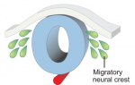 The neural crest is a uniquely vertebrate cell type and has been well studied in a number of model systems. Here, Bariga, Trainor, Bronner and Mayor discuss species-specific differences in neural crest development, urging the community to consider these differences and highlighting the need for further research in complementary systems. See the Spotlight on p.
  Sensory hair cells are mechanoreceptors of the auditory and vestibular systems and are crucial for hearing and balance. Auditory hair cells in adult mammals are unable to regenerate whereas hair cells in the chick cochlea and the zebrafish lateral line are, prompting studies into the factors that regulate hair cell development and regeneration in various species. Here, Cheng and co-workers review these studies. See the Review on p.
 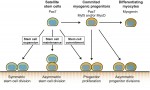 Muscle stem cells, termed satellite cells, are crucial for skeletal muscle growth and regeneration. Here, Rudnicki and colleagues review recent discoveries of the intrinsic and extrinsic factors that regulate satellite cell behaviour in regenerating and degenerating muscles. See the Review on p.
  (No Ratings Yet)