The community site for and by
developmental and stem cell biologists

When the mind is given wings…

Posted by , on 14 March 2014

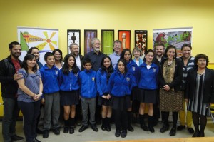
In the beginning of 2014 (January 5-17th) we enjoyed one of the most fascinating experiences professionally and personally. Under the guidance of Dr. Roberto Mayor and a teaching staff that enlightened and motivated us at all times, a wonderful group of 21 students accomplished the 2014-International Course on Developmental Biology at the CIMARQ in Quintay-Chile. Students, mostly from Latin-American countries and some from Europe and the United States, participated in a unique and rewarding experience.

We could dedicate more than a hundred pages talking about the unbelievably high quality of lectures and discussions, the laboratory practices, and the professors themselves and that still wouldn’t be enough to describe the positive impact of this course on our careers. While we enjoyed the beauty and peace of that place during those long and exciting days a prerequisite for every top international course, we were blown away by the warmth of our hosts – and idols – in developmental biology, listening to them impart their expertise and sharing their personal experiences; how they became what they are, the hard decisions and sacrifices they made in order to follow their passion for this vocation.

We had the unique chance to interact with internationally renowned developmental biologists as they shared with us their insights on a wide range of topics. Everyone in their own manner taught us valuable lessons which will be helpful for our own research projects now and in the future. We were also encouraged to look beyond model animals and to go out and think about the fundamental questions in the field of developmental biology from a new perspective, to consider the depth of diversity in the developmental process and the forms it generates, to appreciate the interaction of an organism’s development with its natural habit and to integrate ideas not classically thought to be part of the field. These different views gave us the opportunity to understand development in a broad, holistic manner.

Moreover, we were given the gift of motivation. Motivation to follow our ideas, our instincts, to go into battle with our thoughts and try to give new insights to our projects. As young scientists, we spend our days in the lab trying to fulfill our desires and expectations, giving our absolute best for our projects, trying to push ourselves to be the best researchers we can; and it is not a minor fact that as Latin American students most of us have to deal with great challenges to ensure that our work is up to the right standards. For that reason, the encouragement that we received during the course from all the faculty members was a turning point in our careers. It made us realise that with hard work and enthusiasm, there is no goal that we cannot accomplish.

Another keystone of the course was the opportunity to formally present our own work to classmates and teaching staff. We received essential feedback not only about our projects but also on how to deliver and present science to a diverse scientific community, an art that is demanded of all scientists. This was certainly a rewarding experience for everyone, no doubt about it. Furthermore, we now have a better idea of the research that young Latin-American developmental biologists are doing and how we can network with other researchers and classmates around the world.

An addition to this outstanding experience was the mini-symposium about Developmental Biology which gave us the opportunity to take part in the life of the scientific community in Chile. High-quality lectures gave shape to this event as Chilean and international experts shared their research and thoughts in a delightful atmosphere. The symposium truly completed our experience.

We strongly believe that one of the most powerful tools we have in science is our desire to progress together as a community because we certainly know that the whole is greater than the sum of its parts and we choose that principle as a way of doing science. This course is tangible proof of that, bringing state-of-the-art technology and first class teaching to Latin American students and allowing us to take advantage of this every single day so that we all develop as a group and as researchers. We generated strong bonds and a great sense of community. This course gave to Latin America (and other parts of the world) a network of young developmental biologists that share each other’s interests, aspirations and passions.

We have learned the most fascinating strategies, pathways, and molecular principles for patterning of current and potential model organisms in developmental biology, and during our lab practices we actually heard some of the beautiful chords of their melodic development. However, even more interesting was to personally experience one of the most amazing biological processes; the development of wings in our mind, allowing us to see and think further and wider than we could have ever imagined.

Finally, we want to share a beautiful gift from our inspirational teacher Scott Gilbert, who sent to us this quote that he rescued from Pablo Neruda’s home in Isla Negra: “There is nothing more beautiful than something that comes into being, takes shape right in front of us. There is a rigor in the materials that prevents excess folly…” This is a good epigram for developmental biology.


Post by all the Students – class 2014.
International Course on Developmental Biology
Quintay – Chile


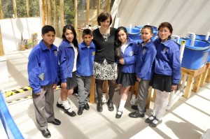
The authors of the post!
Picture by Emilio Lanna. From left to right: Jaime Espina Hidalgo , Gabriela Edwards, Maria Kotini, Kevin Leclerc, Francis Hervas, Santiago Cerrizuela, Gonzalo Aparicio, Daniel Smith, Elías Barriga, Ailin Buzzi, Emilio Lanna, Marcela Arenas G., Gloria Slattum, Gabriel Cavalheiro, Roberto Sanchez, Diego Rojas, Carla Grade, Angie Serrano, Dalmiro Blanco Obregón, Lucimara Sensiate, Guadalupe Barrionuevo, Paul Andrew.


Picture by Emilio Lanna. From left to right: Jaime Espina Hidalgo , Gabriela Edwards, Maria Kotini, Kevin Leclerc, Francis Hervas, Santiago Cerrizuela, Gonzalo Aparicio, Daniel Smith, Elías Barriga, Ailin Buzzi, Emilio Lanna, Marcela Arenas G., Gloria Slattum, Gabriel Cavalheiro, Roberto Sanchez, Diego Rojas, Carla Grade, Angie Serrano, Dalmiro Blanco Obregón, Lucimara Sensiate, Guadalupe Barrionuevo, Paul Andrew.

Thumbs up (12 votes)
Loading...

Tags: ,
Categories: Education, Events

Navigate the archive

Use our Advanced Search tool to search and filter posts by date, category, tags and authors.

Join us at the (alternative) careers in science session, BSCB/BSDB meeting 2014

Posted by , on 12 March 2014

The joint Spring meeting of the British Society for Cell Biology and British Society for Developmental Biology is coming up soon, and the Node will be there! Not only will we be there listening to the talks and meeting the community, but we will also be organising the (alternative) careers session. The session will take place on the first day of the conference (Sunday the 16th of March) at 3 p.m. (main lecture theatre). We have a great panel of science professionals who will tell us a little bit about their careers, but who will mostly answer any questions that you may have:


Woolner


Sean photo 2


Katherine profile pic


Dr Shum Prakash


Paul Turner Ph.D cropped


cat profile pic.jpg


Dr Sarah Woolner, Group Leader at the University of Manchester
Sarah obtained her degree in Biological Sciences from the University of Edinburgh in 2000. She did her PhD at UCL, studying on the MRC LMCB four-year PhD programme and carrying out her thesis work with Paul Martin. Sarah did a first postdoc in Bill Bement’s lab (University of Wisconsin-Madison) and a second in Nancy Papalopulu’s lab (University of Manchester). In July 2012, Sarah was awarded a Wellcome Trust/Royal Society Sir Henry Dale Fellowship to establish her own lab in the Wellcome Trust Centre for Cell Matrix Research at the University of Manchester. The major research goal of her lab is to understand how cell division orientation is coordinated across tissues in the developing embryo.


Dr Seán Mac Fhearraigh, Online marketing manager and sales representative at Molecutools Seán received his Bachelor’s in Genetics from Trinity College Dublin and his PhD from University College Dublin. From there Seán, carried onto to do a short post-doc at the University of Cambridge. After some thought, Seán began his career in industry by taking a job a Abcam as their cancer and stem cell marketer. He is currently online marketing manager and sales representative at molecutools.com and founder of the PhD and Post-Doctoral advice website PostPostDoc.com, you can find Seán on twitter with @PostPostDoc1


Dr Katherine Brown, Executive editor of Development

Katherine gained her PhD from the MRC-LMB in Cambridge, and carried out her postdoctoral work at EMBL Heidelberg. Her research expertise is in the field of morphogenesis, using both Drosophila and teleost fish as model systems. In 2008, she moved into a career in scientific publishing – spending three years as a Scientific Editor at The EMBO Journal before taking up her current position as Executive Editor of Development in late 2011.


Dr Shum Prakash, Business Development Manager at Warwick Ventures

Shum received a PhD in Biochemistry and conducted a postdoc in cancer therapy and brain diagnosis. She worked in policy at the European Parliament, Technopolis Ltd and SQW Ltd. Shum joined Warwick Ventures in 2001 and is an MBA qualified business development manager in technology transfer, taking a broad range of technologies from concept to royalty generating commercial licences and to trading spin off companies.


Dr Paul R Turner, Field Application Specialist for Affymetrix

Paul did a PhD in Cell Biology and Biophysics at the University of Connecticut and a postdoc at the University of California Berkeley. He was a group leader in academia between 1991 and 1999, and has worked in industry (Oxford Glycosciences and GRI) since. Paul has worked as a Field Application Specialist for Affymetrix (Panomics) since 2006.


Moderator- Dr Catarina Vicente, the Node community manager
Cat completed her PhD in Cell Biology at the University of Oxford, researching centrosomes in Drosophila with Prof Jordan Raff. She is now the community manager of the Node, a community blog for developmental biologists and related fields launched by the journal Development.


Please come along and join us! Even if you didn’t sign up for this session when you registered, you are still very welcome to join! And if you are interested in alternative careers in science, you may also want to read some of the posts in the alternative career series that featured on the Node last year. If you are not attending the conference, do follow the official Twitter hashtag: #cbdb14


Thumbs up (2 votes)
Loading...

Tags: , , ,
Categories: Careers, Events

PhD position available to study regulation of macrophage migration in Drosophila

Posted by , on 11 March 2014

Closing Date: 15 March 2021

node banner

PhD position available to study regulation of macrophage migration in vivo using Drosophila

Organisation: Department of Infection and Immunity, The Medical School, University of Sheffield

Supervisor: Dr Iwan Evans

Studentship starting: 1st October 2014

Application Deadline: 18th April 2014

Project:

Inappropriate immune cell responses cause or exacerbate a wide range of human diseases including autoimmunity, atherosclerosis, cancer and chronic inflammatory conditions. An important function of the white blood cells known as macrophages is to remove and degrade cells undergoing programmed cell death (apoptosis). This process can suppress pro-inflammatory responses of macrophages and is linked to the resolution of inflammation.

Fruit flies (Drosophila melanogaster) contain a population of highly migratory macrophages, called hemocytes. The genetic tractability and unparalleled live imaging possibilities of this system enables us to study the cell biology of apoptotic cell clearance by macrophages in vivo. This project will study how clearance of apoptotic cells affects the subsequent behaviour of macrophages, with a strong focus on regulation of their motility and inflammatory responses. We will also characterise how interactions between apoptotic cells and macrophages influences signalling and phenotype within the latter in order to understand how apoptotic cells can dampen inflammatory responses in vivo. Tissue culture approaches will also be used to test the relevance of mechanistic findings to mammalian macrophages.

This project is ideal for a candidate with strong interests in cell biology and the use of model organisms to study human disease-relevant questions in vivo. Knowledge of cell migration is desirable, but an enthusiasm for science and an enquiring mind is far more important. No prior knowledge of Drosophila is required, since the successful candidate will be given intensive training in the use of Drosophila as a genetic platform to study cell biology in vivo. This will involve a significant amount of dynamic imaging of macrophage behaviour in vivo using confocal microscopy, alongside standard cell biological techniques such as whole mount immunostaining and in situ hybridisation.


For further information see:

I. Evans, P. Ghai, V. Urbancic, K.-L. Tan and W. Wood (2013). SCAR/WAVE-mediated processing of engulfed apoptotic corpses is essential for effective macrophage migration in Drosophila. Cell Death and Differentiation. 20(5):709-20.

I. Evans and W. Wood (2011). Drosophila embryonic hemocytes. Curr Biol. 21(5):R173-4.


Evans lab website


Entry Requirements

Candidates must have a first or upper second class honours degree or significant research experience.


To apply

Interested candidates should in the first instance contact Iwan Evans (i.r.evans@sheffield.ac.uk).

For formal applications, visit: http://www.sheffield.ac.uk/faculty/medicine-dentistry-health/graduateschool/prospectivepg/vacancies/ii

Thumbs up (No Ratings Yet)
Loading...

Categories: Jobs

Free online Life Fantastic CHRISTMAS LECTURES teaching resources

Posted by , on 11 March 2014

Alison Woollard presenting the CHRISTMAS LECTURES.

The 2013 Life Fantastic CHRISTMAS LECTURES® presented by Alison Woollard from the University of Oxford, explored the frontiers of developmental biology and uncovered the remarkable transformation of a single cell into a complex organism.

The three hour long lectures investigated questions such as what do these mechanisms tell us about the relationships between all creatures on Earth? And can we harness this knowledge to improve or even extend our own lives?

The Royal Institution (Ri) has developed a series of online CHRISTMAS LECTURES teaching resources comprised of video clips, facts and questions to help primary and secondary school teachers explore the developmental biology covered by Life Fantastic with their students.

On the Ri’s website teachers will find an overview of each of the eight topics including DNA replication and mutation, proteins, cells and organs, and mitosis and meiosis covered by the clips, a brief summary of each clip, related questions and how the topics link to the curriculum. The pages are intended for use as a prompt to explore these topics further in lessons.
The resources are also available on the Ted-ED and TES websites.

All three Life Fantastic lectures, and a range of previous CHRISTMAS LECTURES from our archives, are available to watch in full and for free on the Ri’s critically acclaimed science video platform, the Ri Channel.

Thumbs up (1 votes)
Loading...

Tags:
Categories: Outreach, Resources, Video

From maps to circuits: Models and Mechanisms for Generating Neural Connections

Posted by , on 11 March 2014

We have an upcoming workshop that I hope will be of interest to members on this node.

From Maps to Circuits: Models and Mechanisms for Generating Neural Connections

28/29 July 2014, Edinburgh UK

http://maps2014.org

Organisers: Stephen Eglen, Matthias Hennig, Andrew Huberman, David Sterratt, Ian Thompson, David Willshaw

Aim of the meeting

Understanding the development of the nervous system is a key challenge that has been approached by both experimental and theoretical neuroscientists. In recent years there has been a gradual move towards the two groups working more with each other. The idea of this workshop is to bring key people together who have shown an interest at combining theoretical and experimental techniques to discuss current problems in neuronal development, and plan future collaborative efforts.

Time at the end of each day of the workshop will be devoted to a group discussion about questions that have been raised during the day to identify possible research directions and people willing to pursue them.

Speakers: Tom Clandinin (Stanford), Michael Crair (Yale), Irina Erchova (Cardiff), David Feldheim (UC Santa Cruz), Geoffrey Goodhill (U Queensland), Robert Hindges (Kings College London), Sonja Hofer (Basel), Hitoshi Sakano (U Tokyo), David Wilkinson (NIMR, London), David Willshaw (Edinburgh), Fred Wolf (Gottingen).

This meeting is supported by Cambridge University Press, Company of Biologists, Gatsby Charitable Foundation, Institute for Adaptive and Neural Computation, Wellcome Trust.

Thumbs up (1 votes)
Loading...

Categories: Events

Growing older gracefully – a review of the 10th edition of Developmental Biology

Posted by , on 9 March 2014

This article was first published in Development, and was written by Timothy T. Weil, Department of Zoology, University of Cambridge.


Gilbert Cover.6In the age of Google and Wikipedia, what is the role of the textbook? When revising lecture notes, what motivates a student to pull a clunky book off the shelf, rather than hitting the keyboard and accessing millions of search results in tenths of a second? The question for today’s educator might actually be how to steer undergraduates to the best-suited, most applicable source. When teaching developmental biology, the 10th edition of Developmental Biology by Scott F. Gilbert provides an elegant solution to this conundrum.

Any text in its 10th edition is likely to have had a great deal of success, and Gilbert is no different. The work is comprehensive, with all the expert detail and beautiful data that have become synonymous with his book. While online searches and primary articles can be very intimidating to students and often compound pre-existing confusion, Gilbert works well as an entry point into the vast literature on all the topics covered. The text can function both as a general tool that can be read chapter by chapter, and as a reference for specific questions. This dependable and friendly text enables students to acquire quality basic information, and subsequently directs them smoothly to the primary literature for further exploration.

Like a prologue to a play, the first pages of the text set the scene and introduce the main characters, relationships and drama that motivate the action to follow. Gilbert’s 10th edition is presented in four parts: Questions, Specification, The Stem Cell Concept and Systems Biology. This structure remains mostly unchanged from the previous version; a pragmatic reorganisation that was introduced between the 8th and 9th editions. Each part opens with an introduction in review-like style and standard. These prefaces work well, placing the information to be presented in context, as well as informing and exciting the reader as to why it is important.

The book is further divided into 20 chapters that are well organised, easy to navigate, comprehensive and enriched with primary images and effective diagrams (an impressive 694 illustrations in 719 pages). Within the four parts, the chapters are linked with short, concept-driven openings and end with concluding remarks in the ‘coda’ section, creating a cohesive quality to the book. Also included at the end of each chapter is a ‘snapshot summary’, suggestions for further reading and directions to the online resources that are provided as part of the book package. Although these are expected components of any top textbook, Gilbert executes them extremely well. Throughout the chapters, there are stand-alone sections entitled ‘Sidelights & Speculations’, such as ‘The Nonequivalence of Mammalian Pronuclei’, ‘BMP4 and Geoffroy’s Lobster’ and ‘Transposable DNA Elements and the Origins of Pregnancy’. These vignettes have a mini-review quality to them and are good launch points for small group discussions.

Notably, the 10th edition has considerable new content and references, helping to maintain its position at the leading edge of available textbooks. This includes, but is by no means limited to, content on microRNA-mediated gene silencing, a new Crepidula (sea snail) fate map, epigenetic mechanisms of X inactivation, new ideas of neural tube closure, epithelial-to-mesenchymal transitions in cancer and developmental plasticity due to climate change.

For undergraduate course designers and lecturers, it is useful that the text is question driven, and includes many techniques ranging from classical transplantation and genetic screens to modern molecular networks and super-resolution microscopy. This provides the reader with the necessary information to think about the data as the original researcher did – an essential component in the education of young scientists.

Inherently, however, a textbook is out of date as soon as ink hits paper. It is therefore unfair to criticise the book on failing to include recent advancements, such as the CRISPR/Cas technology for genome engineering in Drosophila and other species. However, these limitations must be noted when considering the place of textbooks, as education inexorably moves towards a paperless existence.

The book ‘extras’ are an attempt to bridge the gap between paper and screen. These include the companion website www.devbio.com – self-described as a ‘museum’ with different ‘exhibits’ that ‘enrich courses in developmental biology’ – and vade macum3, a ‘laboratory manual’ that ‘helps students to understand the organisms discussed and prepare for the laboratory’. Both supplement the text, but are not essential to the book experience. They seek to provide an interactive avenue for students to explore, but when competing against the web are unlikely to become the first point of call for a student. However, they do offer an additional resource for instructors to enhance their lecture material with some available images and short videos. Moreover, by contacting the publisher, lecturers who confirm adoption of the text as part of their course may be granted access to ‘The Instructor’s Resource Library’. This includes images and presentation documents of all figures, tables and videos found in the text. This is a windfall for new lecturers and established educators looking to refresh their material.

Beyond the scientific realm, the text displays Gilbert’s ability to wear other literary hats, keeping the reader interested and engaged. He acts as historian as he brings alive the rich tapestry of developmental biology research; as columnist when relating the science to cultural quotes from the likes of T. S. Eliot, Steve Jobs, Emily Dickinson and Frank Lloyd Wright to name but a few; and finally as comedian with a lighthearted section on the website including ditties such as ‘The Histone Song’, links to YouTube videos and amusing articles.

Altogether, Developmental Biology by Gilbert is a classic and fundamental text. At £52.99 (RRP €63.58, $139.95), it is worth considering whether the 10th edition is a necessary upgrade. For general biology students, older editions will likely suffice. For lecturers or aficionados, the new content is nice, but not essential, especially for anyone owning the 9th edition. However, if you want an excellent text for teaching and learning developmental biology, the 10th edition is an ideal resource.

In the social media-dominated world of today, the future of the traditional course textbook is uncertain. The prospect of a continuously updated, interactive online ‘book’, complete with embedded links to primary sources, live movies and interactive images, is appealing. Still, at present there is no substitute for the well-written, accurate and engaging reference book exemplified by the 10th edition of Developmental Biology by Gilbert.


Developmental Biology, 10th edition by Scott F. Gilbert. Sinauer Associates (2013), 750 pages. ISBN: 9780878939787. $139.95 (hardback).

International Edition: Palgrave Macmillan. ISBN: 9781605351735. £52.99, €63.58.

Thumbs up (7 votes)
Loading...

Categories: Book Reviews

Best of both – balancing research and outreach

Posted by , on 7 March 2014

I have been trying to pin-point when exactly I became interested in science outreach. The earliest I can think back is when we had to dissect Helix aspersa, the garden snail, in our undergraduate zoology practical. It was a slimy business. Our lecturer had asked us to remove the specimen’s body from its shell. It popped out in a spiral, covered in mucus. I took it apart carefully, holding my breath and trying to keep a steady hand whilst unravelling its insides with my shiny new dissecting instruments. As I uncovered its digestive system, its muscles and its ‘love dart’, incision by incision my initial feeling of disgust turned into deep fascination.

A few weeks later I was looking for a present for my little cousin. The book “Die Schnecke” (“The Snail”) seemed like the perfect choice. We read it together, sitting on the ground and admiring the illustrations. I guess that a passion for science communication has always been a big part of my love for science.

Thanks to my supervisors and my University I have been able to develop this passion into an official part of my job. My last post-doc position included a day per week dedicated to outreach, funded by my Faculty. In 2012 I became the University’s first Research and Science Communication Fellow. Now my time is split between plant cell biology and science communication, with a bit of teaching sprinkled on top.

The split is not mathematically accurate and I do not work exactly 2.5 days per week on each. Rather, things come in phases. March is extremely busy with the Oxfordshire Science Festival, and our Brookes Science Bazaar of which I am now the lead organiser. Last year Dr Niall Munro, a lecturer in American Literature, and I developed a training programme for early career science and humanities researchers. With input from a science journalist and poet, our participants paired up to explore interdisciplinary ‘Visions of the Future’, and presented their work on a theatre stage.

Our stall at the Oxfordshire Science Festival 2013 launch event in Oxford City Centre.

I always have several smaller, long-term projects running on the side, such as our DNA gel electrophoresis loan kit scheme or our partnership with the Oxford Academy. Between busy periods I focus on research and restrict my activities to ‘one-offs’ like SciBar or Science Showoff, and social media. I also run internal and external training sessions for researchers and mentor students and scientists who want to dip their toe into science communication.

Sometimes people ask me if I prefer science or science communication. I reply that both have their upsides and downsides. I love the excitement of coming up with hypotheses and designing experiments to test them. But in science, progress tends to be slow. Plants need to grow. Experiments need to be repeated or suddenly stop working for no obvious reasons. This can be very frustrating. When I put hard work and long hours into science communication projects, I know that I will (usually!) get a good outcome. Without exception, all of my projects have been extremely rewarding. I often joke that outreach keeps me sane because it is a positive balance to the constant stream of failures and rejections in science. Sometimes however things happen too fast or sudden and I need to react quickly, for example when two kids are starting to fight in our workshop or volunteers drop out at the last minute.

Juggling research, science communication and teaching is difficult, but not unique to my position. Like many other early career researchers, I am still learning how to squeeze productive bursts into an increasingly fragmented work day. Being able to say ‘no’ is as much an important skill as knowing how to perform a scientific method, and I am getting better at it. I also try to regularly pause and assess – in all areas of my job – whether I am working too much in the ‘urgent, but not important’ quadrant, and not enough in the ‘important, but not urgent’ one.

So how do you get started with outreach? My main advice would be to start small. Engage in existing initiatives: Become a STEM Ambassador, volunteer at your local Science Festival, the British Science Festival or the Big Bang Fair. Try out different communication channels (hands-on activities, comedy gigs, science songs or blog posts, just to name a few!), locations (university, pub, theatre, museum, city centre…) and audiences (children or adults). But at the same time, be realistic about your time and your resources. Always keep in mind your audience and what you want to achieve. Read up on evaluation. Collaborate with others. Volunteer as a research group to run a stall at an event. Write an article with a fellow PhD student, or find like-minded people on Twitter. Work as a team to bounce ideas around and split tasks between you. Once you have tested the water, don’t be afraid to think bigger – who knows where it might lead!

Further reading:
National Co-ordinating Centre for Public Engagement – How to do it.
So you want to do a science communication project?


Outreach logo new squareThis post is part of a series on science outreach. You can read the introduction to the series here and read other posts in this series here.


Thumbs up (2 votes)
Loading...

Tags:
Categories: Lab Life, Outreach

Outreach activity – The Animal Pairs Game

Posted by , on 6 March 2014

Everyone loves a game. So here’s a game that flexes that brain muscle, which can be adapted for all audiences, topics and required level of cuteness.

Presenting: the Animal Pairs Game.

I first produced this for a demonstration event at ThinkTank Museum in Birmingham, a Meet the Scientist day themed around evolution and adaptation. The idea was simple: pair up the most similar animals. So, among the laminated cards, each with striking images and brief descriptions, were two birds, two mammals, two insects and many other animal groups. Among them were animals from similar ecological niches with strikingly similar appearances, yet differing classification, such as the European hedgehog and the echidna; and similar animals with big differences in appearance, such as the hedgehog and a polar bear — one small, one big, both from very different habitats, yet both mammals. Among them, also, were a number of wildcards to provoke critical thinking: should the archaeopteryx, for example, pair with a bird or a reptile?

The full list of creatures was a veritable menagerie intended to provoke wonder and to broaden horizons about the natural world, and linked as much as possible with the museum’s taxidermy collection. Here, pre-paired, are what I presented:

Echidna – Platypus (Monotremes)
Hedgehog – Polar bear (Mammals)
Nautilus – Snail (Molluscs)
Turtle – Icthyosaur (Reptiles)
Fish – Shark (Fish)
Kakapo – Blue-footed booby (Birds)
Crab – Mite (Arthropods)
Bryozoa – Worm (Spiralia)
Butterfly – Beetle (Insects)
Coral – Sea anemone (Cnidaria, with a silent c if you please)
And the wildcards: the Archaeopteryx, Hagfish and Horeshoe crab

As you can see, many of them were pretty difficult, but the beauty of the activity was that cards could be removed for different audiences, and others reclassified (you could use a wider deuterostome/protostome classification, for example, or the lophotrochozoa/ecdysozoa split). You could make a big deal about living fossils, if you fancied, or just tell tales about your favourite animals. All in all, it had many children captivated, and kept their attention for a lot longer than other activities I have demonstrated in the past.

The activity could be tailored for any subject. For developmental biology, for example, you could stick with the protostome/deuterostome divide and ask which develop with a dorsal (like our spines) or ventral (like the Drosophila ventral nerve cord) nervous system, or which form mouth first… or alternative openings? Maybe you could use pictures to signify how similar the earliest stages of vertebrate embryology are? Plenty of developmental biology concepts could be introduced with weird and wacky pictures and a game to pair them, particularly for audiences who would never have seen anything on this topic before.

Here’s the downside: the game requires a lot of ongoing explanation, even for older children. I retired the bryozoa card early, for example, and found myself constantly explaining the differences between — on the surface, rather similar — creatures. But the reward for doing so was undivided attention, and evident fascination. To avoid this downside, consider carefully the details written on the cards below the pictures, and have clear examples on show. Alongside the activity I presented a poster of the tree of life, which I would recommend.


Also read Simon’s outreach post on his internship with the Naked Scientists, bringing developmental biology to the radio.


Outreach logo new squareThis post is part of a series on science outreach. You can read the introduction to the series here and read other posts in this series here.


Thumbs up (2 votes)
Loading...

Tags:
Categories: Outreach, Resources

Generation of Embryoid Bodies: a great tool to study vascular development

Posted by , on 4 March 2014

Hello, my name is Helena and I am a PhD student within the Vascular Signalling Laboratory led by Mariona Graupera in the Bellvitge Biomedical Research Institute (IDIBELL) in Barcelona. It has been 4 years since I started my project in the lab, and before finishing my thesis research, we thought it was a good moment to explore another vascular signalling laboratory and learn from their knowledge and techniques. I just returned from a stage in Dr. Holger Gerhardt’s laboratory in London, funded by the Development Travelling Fellowship from the Company of Biologists.

During my visit at Dr. Gerhardt’s lab I have learned one of the most useful and interesting techniques in the vascular biology field: the generation of embryoid bodies. The embryoid bodies are three-dimensional aggregates of pluripotent stem cells that after stimulation with the vascular endothelial growth factor A (VEGF-A) can be differentiated into endothelial cells and generate vascular sprouts that will grow in between two collagen layers (figure 1).

Dr. Gerhardt’s team has helped me to create embryoid bodies generated from stem cells deficient of my protein of interest. This technique has offered me an excellent ex vivo approach to study in greater detail the role of my protein of interest in sprouting angiogenesis. The experiments that I have carried out during my stage in London will very nicely complement the in vivo work that I have been doing for the last years with the study of the retinal mouse vasculature. Besides the exciting experiments that I have done in London, I have also had the opportunity to meet and interact with different professionals involved in the vascular signalling field that have shared with me their knowledge.

Apart from the academic experience, my personal experience in London has also been fantastic. London has been a great city to spend these 3 months. It is a huge city with so many places, parks, markets and experiences to discover. Of course, compared to Barcelona, the weather was not the best thing to remember but I was lucky that I could enjoy quite a few sunny days!

This experience has been a very nice way to finish my PhD period and I sincerely thank to the Company of Biologists their support.

figure 6 EB

Thumbs up (7 votes)
Loading...

Tags: , ,
Categories: Funding, Research

In Development this week (Vol. 141, Issue 6)

Posted by , on 4 March 2014

Here are the highlights of the current issue of Development:

Auxin biosynthesis: the root of xylem patterning

DEV103473F1In the Arabidopsis root, xylem is organised into a central file of metaxylem that is flanked by protoxylem. Xylem fate is determined by HD-ZIP III transcription factors; high levels promote metaxylem formation whereas low levels specify protoxylem. The factors that downregulate HD-ZIP III levels are known but those that promote HD-ZIP III expression have remained elusive. Yunde Zhao, Ykä Helariutta, Jan Dettmer and colleagues now show that auxin biosynthesis promotes HD-ZIP III expression and metaxylem specification in Arabidopsis (p. 1250). The authors first isolate mutants that display defective xylem patterning and HD-ZIP III gene downregulation. These mutants harbour mutations in the gene encoding TRYPTOPHAN SYNTHASE BETA-SUBUNIT 1. Tryptophan is a precursor in the auxin biosynthesis pathway and, accordingly, the mutants exhibit aberrant auxin levels. They also show that metaxylem development and HD-ZIP III levels are defective in other auxin biosynthesis mutants. Based on their findings, the authors propose that tryptophan-dependent auxin biosynthesis is required for metaxylem formation.


From Hippo to planarians

DEV101915F2The transcriptional co-factor Yorkie (Yki; YAP in vertebrates) is a key effector of the Hippo signalling pathway and has been implicated in growth control and patterning, as well as stem cell regulation and regeneration, in flies and vertebrates. Now, on p. 1197, Alexander Lin and Bret Pearson investigate the role of Yki in planarian flatworms. The researchers report that planarians have a single orthologue of yki and, using RNAi, they show that yki carries out pleiotropic functions. For example, they report that ykiis required for homeostasis of the planarian excretory system, which is analogous to the vertebrate kidney. The researchers also show that, in contrast to its role in flies and vertebrates, yki functions to limit stem cell proliferation and hence the size of the stem cell population. In addition, yki plays a role in axial patterning, where it acts synergistically with Wnt signalling to supress head formation. These, together with other findings, demonstrate that yki plays diverse yet non-overlapping roles in planarian biology.


Following the leader

DEV101675F3Collective cell migration occurs in many developmental contexts but a full understanding of this process has been hampered by a lack of quantitative analyses in 3D in vivo contexts. Using the zebrafish lateral line primordium as a model, Darren Gilmour and co-workers set out to tackle this problem (p. 1282). The researchers develop a method to simultaneously live-label microtubules, centrosomes and nuclei, allowing them to map cell polarity and orientation across the migrating population. Using this method, they identify the transition between leader cells and followers within the collective and show that this transition is marked by changes in cell-cell adhesion; cadherin 2 is expressed across the tissue but is only assembled into adherens junctions (AJs) in the transition zone and further down the leader-follower axis. A tandem fluorescent protein timer-based approach reveals that AJs become progressively more stable along the leader-follower axis. Finally, they show that AJ assembly, but not maintenance, requires dynamic microtubules, revealing a key role for microtubules during leader-to-follower transitions within migrating collectives.


Dbx1 crosses the (mid)line

DEV102327F5During nervous system development, navigating axons ‘decide’ whether or not to cross the midline. Various factors that influence axon guidance and midline crossing have been identified but it remains unclear if any one transcription factor can drive the complete midline crossing transcriptional programme. Here (p. 1260), Yasuyuki Inamata and Ryuichi Shirasaki report that a single homeodomain transcription factor, Dbx1, assigns midline-crossing identity at the progenitor stage in mice. The researchers show that Dbx1 is expressed in a subset of neural progenitors in the dorsal midbrain. Lineage tracing demonstrates that midbrain commissural neurons, which cross the midline, are generated selectively from Dbx1-positive progenitors. Furthermore, gain- and loss-of-function experiments show that Dbx1 is necessary and sufficient for midline crossing. Finally, the authors show that Dbx1 controls a molecular programme that controls the expression of Robo3, an essential regulator of midline crossing, on commissural neurons while repressing the ipsilateral neuron genetic programme. These findings reveal an unanticipated regulatory layer within the transcriptional cascade that controls nervous system wiring.


Lymphangiogenesis: of mice and fish

Fig1 finalIn mice, the formation of lymphatic vessels (lymphangiogenesis) requires the homeodomain transcription factor Prox1. Here, Stefan Schulte-Merker and colleagues examine whether the role of Prox1 is conserved in zebrafish (p. 1228). Using a novel transgenic reporter line, the researchers show that, in contrast to the situation seen in mice, zebrafish Prox1 is initially not expressed in all lymphatic precursor cells and reliably marks this population only during later stages of lymphangiogenesis, arguing against a role for Prox1 in lymphatic specification. In addition, targeted mutagenesis demonstrates that lymphangiogenesis can proceed in the complete absence of Prox1. Finally, they show that the functionally related transcription factors Coup-TFII and Sox18, which are implicated in lymphatic specification in mice, are also dispensable for zebrafish lymphangiogenesis. The authors conclude that an alternative lymphatic specification mechanism is present in zebrafish and propose that differences in the timing of lymphangiogenesis between mice and fish can explain this divergence.


Fresh air in the mesenchyme

Figure_1The mesenchymal compartment of the lung plays a crucial role during lung development but, unlike its epithelial counterpart, its regulation is largely uncharacterised. Now, Gianni Carraro, Saverio Bellusci and colleagues report that miR-142-3p modulates WNT signalling to balance mesenchymal cell proliferation and differentiation during mouse lung development (p. 1272). Using microarray analyses, the researchers identify miR-142-3p as highly expressed in the embryonic lung mesenchyme. Importantly, loss-of-function assays demonstrate that miR-142-3pregulates cell proliferation specifically in the mesenchyme; in the absence of miR-142-3p, progenitor cells prematurely differentiate. They also show that miR-142-3p binds to and regulates the expression of mRNA encoding APC, a negative regulator of WNT signalling. Accordingly, miR-142-3p knockdown can be rescued by activating WNT or reducing APC expression in the mesenchyme. Based on their findings, the authors propose that miR-142-3p adds an extra layer of control to the WNT-FGF feedback loop that operates in the lung mesenchyme to correctly balance cell proliferation and differentiation.


PLUS…


Growing older gracefully – a review of the 10th edition of Developmental Biology

Gilbert Cover.6First published in 1985, Developmental Biology by Scott F. Gilbert has been an influential textbook for a generation of scientists. Here, Timothy Weil provides a brief review of the latest (10th) edition of the series. He comments on updates to the text, highlights details of particular interest to lecturers, and compares this book with other resources available in the internet era. See the Spotlight article on p. 1177


RNA polymerase II pausing during development

Pol II pausingThe transcription of developmental control genes by RNA polymerase II (Pol II) is commonly regulated at the transition to productive elongation, resulting in the promoter-proximal accumulation of transcriptionally engaged but paused Pol II. In their poster article, Bjoern Gaertner and Julia Zeitlinger review the mechanisms and possible functions of Pol II pausing during development. See the Development at a Glance article on p. 1179


Shared signaling systems in myeloid cell-mediated muscle regeneration

muscle regenerationMuch of the focus in muscle regeneration has been placed on identifying and delivering stem cells to promote regenerative capacity. As these efforts have advanced, we have learned that complex features of the microenvironment in which regeneration occurs can determine the success of these approaches . Here, James Tidball and colleagues discuss how myeloid cells can influence muscle regeneration, focussing on how processes in muscle and myeloid cells are co-regulated. See the Review on p. 1184


Thumbs up (1 votes)
Loading...

Categories: Research