The community site for and by
developmental and stem cell biologists

Open study hopes to put STAP in the past

Posted by , on 12 May 2014

This article is a re-post of an article published at the F1000Research blog on the 8th of May, 2014. Eva Amsen is the outreach director of F1000Research.


Many of you will have been following the STAP stem cell saga: In January, researchers from Japan announced in Nature that they had produced induced pluripotent stem cells (iPS cells) by bathing somatic cells in acid. Other researchers were sceptical of these claims, and tried to reproduce the work. One of those scientists, Kenneth Lee of the Chinese University of Hong Kong, liveblogged his attempts on ResearchGate.

It soon became apparent that one of the underlying problems was that not a lot was known about the experiments in the original paper. Not all data was available, some data were potentially incorrect, and the protocol appeared to be insufficient to reproduce the work.

Lee pushed on, though, and using an updated protocol he systematically kept track of everything he did, and openly discussed it with others.

Today, F1000Research has published the full summary of Lee’s work, with all underlying data sets. Using white blood cells isolated from the spleen of neonatal mice – the same cells used in the original study – as well as lung fibroblasts, Lee was unable to replicate the original findings.

 
STAP

No iPS cell markers after acid treatment. Image from article.


Lee’s article has undergone a pre-refereeing check, and has now been sent to peer reviewers, whose comments you will be able to read underneath the article as and when they come in – with reviewer names. Once the article passes peer review (either in this version or after revisions), it will be indexed in PubMed and other external databases.

We use this completely transparent process for all articles we publish in F1000Research, and we believe that this particular case very clearly shows the benefit of a transparent system over the more traditional approach that has inevitably led to the ongoing problems following the original article in Nature: you will all be able to see what the invited reviewers think about Lee’s article, you can leave your own comments as well, and you can track any new incoming referee reports or comments on the paper by clicking “Track” on the article page.

You can also download the associated data sets to do your own analysis. We know that some other stem cell researchers have tried to replicate the acid bath experiments – now you can see how your data compare to those of the Lee lab. (And if you’d like to publish your own findings as a short Data Note, we’re currently waiving the article processing charge on those. Find out more here.)

Stem cell science has suffered from a closed publishing system perhaps more than many other disciplines, and it’s time to open up.


[You can find Lee’s article here, and download the associated press release (pdf) here.]


Thumbs up (1 votes)
Loading...

Tags: ,
Categories: Discussion, Research

Navigate the archive

Use our Advanced Search tool to search and filter posts by date, category, tags and authors.

Grasping tendon development with the zebrafish

Posted by , on 9 May 2014

by Jessica Chen and Jenna Galloway


Animals can contort their bodies into a diversity of movements: running, jumping, climbing, and swimming to name a few. All of these movements are possible because tendons transmit the force produced by the muscles to the bones. Most of us do not pay much attention to our tendons and ligaments until something happens to them. Sports and repetitive motion injuries are very common yet are complicated by slow and limited repair. Surprisingly, very little is known about how tendons and ligaments form and organize to make the appropriate connections within the musculoskeletal system, and then, maintain and repair themselves in the adult. Part of our limited knowledge concerning their developmental program had been due to the absence of early markers at stages that preceded the morphological detection of tendons. The identification of the transcription factor Scleraxis (scx)as a robust marker of tendon and ligament progenitors provided the means to gain an understanding of the molecular regulators of tendon cell induction and organization (Schweitzer et al., 2001). With these questions in mind, we developed the zebrafish as a model to study tendon biology in our recent paper in Development.

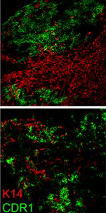
Why study zebrafish and do they have tendons? When we began these studies, we were often asked this question, and itforced us to consider if zebrafish would be an appropriate model to study tendon and ligament biology. Clearly, the forces fish experience in an aquatic habitat are much different than those felt by terrestrial land animals. As such, the tendons in these diverse species, which perform very different movements, could also be different. Previous molecular studies of zebrafish tendon tissues have focused on the myosepta, which connects the muscle segments along the body axis enabling undulatory swimming (Bassett et al., 2003; Charvet et al., 2013; Kudo et al., 2004). The myoseptal tissue functions as a tendon in transmitting force necessary for swimming, and we found that it expresses many tendon markers during developmental stages. At these stages, however, it primarily connects muscle to muscle, and in adult zebrafish, its structure is not similar to the linear tendons of mammals (Charvet et al., 2011; Summers and Koob, 2002). In contrast, we focused most of our studies on the cranial region when we began examining tendon markers in zebrafish embryos. We concentrated on this anatomical location for two principal reasons: here, cartilage and bone are primarily found developing in close proximity to muscle, and second, the pressure for prey capture and feeding would require a functioning musculoskeletal apparatus at very early stages. Indeed, it was in the cranial regions that we found the co-expression of many tendon markers, including scleraxisa, and also where, in adults, the tissue was similar on the ultrastructural level to that of the linear tendons of mammals.

77h_ventral_scxa-xirp2a-DAPI-2
In demonstrating that the zebrafish cranial tendon populations are homologous to their mammalian counterparts, we have expanded our ability to study this tissue in the context of musculoskeletal patterning. Although our work was the first to molecularly describe the head tendons and ligaments in zebrafish, the cranial musculoskeletal anatomy and functional morphology of ray-finned fishes has been studied for over a century (reviewed in (Ferry-Graham and Lauder, 2001)), and models of feeding mechanics in adult fish have detailed cranial tendon and ligament attachments in the context of their role during jaw movements (Liem, 1967; Westneat, 1990). During ontogeny, efficient feeding during developmental stages is linked to survival and thought to be evolutionarily advantageous (Houde and Schekter, 1980). We found it striking that the regions with robust co-expression of tendon markers coincided with pivotal points of force transmittance during larval feeding (Hernandez et al., 2002). Given the diversity of craniofacial morphology in teleosts, it would be interesting to understand how tendon and ligament progenitor induction and organization may play a role in shaping the cranial musculoskeletal anatomy. It is known that the neural crest-derived connective and skeletal tissues pattern the cranial muscle attachments in avian systems (Noden, 1986; Noden, 1988). Our work suggests that cartilage may have a role in tendon organization, and previous studies have found that a disruption to cartilage development results in distorted muscle shapes (Yan et al., 2002). Together, these results underscore the importance of tendon-cartilage interactions in musculoskeletal patterning, and future work in the zebrafish will begin to dissect the mechanisms underlying these processes.

Ultimately, we believe that the fish will provide new avenues for studying tendon and ligament biology in a powerful vertebrate genetic system. Findings from chemical screens in zebrafish have already demonstrated the potential for clinical translation in the treatment of a variety of human disease and developmental conditions (Bowman and Zon, 2010; Kaufman et al., 2009). Identifying the pathways that regulate tendon progenitor cell induction, growth, differentiation, and the formation of the attachment sites has relevance in the clinical setting where poor healing, scar tissue and high failure rates at the tendon-bone interface are quite common. The zebrafish system has the potential to not only expand our knowledge of the mechanisms underlying tendon formation and organization through the use of live-imaging and screen based approaches, but also the ability through the creation of injury and disease models and the development of drug discovery platforms to impact clinical therapies.


References

Bassett, D., Bryson-Richardson, R. J., Daggett, D. F., Gautier, P., Keenan, D. G., & Currie, P. D. (2003). Dystrophin is required for the formation of stable muscle attachments in the zebrafish embryo Development, 130 (23), 5851-5860 DOI: 10.1242/dev.00799

Bowman TV, & Zon LI (2010). Swimming into the future of drug discovery: in vivo chemical screens in zebrafish. ACS chemical biology, 5 (2), 159-61 PMID: 20166761

Charvet, B., Guiraud, A., Malbouyres, M., Zwolanek, D., Guillon, E., Bretaud, S., Monnot, C., Schulze, J., Bader, H., Allard, B., Koch, M., & Ruggiero, F. (2013). Knockdown of col22a1 gene in zebrafish induces a muscular dystrophy by disruption of the myotendinous junction Development, 140 (22), 4602-4613 DOI: 10.1242/dev.096024

Charvet, B., Malbouyres, M., Pagnon-Minot, A., Ruggiero, F., & Guellec, D. (2011). Development of the zebrafish myoseptum with emphasis on the myotendinous junction Cell and Tissue Research, 346 (3), 439-449 DOI: 10.1007/s00441-011-1266-7

Ferry-Graham, L., & Lauder, G. (2001). Aquatic prey capture in ray-finned fishes: A century of progress and new directions Journal of Morphology, 248 (2), 99-119 DOI: 10.1002/jmor.1023

Hernandez, L., Barresi, M. J., & Devoto, S. H. (2002). Functional Morphology and Developmental Biology of Zebrafish: Reciprocal Illumination from an Unlikely Couple Integrative and Comparative Biology, 42 (2), 222-231 DOI: 10.1093/icb/42.2.222

Houde, E., & Schekter, R. (1980). Feeding by marine fish larvae: developmental and functional responses Environmental Biology of Fishes, 5 (4), 315-334 DOI: 10.1007/BF00005186

Kaufman, C., White, R., & Zon, L. (2009). Chemical genetic screening in the zebrafish embryo Nature Protocols, 4 (10), 1422-1432 DOI: 10.1038/nprot.2009.144

Kudo, H., Amizuka, N., Araki, K., Inohaya, K., & Kudo, A. (2004). Zebrafish periostin is required for the adhesion of muscle fiber bundles to the myoseptum and for the differentiation of muscle fibers Developmental Biology, 267 (2), 473-487 DOI: 10.1016/j.ydbio.2003.12.007

Liem KF (1967). Functional morphology of the head of the anabantoid teleost fish Helostoma temmincki. Journal of morphology, 121 (2), 135-58 PMID: 6034528

Noden, D. (1986). Patterning of avian craniofacial muscles Developmental Biology, 116 (2), 347-356 DOI: 10.1016/0012-1606(86)90138-7

Noden DM (1988). Interactions and fates of avian craniofacial mesenchyme. Development (Cambridge, England), 103 Suppl, 121-40 PMID: 3074905

Schweitzer R, Chyung JH, Murtaugh LC, Brent AE, Rosen V, Olson EN, Lassar A, & Tabin CJ (2001). Analysis of the tendon cell fate using Scleraxis, a specific marker for tendons and ligaments. Development, 128 (19), 3855-66 PMID: 11585810

Summers, A., & Koob, T. (2002). The evolution of tendon — morphology and material properties Comparative Biochemistry and Physiology Part A: Molecular & Integrative Physiology, 133 (4), 1159-1170 DOI: 10.1016/S1095-6433(02)00241-6

Westneat, M. (1990). Feeding mechanics of teleost fishes (Labridae; Perciformes): A test of four-bar linkage models Journal of Morphology, 205 (3), 269-295 DOI: 10.1002/jmor.1052050304

Yan YL, Miller CT, Nissen RM, Singer A, Liu D, Kirn A, Draper B, Willoughby J, Morcos PA, Amsterdam A, Chung BC, Westerfield M, Haffter P, Hopkins N, Kimmel C, & Postlethwait JH (2002). A zebrafish sox9 gene required for cartilage morphogenesis. Development, 129 (21), 5065-5079 PMID: 12397114

Thumbs up (1 votes)
Loading...

Tags: , , ,
Categories: Research

Intern, Journal of Cell Science

Posted by , on 8 May 2014

Closing Date: 15 March 2021

Journal of Cell Science is looking for an enthusiastic intern who wishes to gain experience in science publishing.

Journal of Cell Science publishes primary research articles and a well-regarded front section of reviews and topical comment. Joining an established and successful team, including Academic Editor-in-Chief Michael Way, the internship offers an ideal opportunity to gain in-depth experience on one of the major journals in the field of cell biology. The intern will work alongside an experienced Executive Editor in our Cambridge offices.

The intern’s core responsibilities will include:

  • Representation of the journal at scientific conferences and within the wider scientific community, with a view to promoting the journal and commissioning new front-section content
  • Conducting interviews with early-career scientists
  • Writing short summary pieces to highlight the key findings of some of the journal’s research articles
  • Creative involvement in the journal’s development and marketing activities

The internship will last for 9 months at a salary of £15,000 pro rata. Applicants will have a PhD in cell biology or a related field and a broad knowledge of cell and molecular biology. Excellent time management, organisational and communication skills are essential, as are enthusiasm and self-motivation. Previous editorial experience is not required.

The Company of Biologists (www.biologists.com) is a not-for-profit organisation that publishes the three well-established, internationally renowned journals Journal of Cell Science, Development and The Journal of Experimental Biology, as well as the two newer Open Access journals Disease Models & Mechanisms and Biology Open. The organisation has an active programme of charitable giving for the further advancement of biological research, including travelling fellowships for junior scientists and contributions to academic societies and conferences.

To apply, please email your CV and a covering letter, quoting reference JCSINT2014, to Miriam.Ganczakowski@biologists.com. Candidates must be able to demonstrate their entitlement to work in the UK. Please direct informal enquiries to Miriam on 01223 426 164.

Thumbs up (No Ratings Yet)
Loading...

Tags: , , ,
Categories: Jobs

How The Company of Biologists helps the scientific community

Posted by , on 8 May 2014

As most of you will know, the Node is run by The Company of Biologists, a not-for-profit publisher of several journals, including Development. The Company of Biologists exists to benefit the scientific community, and as such all profits are given back to the community in different ways: by sponsoring meetings and societies, funding travelling fellowships and, most important for those reading this, allowing the Node to exist!

At the recent joint meeting of the British Society for Cell Biology and British Society for Developmental Biology, the company interviewed several researchers based in the UK that are associated with, and benefit from, the activities of the company. Watch the short video below for a quick snapshot of how The Company of Biologists helps the scientific community attending the Warwick meeting:


You can find more information about The Company of Biologists, and how you can apply to their grants and fellowships, by visiting their website.

Thumbs up (1 votes)
Loading...

Categories: Video

Good guilt

Posted by , on 7 May 2014

Seán Mac Fhearraigh, from PostPostDoc, recently asked me to write a post about my experiences outside academia. Because I don’t have a lot of experience yet (I have only been working as the Node community manager for less than a year) I decided instead to focus on what I did during my PhD that helped the transition. I have reposted the article here as I thought it might of interest to the Node readers, but you can also read the article in its original page at PostPostDoc.


I spent a lot of my PhD feeling guilty. Guilty that I wasn’t working hard enough. Guilty that I wasn’t reading enough papers. Guilty that I might not be thinking hard enough about my experiments. And most importantly, guilty of all those times when I wasn’t in the lab doing experiments. At my undergraduate graduation party one of my lecturers, finding out that I was going to do a PhD, gave me some advice: ‘the PhD is a marathon, not a sprint’. I interpreted this as: make sure you take it easy and do other stuff during your PhD, or otherwise you will run out of energy before the end.

Following her advice, I made sure that experiments were not everything. During my PhD I was involved in all sorts of other activities: I ran a lecture series, I organised a small conference, I ran my lab’s twitter account, just to name a few. This followed on from some of the projects I had been involved with during my undergraduate degree, such as writing a blog or recording short science radio programmes.  To be fair, all these projects were related with science, but they were not happening in the lab, and were not going in my thesis. I did all these projects because I enjoyed them, and also because they gave me a sense of achievement. Most of my experiments lasted several months from the point I started collecting my sample, to the time I could actually image something, and were all consuming- I could only really deal with one mutant at a time. With each experiment taking that long, I needed to be involved in something with a slightly shorter time frame for the sake of my own mental health. Yet, despite all these good reasons, that nagging feeling of guilt was always there.


CRUK stallCell division and cancer stall at a CRUK open day


When I reached the last year of my PhD, the obvious question of what to do next came to the forefront. I actually applied for a few postdocs, but as I discussed projects it became obvious that I had no wish to continue in the lab. The thought of starting a project all over again, in fact, of having to do even one more PCR, just depressed me. But if I wasn’t going to be a postdoc, what would I do? I remembered the words of Sarah Blackford, who I met at a careers session. Sarah said that she had decided to leave the lab because she enjoyed everything that she did as a scientist, except for the experiments. A bit of soul searching showed that this was exactly the same for me. I did enjoy the time during my PhD- just not doing the actual lab work! In fact, what I really enjoyed was doing all those things that had made me feel guilty!

I started searching for jobs, and soon realised that there were some out there that involved all the things that I enjoyed. They required someone with scientific training, involved interactions with scientists, and needed someone enthusiastic with interest in social media, writing, and so on. My worry was now whether I had enough experience. It must be said that PhD students tend to underestimate their own employability. We actually have many transferable skills- we can write, we can present, we can work in a team and be organised, and all of this under pressure. And we can show without a doubt that we can carry a project through to the end. We have a doorstopper (read: thesis) to prove it! But the most decisive criteria in getting my current job must have been all those extra activities and projects. Yes, not a comprehensive portfolio, but enough experience to show my ability to do my current job- community manager of a science blog, and responsible for the social media presence of a journal.

In retrospect, I was silly to feel so guilty about my extra projects and activities. I don’t recommend that you lazy about during your PhD if you want to finish it, but I would strongly encourage any students to get involved in other projects outside the lab. If you find at the end of your PhD that academia is not for you, maybe those projects will help you discover what else you enjoy doing, and may just give you that extra advantage when you apply for your first job. And if nothing else, the sense of achievement you get might help you deal with the ups and downs of science.


Thumbs up (1 votes)
Loading...

Tags:
Categories: Discussion, Lab Life

Crick Quantitative Biology Conference – June 5th

Posted by , on 6 May 2014

The NIMR are hosting the Crick Quantitative Biology Conference on June 5th. This will cover a broad range of interdisciplinary areas of research including tissue growth, cell mechanics, imaging, gene-networks, evolution etc.  Please see the attached poster for details including a link to the online (free) registration page.


QBIOPOSTER

Thumbs up (No Ratings Yet)
Loading...

Categories: Events

Outreach Activity – Smartphone to Microscope Conversion

Posted by , on 6 May 2014

Development is a fascinating process that few people have a chance to see, let alone photograph! We recently participated with other scientists from the Crick Institute at a Science Museum Lates in London in February. For our activity, we built these inexpensive platforms that convert a user’s smartphone into a microscope screen.

We provided zebrafish embryos at different developmental stages for visitors to visualize and photograph. We showed visitors how to use the simple platforms that include a lens from a laser pointer for magnification. Once visitors had their phones lined up on the platform, they were able to view individual cells in early stage embryos or structures like the eye, brain, and heart in older specimens. We described the process of development from cell divisions to cell movements, gastrulation and segmentation. We found that the best practice was to mount embryos in sealed, agarose-coated petri dishes. This kept the embryo medium and zebrafish contained and the dishes were easy for visitors to manipulate. We also found that focal plane could be slightly different for various phones; it was helpful for visitors to remove phones from their cases.

The Francis Crick Institute - Science Museum Lates

Photo credit: Thomas S. G. Farnetti/Wellcome Images

 
Visitors were very excited to be able to take images like these with their smartphones. The event produced several shares on social media sites like Twitter and Instagram of pictures of zebrafish.

Eight cell stage zebrafish embryo #scicomm #microscopy

Photo credit: Alexis Webb


These microscope platforms are affordable and portable, making them suitable for demonstrations in schools and classrooms. Because they don’t require any power, they can be used outdoors or in areas that do not have electrical outlets. Imaging other types of live specimens is also possible. We hope that other researchers interested in showing off their model organism will consider using this type of low-tech, but high reward activity!


Outreach logo new squareThis post is part of a series on science outreach. You can read the introduction to the series here and read other posts in this series here.


Thumbs up (2 votes)
Loading...

Tags: , ,
Categories: Outreach, Resources

In Development this week (Vol. 141, Issue 10)

Posted by , on 6 May 2014

Here are the highlights from the current issue of Development:


Sara sorts out stem cell asymmetric division

FigureAdult stem cells play crucial roles in tissue homeostasis, giving rise to both new stem cells and differentiating daughter cells. The generation of these two cell types often involves the asymmetric distribution of cell fate determinants, but how these factors are partitioned asymmetrically has been unclear. Now (p. 2014), Chrystelle Montagne and Marcos Gonzalez-Gaitan reveal a role for Sara endosomes in the asymmetric division of Drosophilaintestinal stem cells (ISCs). Using live imaging of the adult fly midgut, the researchers first show that Sara endosomes, which are characterised by the localisation of the endosomal protein Sara, are unequally partitioned during ISC divisions, being preferentially targeted to the presumptive differentiating cell. They further examine the distribution of Notch and Delta, which have been implicated in regulating ISC fate, and show that both Notch and Delta traffic through Sara endosomes and, accordingly, are also asymmetrically dispatched to the differentiating cell. Importantly, they demonstrate that midgut homeostasis is perturbed in Sara mutants; the number of ISCs in the midgut is significantly higher in Sara mutants than in controls, indicating that Sara endosomes play a central role in assigning ISC fate. These, together with other findings, uncover a cell-intrinsic endosomal-based mechanism for regulating cell fate and asymmetric cell division.

WNT takes a free ride

FigureIt is widely accepted that, in amniotes, WNTs secreted by the dorsal neural tube form a concentration gradient that regulates somite patterning and myotome organisation. Here, Olivier Serralbo and Christophe Marcelle challenge this assumption and uncover a novel mode of long-range WNT signalling in which WNTs are delivered to their target sites by migratory neural crest cells (p. 2057). The researchers first show that WNT1 protein is present on the surface of early migrating neural crest cells (NCCs) in the chick embryo. Furthermore, they demonstrate that the migration of NCCs is required for correct myotome organisation and for the WNT1-dependent activation of WNT11 in a somite derivative known as the dorsomedial lip (DML). These processes, in turn, are dependent on expression of the heparin sulphate proteoglycan GPC4 by NCCs; knockdown of GPC4 in NCCs, but not in DML cells, causes a reduction in WNT11 expression in the DML, highlighting a crucial role for GPC4 in donor but not receiving cells. Overall, these findings suggest a model in which WNT proteins are loaded onto migratory NCCs and are physically delivered to the receiving cells of the DML in a GPC4-dependent manner.


ELAVating insight into Hox RNA processing

FigureHox genes play a crucial role in assigning cellular identities along the anterior-posterior axis of animal bodies. Hox gene expression can be regulated via transcriptional mechanisms and recent studies have also uncovered a regulatory role for Hox RNA processing, yet the mechanisms underlying this regulation remain unknown. Now, Claudio Alonso and colleagues identify the neural RNA-binding protein ELAV as a key regulator of Hox RNA processing in the Drosophila embryonic central nervous system (p. 2046). The researchers use the Drosophila Hox gene Ultrabithorax (Ubx) as a gene model for investigating RNA processing and discover that elav mutants produce patterns of Ubx alternative splicing and polyadenylation that are distinct from those observed in wild-type embryos. They further demonstrate that ELAV binds directly to discrete elements within Ubx RNA. In the absence of elav, they report, Ubx mRNA and protein levels are reduced, whereas nascent Ubx RNA transcripts accumulate, suggesting that ELAV-dependent processing of Ubx RNA is able to fine-tune the levels of Ubx expressed. Furthermore, an analysis of the cellular pathways affected in elav mutants reveals a role for ELAV in Hox-dependent apoptosis. The authors thus propose a model in which ELAV modulates Hox RNA processing, expression and function in order to regulate local programmes of neural differentiation.

Ssm1b: a novel modifier of DNA methylation

FigureA locus in mice known as strain-specific modifier 1 (Ssm1) has previously been shown to be responsible for the strain-dependent methylation of E. coli gpt-containing transgenic sequences. Now, Ursula Storb and co-workers identify the Ssm1b gene that underlies this phenotype and characterise its expression in early mouse embryos (p. 2024). Through extensive mapping studies, the researchers identify Ssm1b as a KRAB-zinc finger gene that is located on distal chromosome 4. They further demonstrate that Ssm1b is expressed in early embryos up until embryonic day 8.5 and, in line with this, its target transgene gains partial methylation by this stage. The Ssm1b gene lacks the conserved transferase sequence present in all DNA methyltransferases, but the researchers demonstrate that Ssm1b mediates transgene methylation via the de novo methyltransferase Dnmt3b. By contrast, they report, the methylated DNA-binding protein Mecp2 is not involved in Ssm1b-dependent DNA methylation. These findings, together with preliminary analyses of Ssm1b function, uncover a novel gene and highlight the existence of a new family of genes that can initiate DNA methylation and chromatin modification and hence are likely to be involved in the epigenetic control of early development.


Plus…


Adult neurogenesis: mechanisms and functional significance

1983
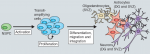
Adult neurogenesis has been implicated in physiological brain function, and failing or altered neurogenesis has been associated with a number of neuropsychiatric diseases. Simon Braun and Sebastian Jessberger provide an overview of the mechanisms governing the neurogenic process in the adult brain and describe how new neurons may contribute to brain function in health and disease. See the Development at a Glance poster article on p. 1983


Apical constriction: themes and variations on a cellular mechanism driving morphogenesis

1987Apical constriction is a cell shape change that promotes tissue remodelling in a variety of contexts. Martin and Goldstein review the cellular machinery required for apical constriction and discuss how it can be tunedto regulate apical constriction in diverse cellular contexts. See the Review article on p. 1987


Cell migration: from tissue culture to embryos

1999
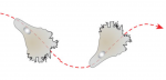
Cell migration is a fundamental process that occurs during embryo development. Here, Concha and colleagues review the guidance principles of in vitro cell locomotion and examine how they apply to examples of directed cell migration observed in vivo during development. See the Review on p. 1999

Thumbs up (No Ratings Yet)
Loading...

Categories: Research

A simple step to reverse ageing

Posted by , on 3 May 2014

april 2014

How great would it be if we knew how to reverse ageing and turn old organs into young ones? Actually, this might not be as crazy as it sounds. As a matter of fact, a team of scientists managed to regenerate the thymus in old mice and observe what closely resembles the juvenile thymus!

The thymus is a key organ of the immune system as it is where T cells, major actors of one’s immunity, develop and mature. In normal healthy people, the thymus degenerates with age (a process called thymic involution) and this results in a decline of the immune system function. Since our immune system is what protects us against diseases, it is evident that being able to restore the function of the thymus in elder people would be very beneficial.

Interestingly, in this study recently published in Development, Bredenkamp and colleagues achieved thymus regeneration in old mice. They observed a juvenile-like thymus after using genetic engineering to force the expression of the protein foxn1 in the aged thymus.

In this picture, you can observe the cortical thymic epithelial cell marker CDR1 in green and the medullary thymic epithelial cell marker keratin 14 (K14) in red. The cortex and the medulla are distinct regions of the thymus and they deteriorate during thymic involution. This results in reduced distinction between cortex and medulla as observed in the bottom picture taken in 24 months old mice. However, when foxn1 is over-expressed in the same 24 months old mice, you can observe that the clear distinction between cortex (green) and medulla (red) is restored, indicative of thymus regeneration.

In addition to the restoration of thymic architecture, the authors show that the regenerated organ has an increased T cell output and a gene expression profile similar to the juvenile thymus.

Most amazingly, apart from being able to “reverse ageing” in the thymus, they show that this regenerative process relies on the over-expression of a single factor (foxn1), making it a lot simpler than one might have anticipated! Thus, this study brings a new provocative concept that will most likely have a broad impact for regenerative biology.


Picture credit:

Bredenkamp, N., Nowell, C., & Blackburn, C. (2014). Regeneration of the aged thymus by a single transcription factor Development, 141 (8), 1627-1637 DOI: 10.1242/dev.103614

Thumbs up (1 votes)
Loading...

Tags: , ,
Categories: Research

6th Young Embryologist Annual Meeting

Posted by , on 2 May 2014

ImageJ=1.48a

6th Young Embryologist Annual Meeting
Friday
27th June 2014
JZ Young LT, Anatomy Building, UniversityYEN2014_small College London

Last call for abstracts

The 6th Young Embryologist Meeting aims to bring together developmental biologists from across the UK and abroad to discuss their work at a leading UK institution by presenting a talk or a poster. This year, we are pleased to include The Sammy Lee Memorial Lecture presented by Professor Bill Harris (University of Cambridge). We will also have a Q&A session about ‘Careers in Science’. In addition, prizes will be awarded to the best talks and posters, including a medal in memory of Sammy Lee

This unique meeting is run by PhD students and Post Docs at UCL, KCL, NIMR and  Imperial and aims to create a more interactive research community. Everyone with an interest in developmental biology is welcome to attend this free meeting!
 
We hope the meeting will provide a platform for young researchers (preference is given to PhD and Post Docs) to present their work in a series of 20 minute talks and in the poster session. We will favour diversity of topics and models in the field of embryology.
 
Our main goal is to generate a space where students, postdocs and PIs can discuss a wide variety of topics in developmental biology in a relaxed setting. Ultimately, we would like to strengthen  the Young Embryologist Network to enhance the research environment for embryologist, so please come!
 
Registration is free and can be done here: http://yen2014.eventbrite.co.uk
If you are interested in giving a 15 minute talk or presenting a poster please submit your abstract by clicking here
Deadline for submission is May 19th 2014
 
More information can be found on our website: http://www.youngembryologist.org/
If you want to get more out of your research, meet fellow researchers and get new ideas then join the Young Embryologist Network (YEN) by emailing: youngembryologistnetwork [at] gmail.com
Thumbs up (1 votes)
Loading...

Categories: Events