The community site for and by
developmental and stem cell biologists

Giving scientific ideas a voice (and a video)

Posted by , on 20 May 2014

Explaining new scientific concepts can be a daunting task for anyone involved in outreach. We are constantly trying to come up with ways to explain, show and describe theories and ideas step by step. I’ve recently stumbled across a new app (albeit only available on ipad) called Adobe Voice that could help out scientific communication.


Describing scientific research is like telling a story and Adobe Voice is an app made for just that. It combines on screen images of your choice with your own voice over in an elegant yet simple manner to guide the viewer through a thought process. It has many pre-set themes and background songs to choose from to help personalise your video, and I’m yet to find a combination that doesn’t look professional!


In developmental biology, visualisation of our concepts and ideas helps explain them, as description alone doesn’t convey the beauty of what we study everyday. These days, videos of the first hours of an embryo’s life are almost always included in any speaker’s presentation on development so why shouldn’t we extend this visualisation to our outreach? If, like me, you are not always able to visit as many schools as you’d like, creating these videos with adobe voice means you can send a part of yourself into classrooms virtually!


With Adobe Voice I have started what will hopefully be a mini-series resource that schools can pick up on. I’m focusing on the first ideas of genetics for a Key Stage 4 class (starting their GCSE curriculum). My first video took maybe an hour from concept to completion – I spent most of that learning how to draw a monkey – and I intend to make many more already.


With so many beautiful images available from developmental biology research, the field is made for showing off and these videos are a simple way of doing it justice. I’ve included my first video above and a link to my blog below where I’ll be putting up further videos over the coming weeks. If you have any suggestions for other videos I could make or other topics to cover please let me know, all feedback is welcome for this new endeavour into the video world!


www.genesandeverythinginbetween.wordpress.com


NOTE 17/06/2014:

The second video in this mini series I’m creating is now up! I’ve played around with the sound settings a bit and used only images included in the app itself to see how far I could stretch its applications to teaching scientific ideas.


FINAL outreach logo

This post is part of a series on science outreach. You can read the introduction to the series here and read other posts in this series here.


Thumbs up (2 votes)
Loading...

Tags:
Categories: Outreach, Resources, Video

Navigate the archive

Use our Advanced Search tool to search and filter posts by date, category, tags and authors.

Funded PhD position studying developmental migration of Drosophila immune cells

Posted by , on 20 May 2014

Closing Date: 15 March 2021

FULLY FUNDED PhD POSITION
Regulation of immune cell migration during development in Drosophila

Seeking highly motivated, intelligent and creative student with lab experience and either a bachelor’s or a master’s degree to join a research group studying cell migration at IST Austria for their PhD.

The developmental cell biology team of Professor Dr. Daria Siekhaus is an ambitious new group at IST Austria working in the field of invasive migration of immune cells using Drosophila melanogaster. We genetically identify mutants that affect cellular barrier penetration in the embryo, and characterize them molecularly, cellularly and by live imaging. To complement our international team (working language is English), we seek applications for a graduate student to work on a project involving a mutant we have identified in a lipid metabolism enzyme that has effects on specific aspects of migration, apparently in penetrating a tissue barrier. We seek to understand how this protein functions during migration. The project would involve further genetic characterization of this mutant, live imaging of its migratory behaviors, and genetic and biochemical investigations of its function. This mutant may have a relationship to a known human disease gene and exploring that would also be part of the project as well as an investigation of its roles in vertebrate immune cell function in collaboration with the Sixt group.

IST Austria is a new dynamic interdisciplinary institute that seeks to bring the American graduate student model to Europe.

Must interview by early July 2014 and start in mid September 2014. Would present and defend a thesis proposal in a qualifying exam by the following December, and graduate within three to four years from that point.

For more information about IST Austria and the Siekhaus group please visit:

http://ist.ac.at and http://ist.ac.at/research/research-groups/siekhaus-group/
To apply:

This funded graduate student position is available to any nationality.  To apply and to ask any questions send an email to Dr. Daria Siekhaus (Daria.siekhaus@ist.ac.at).
Include in application:

-CV and official transcript of Bachelors and Master’s (if applicable) grades

-Personal statement (up to 2 pages) about your scientific experience, interests and career goals

– Names and contact information of three people who can write a letter of recommendation

-Any other relevant information


References:

Siekhaus et al, Nature Cell Biology (2010) http://www.ncbi.nlm.nih.gov/pubmed/20495554

Cho, Keyes, … Krasnow (2002) http://www.ncbi.nlm.nih.gov/pubmed/11955438

(Review) Williams MJ J Immuno (2007) http://www.ncbi.nlm.nih.gov/pubmed/17404248


Thumbs up (No Ratings Yet)
Loading...

Categories: Jobs

Woods Hole images 2014 round 1- vote for a Development cover

Posted by , on 19 May 2014

Every year we give the Node readers the chance to choose their favourite images from a selection of great pictures taken by the students of the Woods Hole Embryology course. Last year you chose 4 beautiful images that featured in the cover of Development, and now is time to make your choice again!

Winners 2013

Below are 4 great images from last year’s course. Vote in the poll at the end of this post to choose the image that you would like to see in the cover of Development! You can see a bigger version by clicking on the images.

Voting will close noon GMT on June 9th.


1. Squid (Loligo pealeii) embryo stained with anti-acetylated tubulin (red), anti-serotonin (green), and DAPI (blue,nuclei). Imaged on a Zeiss LSM 700 confocal. This image was taken by Nathan Kenny (University of Oxford), Kathryn McClelland (Institute for Molecular Bioscience, University of Queensland), and Sophie Miller (University of Cambridge).


2. Annelid (Capitella teleta) larvae (stage 8), stained with Phalloidin (cyan, F-actin), anti-acetylated-tubulin (yellow), anti-FMRFamide (red) and Hoechst (purple, nuclei). Imaged on a Leica SP8 confocal. This image was taken by Poulomi Ray (Clemson University).


3. Live late stage embryo of a squid (Loligo pealeii). Imaged with a Zeiss SV8 and a SPOT Flex camera. This image was taken by Brijesh Kumar (Indian Institute of Technology, Kanpur).


4. Eye of a late stage squid (Loligo pealeii) embryo stained with Phalloidin (green, F-actin), anti-acetylated tubulin (red), and DAPI (blue, nuclei). Imaged on a Zeiss LSM 780 confocal. This image was taken by Ezgi Kunttas (Carnegie Mellon University).


Thumbs up (1 votes)
Loading...

Tags: , , ,
Categories: Images

(Developmental) Biology around the internet- May 2014

Posted by , on 19 May 2014

Here is May’s round-up of some of the interesting content that we spotted around the internet:


News & Research:

– This month was a sad one for science, as two renowned  scientists passed away: Robin Holliday and developmental biologist Julian Lewis.

– The Royal Society announced their new fellows list, including several developmental biologists: Cliff Tabin, Liam Dolan, Mandy Fisher and Martin Johnson. EMBO’s list of new members also included developmental biologists.

– Recent papers in Science and Nature Medicine examined the rejuvenation effect that blood from young mice has on old mice

Nature reviewed the book Embryos under the microscope‘, an overview of the history and science of embryology by J.Maienschein

– The Wellcome Trust announced that in 2016 the first volunteers will receive blood cultured from stem cells.

– And do you like science and beer? The Pint of Science festival is kicking off this week (19th-21st of May) in several countries around the world. If you are attending and would like to write about it for the Node, get in touch!


Weird & Wonderful:

– Buzzfeed collated some fantastic science tattoos.

– Check out this segmentation cake to honour David Ish-Horowicz

– And on the topic of cakes, how about an evolutionary tree of cakes and biscuits?


Beautiful & Interesting images:

– As the weather warms up, time for strawberry mitosis.

– And this comic reveals what the audience is really thinking when you give a talk:  

Videos worth watching:

Enrico Coen talks about about his book ‘Cells to Civilizations’.

– Undergraduate students at a science communication course created this short video that uses the cartoon Pokemon to explain concepts of evolution.

– Here is an excellent video introducing DNA and the genome to a lay audience.

– And finally the MPI-CBG Institute in Dresden made their own video to the sound of the song ‘Happy’ (spot the dancing directors!):


Keep up with this and other content, including all Node posts and deadlines of coming meetings, by following the Node on Twitter.

Thumbs up (1 votes)
Loading...

Tags:
Categories: News, Video

Use it or Lose it: Insights on neuronal connections in the vertebrate retina

Posted by , on 18 May 2014

This is the final post from our developmental neurobiology seminar this semester. Two students wrote about our discussion of the importance of neuronal activity during synaptogenesis and their professor combined and edited the pieces. As usual, we focused  on development in the vertebrate retina. Hope you’ve enjoyed our contributions, we’ve enjoyed sharing our new-found understanding.


“Use it or lose it.” One of my mother’s favorite phrases, applicable to any number of situations. Fail to practice piano? Don’t use a toy for a while? Realize you’ve forgotten your high school Spanish? “Use it or lose it.”

The concept of “use it or lose it” can also be applied to synaptogenesis and neural circuit development (although I doubt my mother was thinking of this when she used the phrase). The idea of Hebbian connections – that synapse formation and elimination is dependent on activity – has been primarily supported by studies of the neuromuscular junction [1]. In general, input neurons converging on dendrites differ in synaptic activity, resulting in more active inputs outcompeting less active inputs for dendrite connection. Inputs with less active synaptic regions are ultimately eliminated [2],[3]. Essentially, the less active neurons don’t “use it” and so they “lose it”, with “it” being an electrochemical connection to a neighboring neuron.

Several years ago, investigations of developing connections in the vertebrate retina suggested that this idea of “use it or lose it” might not be the sole mechanism employed within the developing nervous system. Our class recently read and discussed a paper by Kerschensteiner et al. (2009) [4]. This detailed study suggested that, in contrast to the classical model, some neuronal connections are formed and maintained in the inner plexiform layer independently of synaptic activity.


Anatomy and Physiology Background: ON and OFF cells in the Inner Plexiform Layer

When light hits the mammalian eye, it changes the electrical properties of photoreceptors so that their downstream partners, bipolar cells, deliver a message to retinal ganglion cells (RGCs), which in turn relay information about visual cues to the brain (see Figure 1). The middlemen in this process, bipolar cells, can be depolarized when light hits photoreceptors (ON cells) or hyperpolarized when light hits photoreceptors (OFF cells). Bipolar cells communicate with RGCs by forming synapses with RGC dendrites in the inner plexiform layer (IPL). This layer is stratified primarily into two sections, an outer layer and an inner layer, where RGCs synapse with OFF and ON bipolar cells, respectively. There are three main types of RGCs – those that only synapse with ON or OFF bipolar cells (monostratified RGCs), as well as RGCs that synapse with both ON and OFF bipolar cells (bistratified RGCs).

WongFig1-1

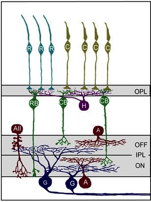
Figure 1: Model of primary cell types and their connection patterns in the mouse retina. First, Photoreceptors (teal rods (R) and yellowish cones, (C)) synapse with horizontal cells (purple) and bipolar cells (RB and CB, green) in the outer plexiform layer (OPL). Next bipolar cells synapse with ganglion cells (G, blue) in the inner plexiform layer (IPL). Amacrine cells (red) also synapse with both ganglion cells and bipolar cells in the IPL. Notice the stratification within the IPL based on the ON/OFF status of the bipolar cells. (Reproduced for educational purposes from Development of cell types and synaptic connections in the retina (http://webvision.med.utah.edu/).

During retinal development, RGCs form stratified dendritic arbors with synaptic connections to either exclusively ON bipolar cells (ON RGCs), exclusively OFF bipolar cells (OFF RGCs), or ON and OFF bipolar cells (ON-OFF RGCs). In the latter, RGC dendrites form synapses with ON and OFF bipolar cells such that ON and OFF inputs reside on separate laminar arborizations (e.g., Figure 1). Typically, ON and OFF bipolar cells form similar numbers of synapses with RGC dendrites, regardless of whether the RGCs are monostratified (ON or OFF) or bistratified (ON-OFF).


Neuronal Activity Determines Synapse Density within ON and OFF Layers of the IPL

To test whether this classical “use it or lose it” model for developing neuronal connections held true in the retina, Kerschensteiner et al. created a new transgenic line of mice that were unable to release glutamate from their ON bipolar cells, which is crucial for signaling to RGCs. The authors made a transgenic mouse that coupled the transcription of TeNT, a bacterial protease that inhibits vesicle fusion (which is necessary for presynaptic signaling), with the promoter for mGluR6, a glutamate receptor expressed in ON bipolar cells but not in OFF bipolar cells. Thus, ON cells could not communicate with the RGCs, while OFF cells could. If the classical model explains the development of connections between bipolar cells and RGCs, one would expect retinas in the transgenic mice to have no or very few RGC – ON cell synapses, and many RGC – OFF cell synapses.

As expected, the resulting mice exhibited normal receptive field properties from OFF-RGC connections and reduced or absent responses from ON-RGC ones. The dendritic branching and stratification patterns as well as overall connectivity appeared identical in transgenic and wild-type mice. Normal RGC development in the presence of significantly reduced ON bipolar cell activity indicates that partner selection and dendritic stratification of post-synaptic RGCs occur regardless of bipolar cell activity. These data provide evidence that RGCs are programmed to synapse to either ON bipolar cells, OFF bipolar cells, or both, regardless of presynaptic glutamate release.

By quantifying the density of individual postsynaptic sites in RGCs adjacent to ON bipolar cells in transgenic mice, the authors found that loss of glutamate release correlated with fewer mature synapses adjacent to ON bipolar cells (~50% fewer synapses in transgenic compared to wild-type retinas, Figure 2B). In addition, as might be predicted from the “use it or lose it” model, the density of synapses in bistratified RGCs connecting with the silenced ON bipolar cells was decreased relative to the density of synapses in the arbors connecting with active OFF bipolar cells (Figure 2C).


Figure 2

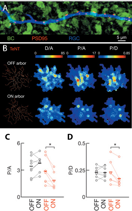
Figure 2: Density of bipolar-RGC synapses changes in response to glutamate release. A) Post-synaptic densities (PSD95, red) form at specific sites along RGC dendrites (blue) when closely apposed to biploar cell termini (green). B) Heat maps of glutamatergic post-synaptic densities for OFF and ON arborizations in bistratified RGCs. C-D) Graphical comparison of data in B. Values from non-transgenic retinas in black, transgenic animals in red. D/A is dendritic density, P/A is post-synaptic puncta per arbor area, P/D is post-synaptic puncta per dendritic length. Images are select panels from Figure 2 in Neurotransmission selectively regulates synapse formation in parallel circuits in vivo, Nature August 2009 (Vol 460, pp. 1016-1020). Kerschensteiner, D., Morgan, J., Parker, E., Lewis, R. & Wong, R. Used with permission of Nature Publishing Group.

When Kirchensteiner and coauthors examined the underlying mechanism for this decreased density, the “use it or lose it” model didn’t seem to hold true. Using time-lapse imaging, the authors showed that the silenced ON bipolar cells eliminated synapses at a rate comparable to wild-type but initial synapse formation was dramatically reduced. These data indicate that glutamate release is necessary for synapse formation and that elimination of synapses may not be directly tied to the activity of presynaptic input.

When the authors examined synapses at the ultrastructural level, they found even more evidence that “use it or lose it” isn’t the best analogy for synaptogenesis in the IPL. They show that ribbon synapses, the synaptic vesicle anchoring structures implicated in rapid neuronal transmission, are more numerous in the silent ON bipolar cells than in the active OFF bipolar cells (or in active ON bipolar cells in wild-type retinas). These intriguing electron micrographs raise the possibility that post-synaptic signaling, perhaps in response to glutamate, limits ribbon synapse formation.

With this study, Kerchensteiner et al. helped elucidate the role of glutamate release in forming synapses between bipolar cells and RGCs. Interestingly, glutamate release regulates how many synapses are formed between bipolar cells and RGCs but not how many synapses are eliminated, contrary to the classical “use it or lose it” model of synaptic formation. In a follow-up study, published in 2011 [5], some of the same authors investigate this teamwork approach to syanpatogensis in the IPL, providing evidence that differential synaptic maturation of axo-dendritic appositions is shaped by presynaptic activity and occurs in a cell type-specific manner. Whether (and how) post-synaptic cells may influence this process still seems to be an open question.

Although the “use it or lose it” analogy doesn’t completely describe what happens during synapse formation in the IPL, it does provide a framework for thinking about what cells need to connect with each other. In the case of bipolar cells and RGCs, it seems that glutamate needs to be used for efficient and robust synaptogenesis or the cells lose out all together, rarely making functional connections.


——————

[1]Kasthuri, N., & Lichtman, J. (2003). The role of neuronal identity in synaptic competition Nature, 424 (6947), 426-430 DOI: 10.1038/nature01836

[2]Wong, R. O. L. & Lichtman, J. W. in Fundamental Neuroscience 2nd edn (eds Squire, L. R. et al.) Ch. 20, 533–554 (Academic Press, 2002).

[3]MILLER, K. (1996). Synaptic Economics: Competition and Cooperation in Synaptic Plasticity Neuron, 17 (3), 371-374 DOI: 10.1016/S0896-6273(00)80169-5

[4] Kerschensteiner, D., Morgan, J., Parker, E., Lewis, R., & Wong, R. (2009). Neurotransmission selectively regulates synapse formation in parallel circuits in vivo Nature, 460 (7258), 1016-1020 DOI: 10.1038/nature08236

[5] Morgan, J., Soto, F., Wong, R., & Kerschensteiner, D. (2011). Development of Cell Type-Specific Connectivity Patterns of Converging Excitatory Axons in the Retina Neuron, 71 (6), 1014-1021 DOI: 10.1016/j.neuron.2011.08.025


Thumbs up (1 votes)
Loading...

Tags: , , , , , , , , ,
Categories: Research

Cosy Science talk- How do cells communicate to build tissues?

Posted by , on 16 May 2014

moon

Cosy Science is an informal pub gathering where scientists talk about their research over a pint of beer (or glass of wine!) with anyone who is interested in listening. It is mostly a monthly event, taking place at The Cittie of Yorke pub in London, and everyone is welcome!


suzanne_eatonAs you may know, The Company of Biologists runs a series of workshops for scientists, and we collaborate with Cosy Science to bring some of the exciting research discussed at the workshops to the general public. Our latest workshop will focus on cell polarity, and one of the participants, Dr Suzanne Eaton (MPI-CBG, Dresden) will be joining Cosy Science next Wednesday (21st of May) to bring developmental biology to the pub! Suzanne will give a short talk about how cells communicate to build tissues, and its implications in regulating size, embryogenesis and cancer. After a short break to refill, the floor will be open for questions and friendly discussion. So if you’re in the area, bring along that friend who always wanted to know what developmental biology is all about, and enjoy an evening of pub science sponsored by The Company of Biologists!


Find out more information at the Cosy Science website


Thumbs up (2 votes)
Loading...

Tags: ,
Categories: Events, News

Postdoctoral Associate in epithelial growth and morphogenesis

Posted by , on 16 May 2014

Closing Date: 15 March 2021

Department of Molecular Biosciences, The Wenner-Gren Institute

Stockholm University is a leading European university offering a multicultural environment in one of the world’s most dynamic capital cities. With more than 60,000 students and 5,000 staff, the University facilitates individual and societal development by providing top quality education that is tightly linked to its internationally recognized research programs.

The Department of Molecular Biosciences, The Wenner-Gren Institute (MBW) unites 30 independent research groups pursuing fundamental questions in molecular cell biology, infection and immunobiology, and integrative biology. The department carries out experimental research primarily investigating the function of genes and cells in tissues and organisms.

Main responsibilities: We are looking for a highly motivated and focused individual to join Professor Christos Samakovlis’s research group. The successful applicant will utilize state-of-the-art approaches aimed at obtaining a fundamental molecular understanding of epithelial morphogenesis, regeneration and malignant growth in Drosophila. The laboratory utilizes genome-wide, tissue-specific RNAi screens, live-imaging and transcriptional profiling. The successful candidate is expected to participate in the comparative analysis of gene function in insect and vertebrate tissues.

Qualification requirements: The applicant must have received a doctoral degree from university in the field of molecular cell biology, developmental biology or genetics. Applicants must have demonstrated productivity in pursueing research in molecular cell biology or bioinformatics. Experience with the Drosophila model system is an advantage. Excellent English language skills, both written and spoken, are a requisite. Further information about the position can be obtained from Christos Samakovlis, christos.samakovlis@su.se

Application:The application deadline is June 13, 2014.

Applications should comprise the following:

  1. CV, including full contact information and date of birth
  2. Personal statement describing research interests (1-2 paragraphs), research
    experience (1–2 paragraphs) and career goals (1-2 paragraphs)
  3. List of 2-3 references, please include name, e-mail address and telephone number Stockholm University is an equal opportunity employer
Thumbs up (No Ratings Yet)
Loading...

Categories: Jobs

Postdoctoral position in lung development

Posted by , on 16 May 2014

Closing Date: 15 March 2021

A postdoctoral fellowship is available for at least three years at the SciLifeLab, Stockholm, Sweden. Science for Life Laboratory (SciLifeLab) is a national center for molecular biosciences with focus on health and environmental research http://www.scilifelab.se/. The center combines frontline technical expertise with advanced knowledge of translational medicine and molecular bioscience.
The position is associated with the laboratory of Professor Christos Samakovlis. Research at the laboratory addresses airway morphogenesis, epithelial regeneration and malignant growth in Drosophila. We utilize genome-wide, tissue-specific RNAi screens, live-imaging and transcriptional profiling. The successful candidate is expected to participate in a comparative analysis of gene function in fly and mouse tissues.

Qualification requirements: Research experience in mouse developmental biology is an important selection criterion and strong interest in genetics and molecular biology is advantageous. Applicants must have demonstrated productivity in pursueing research in developmental or molecular cell biology. Excellent English language skills, both written and spoken, are a requisite.
Additional information can be obtained from Christos Samakovlis, (christos.samakovlis@su.se), and http://www.su.se/mbw/

Application: 
The application deadline is June 13, 2014.

Applications should comprise the following:
1. CV, including full contact information and date of birth
2. Publication list and a personal statement describing research interests (1-2 paragraphs), research experience (1–2 paragraphs) and career goals (1-2 paragraphs)
3. List of 2-3 references, please include name, e-mail address and telephone number

Stockholm University is an equal opportunity employer

Thumbs up (No Ratings Yet)
Loading...

Categories: Jobs

Scientific Copy Editor (Maternity Cover)

Posted by , on 15 May 2014

Closing Date: 15 March 2021

The Journal of Experimental Biology is the leading journal in the field of comparative physiology and publishes papers on the form and function of living organisms, from the molecular and subcellular to the integrated whole animal. We are currently seeking applications for the role of full-time Scientific Copy Editor to provide maternity cover.

The role entails copyediting articles to a high standard, compiling and paginating articles within an issue, overseeing the journal production process and liaising with authors, academic editors and production staff to ensure that articles are published in a timely and professional manner.

Candidates for this position should have a degree (ideally a PhD) in a relevant scientific area, and previous copyediting experience is strongly preferred. Additional requirements include excellent literacy skills, high attention to detail, a diplomatic communication style, good interpersonal and IT skills, a flexible approach and the ability to work to tight deadlines.

This position represents an excellent opportunity within science publishing and offers a competitive salary and benefits. The position is a temporary role for a period of up to 12 months, likely starting in September 2014, and is based in our Cambridge (UK) offices. The post is full time (35 hrs) although part time might be considered.

The Company of Biologists is a not-for-profit organisation and publishes the three internationally renowned, established journals Journal of Cell Science, Development and The Journal of Experimental Biology, as well as the two newer Open Access journals Disease Models & Mechanisms and Biology Open. The organisation has an active programme of charitable giving for the further advancement of biological research, including travelling fellowships for junior scientists and contributions to academic societies and conferences.

Applicants must be eligible to work in the UK and should send a CV along with a covering letter that clearly states their current position and salary expectations, relevant experience and why they are enthusiastic about the post.

Please send applications by email to Miriam.Ganczakowski@biologists.com by 6 June 2014 using the reference JEBMat2014. Informal queries should be directed to Miriam Ganczakowski on 01223 426164.

Thumbs up (No Ratings Yet)
Loading...

Tags: , ,
Categories: Jobs

Sweet Swiss…Zebrafish?!

Posted by , on 15 May 2014

chocolate and Alps

There are several things that could bring me to Switzerland. I guess that the Alps, cheese, and swiss chocolate would be 3 top-choices, I could talk about Swiss army knives and watches too, but I would never assume that the reason for my Switzerland visit would be… zebrafish. Of course not just as the ordinary wildtype zebrafish swimming in rivers of India. Not even the one nowadays available in the pet shops all over the world. I came to Switzerland for very specific (transgenic) zebrafish lines that could allow me to find out the answer for the questions I keep asking myself from the beginning of my PhD study.

Knowledge about the embryonic origin of paired teleost fins is a missing element that would help us to fully understand fin development and the fin to limb transition in evolution. It has been shown before that skeleton of mammalian limbs originates from lateral plate mesoderm; contrarily, it has been thought that rays in fish fins origin from trunk neural crest. Recent publications, including of my PhD supervisor Dr. Tomas Carney (AStar Singapore), have shown that it is not neural crest but paraxial mesoderm that gives rise to the osteoblasts (bone forming cells) in the zebrafish tail. This again rises the question about the origin of skeletal elements in paired fins. We had discussed ideas for possible answers but we were lacking tools to perform informative experiments.


fin section antibody staining

 antibody staining performed on fin section of fish line for which I have traveled all way down to Switzerland

The laboratory of Prof. Christian Mosimann at the University of Zurich works on early cell fate determination of the lateral plate mesoderm, which gives rise to the circulatory system, the kidneys, and the limbs. The Mosimann lab has established unique transgenic zebrafish lines that allow visualization, tracking, and genetic lineage tracing of the developing lateral mesoderm. Prof. Mosimann and Dr. Carney had previously met at the International Zebrafish meeting in Barcelona in 2013, and over a glass of Rioja discussed the possibility to use the Tamoxifen-controlled Cre/lox technique developed by Christian to tackle the lateral plate contribution of the developing fins. So the plan was born to send me to Switzerland. With the financial support from the Company of Biologists who kindly awarded me with a travelling fellowship, I was able to collaboratively visit Zurich.

The University of Zurich and especially people from the Mosimann lab created a welcoming and productive environment to work. Despite the lab’s recent start in spring 2013, the place is buzzing with exciting science. The friendly, energetic atmosphere and unconditional support from all members of Mosimann lab made my stay highly enjoyable and productive. I had a chance to learn new techniques that members of Mosimann lab handled with real expertise. Anastasia Felker, a fellow PhD student in the Mosimann lab, is already an expert in cre:lox techniques while Christopher Hess, another PhD student, critically helped me with the molecular biology side of things. Even members of other laboratories have been ready to help me. I owe big thanks to Claudio Cantu from Basler group who has shared his work space with me and helped me to perform my crucial experiments on sectioned fins. I have gained new work colleges who then became my friends. Despite the serious hours spent in front of a cryostat and on the several fluorescent microscopes, I had ample opportunity to explore Zurich and the finer points of Swiss culture (including chocolate, cheese fondue, raclette, and snowboarding in the Alps). And yes, I now do own a Swiss army knife, and have acquired a similarly versatile new skill set to tackle my biological interests using zebrafish.


farewell raclette

My farewell raclette wth the Mosimann Lab

Thumbs up (3 votes)
Loading...

Tags: ,
Categories: Funding