The community site for and by
developmental and stem cell biologists

Stem Cells in Developmental Biology: a debate at the BSDB

Posted by , on 29 March 2013

See below a posting from our website (http://amapress.gen.cam.ac.uk/) on the discussion that took place at the BSDB on whether to change or not the name of the society to include Stem Cell Biology. Some of the people who have read it have encouraged me to post it here to see what people think and I believe is a good idea (to open it up to discussion). In many ways it follows the spirit of Olivier Pourquie’s editorial at the beginning of the year in Development (http://dev.biologists.org/content/139/1/1.full)

With my wishes for a good Easter

Stem Cells in Developmental Biology: a debate at the BSDB

Last week the BSDB (British Society for Developmental Biology) celebrated its annual gathering at Warwick. Always a good place to go for quality developmental biology which is enhanced by the arrangement of holding the meeting together with the BSCB (British Society for Cell Biology): these days there is much cell biology in developmental biology. One of the BSDB sessions focused on Stem Cells and highlighted the clear connection between this area of research and developmental biology, or so it seems to some of us but ……perhaps not all.

The AGM of the BSDB had, for the second year running, a ballot to change the name of the society to include “stem cells’ in its name. Thus, the proposal was to change the name from “British Society of Developmental Biology” to “British Society of Developmental and Stem Cell Biology”. The proposal had been flagged last year and, after a vigorous discussion, was rejected, but by a narrow margin which allowed the subject to be brought up again to the AGM this year, where it was resoundingly rejected. But read on……….

The discussion preceding the vote was heated and highlighted several misconceptions about research in stem cells which, perhaps, represent some reality.

The ones in favour argued, correctly I think (I was and am in favour of the change) that Stem Cell research is part of developmental biology and that while there is much that has to do with medicine, the links between Developmental and Stem Cell Biology are strong and essential for both fields, that Developmental Biology can bring rigour and direction to Stem Cell Biology and that Stem Cell Biology can bring challenges, new ways and possibilities for Developmental Biology. They (we) argued that, after all, Stem Cell biology has always been part of Developmental Biology, albeit somewhat cryptically. Including Stem Cells in the name of the society is an acknowledgement of the times and can have its benefits because there is no denying that Stem Cell Biology is a central and key element of research these days; including it explicitly in the Societie’s agenda would allow the BSDB to have a strong voice in policy, funding and education in these increasingly influential area of research.

The ones against the change argued that Stem Cell Biology is different from Developmental Biology, that it has a clinical slant which would attract a different crowd to the meeting, force different content in sessions and, overall, distract us from the main business: the workings and evolution of embryos and systems. It was suggested that such a change would alienate several established constituencies within the society that would abandon the group. Mpre significantly, that, eventually, with another vote a few years down the line, Developmental Biology would be booted out of the title for the society to become the British Society for Stem Cell Biology.

As far as a I am concerned this was a missed opportunity. While I appreciate many of the points made by the ‘noes’ , I feel that their arguments are based on fear for a future that will always take over no matter what we do and that, in the long term, there will be consequences from not seeing this. We live in an increasingly corporate world in which lobbies are important and, in the context of our business, provide the basis for funding and policy. For the Society of Developmental Biology Society to have an explicit voice in the Stem Cell community is not just an extension of its natural remit and interests, but it is a way to bring some real science into a field that is increasingly interested in applications without having covered the bases. The foundations of Stem Cell Biology lie in Developmental Biology and it is important that developmental biologists have a say on decision making in that important field. Stem Cell Biology is not exactly, as some people claim, a new area of research (developmental biologists have been working with stem cells and their lineages for years) but it is certainly an area that recently has come of age to carve its own intellectual niche like, in many ways, Developmental Biology did in the 1970s (let us not forget that Developmental Biology is an offshoot of Embryology). It was argued that Stem Cells are born with Till and McCulloch (1964 Proc Nat Acad Sci. 51, 29–36). True but what they were looking at is the question of the origin of the blood, a problem in Developmental Biology whether one likes it or not. At certain places, and stem cell research is one of them, boundaries blur. Is genomics and bioinformatics genetics? Yes it is. In my book, Stem Cell Biology is part, and a very important part of Developmental Biology.

But let us move away from the heart of the question (of course the scientific content) and look, briefly, to the context of the discussion. The boundary, as I have said is blurred, and a situation can develop (and in certain places is happening) that some people, funding bodies, society, come to see Developmental Biology through the eyes of an unbridled Stem Cell Biology. After all, is it not organs out of cells that is the goal of stem cell biology? And is it not understanding these processes the goal of Developmental Biology? Then, what is the difference? The answer is simple, Stem Cell Biology wants to do, Developmental Biology wants to understand. It would be a pity not to bring them together. One can see here history trying to repeat itself: throughout the XVIII and XIX century engineers and inventors were making steam engines with little knowledge of physics, and they worked, but it is when the knowledge of thermodynamics is brought into the frameowork of the engineers that the engines become efficient. The same can be said of computing where, again, it is physics that makes the hardware that we have today. The fundamental science will always help the more applied side and needs it. So, much to be gained from Developmental Biology having a say in Stem Cell Biology. But there is a second more difficult question: what will be the consequences of the agenda of stem cells running that of developmental biology?

I can see a marginalization of model organisms and a biasing of the agenda towards applied science, applied in a trial and error way, rather than in the tradition of Science. I might be wrong in the extreme formulation of these concerns but I am certain that some of this will happen.

In the end, my impression was that the ‘noes’ were afraid, afraid of the future without realizing that he future will happen and that by not seeing the trends and joining them, we shall always be left to mercy of those trends, without a voice to influence them. I worry that model systems that have taught us so much about basic biology will slowly be squeezed into corners because we do not have a voice to explain that flies have stem cells, that stem cells are part of the make up of an organism which cannot be understood outside its context, that stem cells are a problem of evolutionary biology, that stem cells are a linguistic twist of linage analysis and lineage analysis has always been a problem of developmental biology, from Roux and Driesch to Garcia Bellido with Till and McCulloch in the middle. Incorporating Stem Cell Biology in the name of the society would have been a way of having a strong voice in a trend that is rapidly gaining momentum.

We shall see what the future harbours. The BSDB is a strong society which represents a vibrant and engaging community so there is no reason why things will change rapidly. However, one thing is clear: there is a need for the voice of developmental biologists to be heard in the Stem Cell community. A mechanism needs to be found for this. It is necessary as much to have a representation to remind that community where their real roots lie and the benefits of listening to the fundamentals of their field. There is a drift which was, unfortunately, at the heart of many of the speeches for the no, that Stem Cell Biology is more clinically that basic research orientated. One can see how this can be construed but, decisions like the one we have taken will increase this gap and foster this misunderstanding. It would be good (it is always good) to take lessons from history. As the interactions between physics and engineering prove, there is much to be gained from the interactions between a field that tries to find practical solutions and one that explain the causes of the problems. Let us hope that the BSDB can find a way to influence some of the directions of Stem Cell biology. For the moment it is as if two twins have been separated; each with their own mind but with shared genetics.

Thumbs up (6 votes)
Loading...

Tags: ,
Categories: Discussion

Navigate the archive

Use our Advanced Search tool to search and filter posts by date, category, tags and authors.

March news: Unproven stem cell therapy in Italy and an upgraded stem cell toolkit

Posted by , on 28 March 2013

I’ve just sent out EuroStemCell’s monthly newsletter and thought I’d share a couple of the big items that might interest all of you.

It’s been another busy month, with lots of events ranging from schools activities to discussions with policy makers about changes to the EU Clinical Trials Directive. Plus, we’ve given our toolkit of educational and outreach resources a facelift to make it even easier to find the public engagement tool you need for your event.

In Italy stem cells have hit the headlines as the Italian Ministry of Health has overruled regulators to allow use of an unproven stem cell therapy in public hospitals. Stem cell researchers in Italy and beyond have expressed alarm about the decision. We explain the issues and share quotes from leading scientists in our news article.

You can read the full March newsletter on our website. Below a bit more on this month’s big items.

Scientists raise alarm as Italian Government rules on unproven stem cell therapy

An unproven stem cell therapy has taken centre stage in Italy after patients successfully lobbied the Italian government to allow its use in public hospitals. The highly controversial and untested procedure was created by the privately owned Stamina Foundation, but blocked by the Italian Medicine’s Agency, AIFA. Last week’s decision by the Ministry of Health to override AIFA’s block has horrified Italy’s leading stem cell scientists. In a letter to the Ministry, they describe the decision as providing “a dangerous short circuit between patients’ hopes and lucrative commercial practices” of organisations operating outside the “scientific and moral foundations” of medicine. Read more

Our stem cell toolkit has had a facelift!

Our toolkit of downloadable stem cell resources and activities has been growing so fast it’s getting hard to keep up with all the new additions. We launched with just five tools, but that number has since grown to 13 tools for teaching and talking about stem cells, in the classroom, science centre, open day, festival and other educational settings. We’ve had great feedback from teachers and others from around Europe: “[The] toolkit web page … is very easy to use. Everything appeals to me: not only the “surface”, icons and links, but also the contents are very useful and interesting.” Teacher, ItalyRead more

Thumbs up (No Ratings Yet)
Loading...

Tags: ,
Categories: News

On how odd critters can answer important questions

Posted by , on 27 March 2013

Sproing! Sproing! Sproing! If there is one animal that deserves its own cartoon sound, it is the jerboa – a bipedal desert rodent with extraordinarily elongated hindlegs, fused foot bones, and loss of the first and fifth toes. I blogged here from China last spring during the most recent field collection of jerboa embryos, and now I’m excited to share news of the first research article using jerboas to answer a fundamental question in cell biology and skeletal growth and evolution: how do growth plate chondrocytes enlarge, and how do growth plates adjust cell size to contribute to differences in rates of skeletal growth?  I have been derelict about entries, not sure what people would find interesting to read, so there is a lot of backstory that I will try to summarize. If anyone wants to see some of this story fleshed out, I would be happy to take blog post requests :)

I decided to postdoc with Cliff Tabin because of my interest in the limb as a structure to study morphological diversity, but “I am interested in limb diversity” isn’t a focused way to start a project. We spent a few months tossing around ideas for a specific animal to study wherein I laughingly proposed dinosaur, dolphin, and horse. That’s one great thing about Cliff – he took each of my crackhead ideas seriously and never said “no”. That meant my joking suggestion of horses turned to “Here’s your horse” when a student at Harvard introduced us to the jerboas. The jerboa and horse have converged on similar adaptations including loss of toes and fusion and elongation of remaining elements. I am actually starting to address convergence in horses as well since it turns out horse embryology isn’t impossible. But that’s a story for another day.

The early years were focused on acquiring specimens involving multiple trips to China to collect pregnant females and harvest the embryos. The benefit of going to China is that the species there hibernate through the winter and breed almost synchronously in the spring. This means that if we hit the timing right, we can get tons of embryos from a short collection. Unfortunately it takes some trial and error to get that timing right. When you work on a seasonal animal that means it can take several frustrating years to achieve success.

Meanwhile I embarked on an adventure to get the first research colony of jerboas established, and everything I’d read online said they would not take care of their offspring in captivity. It was the tiger mauling at the San Francisco Zoo in 2007 that brought to my attention the Association of Zoos and Aquariums and, more importantly, the International Species Information System. I sent an email to ISIS to ask if they had a husbandry manual or holdings records for any species of jerboa, and they responded with no husbandry manual but with the contact information for 11 zoos and conservation centers that had jerboas in their records at some point since the 1960s. I emailed all of them and got one amazing response from the Breeding Centre for Endangered Arabian Wildlife in Sharjah, UAE. This species of jerboa isn’t endangered, but that whole story and how I learned to raise jerboas is probably also best saved for another entry or this will get ridiculously long.

I started investigating mechanisms of digit loss and mechanisms of rapid skeletal elongation in parallel, but the embryos to study digit loss were in short supply until this last (amazing) collection. Meanwhile, the colony started to breed, and I realized that the significant time window for increased hindlimb elongation was in early postnatal stages – easy to get from my burgeoning colony without decimating the breeding population. I started with an analysis of BrdU labeling index…nothing exciting there. One day as I was looking at sections that were stained with H&E it struck me: those hypertrophic chondrocytes in the jerboa foot are HUGE.

So I dug through the literature and found a paper by Norm Wilsman and colleagues in 1996 that quantified the percent contribution of a number of factors to the daily rate of skeletal elongation (growth in microns per day). They determined that the process of volume enlargement during hypertrophy of the terminally differentiated chondrocytes contributes most significantly to skeletal elongation. Additionally, the size of those hypertrophic chondrocytes varied most between skeletal elements that elongate at different rates within an animal (ie the fast proximal tibia growth plate versus the slow proximal radius).

Hypertrophic chondrocyte size contributes significantly to skeletal growth and to differences in rates of growth, so how is cell size regulated? But that question turns out to be the second question in the pipeline. The first question was “How do these cells get big?” There was some discussion in the literature, primarily from Joseph Buckwalter and Peter Bush, suggesting that chondrocytes may enlarge by cell swelling – a disproportionate increase in cytoplasmic fluid volume. However when I talked to a couple of cell biologists at Harvard, I was met with some resistance. Plant cells swell, but they also have cell walls to contain the pressure. Animal cells don’t swell. If the neurons in your brain swell by as little as 10%, they rupture which causes some serious problems. Regulated volume increase and regulated volume decrease are so important that our bodies have developed mechanisms to stabilize cell volume in cases of shifts in blood plasma osmolarity. But hypertrophic chondrocytes looked really empty in transmission electron micrographs…

I decided to address this question of whether chondrocytes swell and put together a K99 proposal (which missed the payline by one point in the only submission I could make before hitting the 5 year mark). In the lead up to grant submission, Cliff put me in touch with Tim Mitchison in Systems Biology who introduced me to Seungeun Oh in Marc Kirschner’s lab. Seungeun had just finished her PhD in Spectroscopy at MIT and had joined Marc’s lab to apply microscopy methods to questions of cell volume control in the cell cycle. She had the perfect methodology to answer my question – using the retardation of a wavelength of light to quantify cellular dry mass contents. The very first experiment we did was on her old set up at MIT the day before my grant deadline to provide proof of principle. I had dissociated postnatal day 5 mouse tibia chondrocytes, and we carried the dish on the M2 shuttle across town to the basement of MIT. (Isn’t all of MIT a long basement corridor?) One of the first images we took was a perfect cluster of 3 cells of varying sizes showing a small cell, an intermediate cell that was more red on the phase shift heat map, and a third cell that was enormous and less red on the phase shift heat map indicating it hadn’t increased in total mass as would be expected for a larger cell maintaining high density. Even though we still had to collect data from many more samples and show that the cells remained spherical and didn’t just flatten out (which could also decrease the phase delay), we were so excited and gave each other a good high five. That image from the very first day became panel “a” of Figure 1, because we never again saw such a perfect cluster of cells together demonstrating the effect of cell swelling on phase shift in a single image. It’s like they were waving at us with a taunting “You’re right!”

That first day led to 2 ½ years together in a small dark room imaging cells, countless hours of clicking on images in Matlab, writing, re-writing, reworking figures, and discussions of osmolarity and properties of light in the hybrid language of an organismal biologist and a physicist. It was a perfect merging of two disciplines. From that initial observation in the mouse proximal tibia, we went on to discover that there are three phases of chondrocyte enlargement: an initial phase where chondrocytes increase in volume while maintaining a “normal” dry mass density of about 18%, a second phase where cells swell and dilute their dry mass to about 7%, and a final phase where cells continue to get larger but maintain low dry mass density. Larger cells get bigger by extending the third phase, and smaller cells truncate the process that’s shared by all chondrocytes observed in this study. To get at a genetic mechanism controlling this process, we investigated the cell phenotype in animals that were conditional mutants for insulin-like growth factor 1. We found that not only do the growth plate specific differences in cell size disappear, but that igf1 controls the third phase that is most variable in growth plates elongating at different rates. This puts the igf1 pathway in a perfect place to investigate the genetics underlying evolutionary differences in growth rate. Stay tuned.

But for all of the answers this paper provided, it opened a whole new can of question mark shaped worms. How do animals establish different skeletal proportions? (The original question I set out to answer.) How does the cell carry out metabolic functions in the face of a 3-fold decrease in dry mass density? How does the cell regulate its membrane surface area? And a big one – how in the heck do these cells drive all that water in to dilute the cytoplasm? As I’m preparing to set off and establish my own lab, I’m looking forward to where these questions and more will lead.

Thumbs up (6 votes)
Loading...

Tags: , ,
Categories: Research

In Development this week (Vol. 140, Issue 8)

Posted by , on 26 March 2013

Here are the highlights from the new issue of Development:

Molecular map of posterior hypothalamus

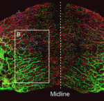
F1.smallThe hypothalamus is a key integrative centre in the vertebrate brain that regulates many essential functions, including homeostasis and stress responses. Several transcription factors that are essential for hypothalamic development have been identified but the production of diverse neuron types in this complex brain region is poorly understood. Here (p. 1762), Andrea Wolf and Soojin Ryu identify the transcription factors required for the specification of two distinct neuron types in the zebrafish posterior hypothalamus. They show that the transcription factor Fezf2 is important for the early development of the posterior hypothalamus. Furthermore, the differential expression of Fezf2, Otp, Foxb1.2 and Sim1a defines distinct subdomains in the posterior hypothalamus during neuronal specification. The neuron types that produce the hypothalamic hormones Vasoactive intestinal peptide (Vip) and Urotensin 1 (Uts1) develop in these different subdomains, they report, and Vip neuron specification requires Otp and Sim1a whereas Uts1 neuron specification requires Fezf2, Sim1a and Foxb1.2. Together, these findings provide mechanistic insights into the generation of neuronal diversity in the hypothalamus.


Afadin: making and shaping tubules

F1.small-1The formation and elongation of polarised epithelial tubules is essential for the structure and function of several metazoan organ systems but the molecular mechanisms that regulate tubulogenesis are largely unknown. Here (p. 1774), Denise Marciano and colleagues provide new insights into tubulogenesis by studying the developing mouse nephron. The researchers show that kidney mesenchymal cells contain Par3-expressing microdomains on adjacent cells. These microdomains coalesce to form a continuous lumen, which elongates by extension and by additional de novo lumen formation. Both lumen formation and elongation require afadin, a nectin adaptor protein that is implicated in adherens and tight junction formation. Using mice that lack afadin in nephron precursors, the researchers demonstrate that afadin is required for the coalescence of Par3-expressing microdomains, which is needed to establish apical-basal polarity and generate a continuous lumen. These results reveal a novel mechanism for lumen formation and morphogenesis in vivo in which afadin plays a central role through its recruitment of polarity and junctional proteins to sites of lumen formation.


Ubiquitylation promotes planar polarity

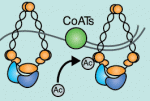
F1.small-2Planar cell polarity depends on the asymmetric localisation of core planar polarity proteins at apicolateral junctions. This asymmetric distribution probably develops through amplification of an initial asymmetry and seems to require the regulation of core protein levels. Now, Helen Strutt, Elizabeth Searle and co-workers (p. 1693) show that two distinct ubiquitylation pathways regulate the junctional levels and asymmetry of core planar polarity proteins in Drosophila. The researchers report that a Cullin-3-Diablo/Kelch ubiquitin ligase complex and the deubiquitylating enzyme Fat facets regulate the levels of the core planar polarity proteins Dishevelled and Flamingo, respectively, at apicolateral junctions but have no effect on the total cellular levels of Dishevelled and Flamingo. Notably, both increases and decreases in the junctional levels of core proteins caused by disruption of the ubiquitylation machinery reduce core protein asymmetry and disrupt planar cell polarity. Thus, the researchers suggest, ubiquitylation maximises the asymmetric localisation of core planar polarity proteins by fine-tuning their levels at junctions.


Tcf7l1 sets the stage for lineage specification

F1.small-3During mammalian embryogenesis, substantial cell proliferation occurs before the establishment of the body plan during gastrulation. Thus, before gastrulation, individual embryonic cells must be pluripotent. In vitro experiments with embryonic stem cells (ESCs) have indicated that the transcription factors Oct4, Sox2 and Nanog are components of a gene regulatory network (GRN) that stimulates self-renewal of pluripotent cells and have identified Tcf7l1 (Tcf3) as an inhibitor of GRN activity. But what is Tcf7l1’s function during embryonic development? To find out, Bradley Merrill and colleagues have been examining embryogenesis in Tcf7l1-/- mouse embryos (see p. 1665). They report that mesoderm specification is delayed in these embryos, thereby uncoupling it from primitive streak induction. Moreover, in vitro, Tcf7l1 activity is necessary to switch the response of pluripotent ESCs to Wnt/β-catenin signalling from self-renewal to mesoderm specification. Thus, the researchers suggest, Tcf7l1 prepares pluripotent epiblast cells in gastrulating mouse embryos for lineage specification and ensures that lineage specification is coordinated with the dynamic cellular events that occur during gastrulation.


β-catenin helps ES cells stick to pluripotency

F1.small-4Canonical Wnt signalling and E-cadherin-mediated cell adhesion are both involved in mouse embryonic stem (mES) cell maintenance. β-catenin (Ctnnb1) is central to both these processes – it mediates the transactivation of Wnt target genes and also connects E-cadherin to the actin cytoskeleton via α-catenin. But which β-catenin function is absolutely required for mES cell self-renewal and pluripotency? On p. 1684, Ignacio del Valle and colleagues investigate this controversial question. The researchers use Ctnnb1-/-Eα mES cells in which the constitutive expression of an E-cadherin-α-catenin fusion protein maintains cell adhesion. Preservation of cell adhesion, the researchers report, is sufficient to promote the leukaemia inhibitory factor (Lif) signalling pathway, which is required for mES cell maintenance, and the transcriptional factor network that controls the mES cell state. They also implicate E-cadherin in the activation of Lif signalling by showing that it stabilises the Lifr-Gp130 co-receptor complex. Together, these findings suggest that only the adhesive function of β-catenin is absolutely required for the propagation of mES cells in culture.


Hopx marks hair follicle stem cells

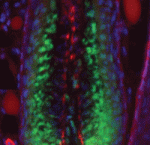
F1.small-5Adult tissue-specific stem cells both self-renew and generate functional progeny. Mammalian hair follicles, which are characterised by cyclical phases of growth (anagen), regression (catagen) and rest (telogen), are an ideal system in which to investigate the homeostasis of an adult stem cell population. On p. 1655, Jonathan Epstein and co-workers show that Hopx, which encodes an atypical homeodomain protein, is specifically expressed in long-lived stem cells in the basal bulge of the mouse telogen hair follicle. Hopx+ cells, they report, contribute to all the lineages of the mature hair follicle. In addition, the researchers identify a previously unknown progenitor population in the lower hair bulb of anagen-phase follicles and show that these Hopx-expressing cells contribute to the cytokeratin 6-positive inner bulge niche cells in telogen that regulate the quiescence of adjacent hair follicle stem cells. Because Hopx expression also marks other adult stem cell populations, the researchers suggest that tissue-specific stem cell populations might share homeostatic mechanisms.


PLUS:


Brassinosteroid signalling

F1b.posterZhi-Yong Wang and colleagues provide an overview of the highly integrated BR signalling network and explain how this steroid hormone functions as a master regulator of plant growth, development and metabolism.

See the Development at a Glance poster article on p. 1615
 

Morphogen transport

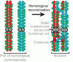
F1morph.largeAlex Schier et al analyze various morphogen transport models using the morphogens Nodal, fibroblast growth factor and Decapentaplegic as case studies. They propose that most of the available data support the idea that morphogen gradients form by diffusion that is hindered by tortuosity and binding to extracellular molecules.

See the Hypothesis article on p. 1621


Thumbs up (No Ratings Yet)
Loading...

Categories: Research

Developmental Biology Teaching Workshop at Darling Marine Center

Posted by , on 22 March 2013

The University of Maine Darling Marine Center will once again host a Developmental Biology Teaching Workshop, June 25-29, 2013.  Openings are still available!

class participantsThis workshop is geared toward university faculty, postdocs, and graduate students interested in teaching developmental biology.  It provides basic hands-on experience with organisms commonly studied in teaching laboratories. These include sea urchins and sand dollars, planaria, Drosophila, chick embryos, Spirostomum, Hydra, Lumbriculus, and flowering plants. Techniques will range from classical microsurgical techniques to fluorescence microscopy and applications of reporter gene technology.

Course Instructors:
Dr. Leland Johnson, Augustana College, SD
Dr. Eric Cole, St. Olaf College, MN
Guest Instructor, Dr. Mark Spiro, Bucknell University, PA

This workshop is the recipient of a Non-SDB Educational Activities Grant from the Society for Developmental Biology.  For more information visit the course website here: http://www.dmc.maine.edu/workshops.html#devbiocourse

Thumbs up (2 votes)
Loading...

Tags:
Categories: Events

New Reviews Editor at Development!

Posted by , on 22 March 2013

523165_10150645671594211_701681310_nDear readers of the Node,

Hi there! My name is Caroline and I am the new Reviews Editor covering all things stem cells at Development. I come to you from the land down under, namely Australia, where I did my PhD on kidney development and stem cells in the lab of Prof Melissa Little at the University of Queensland. Shortly thereafter, I landed myself what I considered to be “the dream postdoc” studying lineage reprogramming and iPSC disease modelling in the lab of Prof Ihor Lemischka at Mount Sinai School of Medicine, New York. USA. But fate was not finished with me yet, and now I find myself in Cambridge, UK, in the hallowed halls of the Development offices. Having moved continents three times in less than two years, I am definitely ready to settle!

I am really looking forward to bringing you the latest and greatest in all things stem cells and development. The stem cell field grows larger by the month but no matter how big it gets, its foundations will always be in development.  The two are inextricably linked. In one sense, the stem cell field can be thought of as applied developmental biology, and this is certainly the case for stem cell therapies. But understanding the fundamental mechanism of how stem cells can give rise to an entire tissue or organism is as much an integral part of developmental biology as any other, and so deserves to be studied in its own right.

I can’t wait to hear more from everyone as my time here goes on. I’m really looking forward to meeting you at all at various conferences and getting your thoughts on stem cells in development! There are big things planned for stem cells in the journal this year so stay tuned!

Thumbs up (8 votes)
Loading...

Categories: News

Report on “The Making of a Vertebrate – RIKEN CDB Symposium 2013”

Posted by , on 22 March 2013

When your model organism of choice is medaka, and you spend your mornings trying to rid your embryos of their tough chorion, you comfort yourself thinking that one day all the hard work will result in A) a paper, and/or, B) a visit to Japan. After all, medaka are Japanese killifish (Oryzias latipes). When the day in which one of these two options unexpectedly becomes true, you realise that medaka is truly the best model organism anybody could work on!

And so it was that a few weeks ago, I got an email from the RIKEN Centre for Developmental Biology in Kobe telling me I had a full travel grant to attend their 2013 Symposium, entitled “The Making of a Vertebrate”, and which would take place in March. The list of speakers was impressive, and the theme selected for this year’s meeting was particularly exciting. Our lab here in Seville works on vertebrate morphogenesis (Juan Martínez-Morales’ lab, at the Andalusian Centre for Developmental Biology) and the Symposium felt like the ideal place to tell our medaka-themed story.

At breakfast on the first day of the Symposium I met the other four lucky people who had also been awarded travel grants. We all came from different corners of the world, and none of us were working in our own countries. We bonded through our nomadic dispositions that morning, visiting each other’s posters in the afternoon, and by the time the sushi was served during the reception dinner that evening, we were plotting the conquest of Kobe.

The Symposium itself had a great start. The first session took on the origins of vertebrates, with a comprehensive overview by Nicholas Holland, asking what the true shape of the “tree of animal life” is.  His introduction was followed by talks on the hourglass model (Naoki Irie) and the evolution of chordates (Billie Swalla), both of which shed light on the question initially proposed by Dr. Holland. Next were the talks on hemichordates, annelids and the ancestry of vertebrates and their body plan. Detlev Arendt showed there is a conserved set of transcription factors, expressed in a specific, serial pattern in cnidarian gastric pouches, coelomic cavities in amphioxus, as well as in annelid mesodermal segments; suggesting that segmentation evolved before the split between cnidarians and bilateria. The final session of the day addressed the origins of pluripotency in vertebrates and its associated transcription factor network, which contains functionally overlapping genes that provide robustness to the network (Hitoshi Niwa) but has also evolved by gene co-option and duplication (Miguel Manzanares). The speakers were all engaging, the discussions insightful, and the organization of the whole day was great, including the best conference food I have ever tasted, which was also beautiful to look at: bento boxes.

Day two began with a fresh look on gastrulation, and I particularly enjoyed Masahiko Hibi’s images showing the timing of dorsal determinant transportation in the zebrafish yolk, and Hidehiko Inomata’s talk explaining how dorsoventral patterning is adjusted to the size of the embryonic axis. The afternoon’s subject was the Genetic Control of Morphogenesis, and included a talk on medaka (Hiroyuki Takeda), so that made it particularly thrilling for me. Dr. Takeda  is using a genome-wide approach to study epigenetic marks on key developmental genes, and finds their DNA-methylation status, as well as their histone modification states, change dramatically in time; suggesting that gene regulation occurs in “steps”.

The last day of the Symposium was all about vertebrate characters and their origins. It began, to my delight, with talk of dinosaurs and beautiful pictures of in situs on penguin embryos, with a compelling argument for the use of chick embryos as models for dinosaur morphogenesis (Koji Tamura). David Kingsley uses sticklebacks to address evolutionary changes, and suggested that evolution has few ways to achieve change, but uses them repeatedly: regulatory mutations, unlike deleterious loss-of-function ones, can be both advantageous and dramatic. The talk by Shigeru Kuratani discussed his lab’s work to clarify the patterning of craniofacial structures. They have successfully bred hagfish and have studied their embryology in minute detail, concluding that there is a “pan-cyclostome embryonic pattern” which is the ancestral programme of craniofacial development of vertebrates, and during evolution gnathostomes have lost or modified components of this pattern. I have mentioned my personal highlights, but would like to state that in general, the speakers were fantastic and it was hard to select only a few for this piece.

After the meeting was over, I made a short trip to Kyoto to see the sights. I am sharing some of the pictures I took during my visit, hoping that those of you who haven’t experienced Japan yet can have a peek at how magical it is. I hope you enjoy them.

Thank you very much indeed (domo arigato gozaimasu) to RIKEN CDB for the travel grant, and for an excellent and memorable meeting.

Finally, now that option B on my list became true, I better get on with making option A (the paper) happen too…our dear medaka are certainly doing their part!

cdb

Images: Fushimi-Inari Shrine, Chion-In Temple, Kinkaku-ji Temple, lunch at a market

Thumbs up (13 votes)
Loading...

Tags: , , ,
Categories: Events

Postdoc and PhD positions: Growth control in Drosophila

Posted by , on 20 March 2013

Closing Date: 15 March 2021

Post-doctoral and PhD positions are available in the lab of Savraj Grewal, University of Calgary, Canada.

(https://www.facebook.com/The.Grewal.Lab).

The main focus of the lab is to study the control of growth using Drosophila as a model system. We use a combination of molecular, genetic and proteomic approaches to investigate the cell-cell signalling pathways and the genetic mechanisms that control how cell, tissue and body growth are regulated during development.

Post-doc applicants with a strong background in developmental biology, genetics, or molecular biology are encouraged to apply. PhD applicants should have a strong undergraduate degree in any area related to the biological sciences. Interested individuals should send a CV, a one-page statement of research interests, and three names of references to grewalss[at]ucalgary.ca. Applications will be accepted on an ongoing basis and positions will remain open until filled.

For more details about our lab, our research and the University of Calgary, please visit our lab web page:

https://www.facebook.com/The.Grewal.Lab


Thumbs up (No Ratings Yet)
Loading...

Tags: , , , , , , ,
Categories: Jobs

CiteAb: a search engine for antibodies

Posted by , on 14 March 2013

This week we’re launching CiteAb – a brand new antibody search engine suitable for Developmental Biologists working with model organisms, including C. elegans, Drosophila, Zebrafish, Xenopus and Chick, as well as mammals. We feature nearly 1 million antibodies, rated by citations.

See http://citeab.com for more details.

This is a new site so we would very much welcome feedback from users of the Node.

Thanks :)

Thumbs up (3 votes)
Loading...

Categories: Resources

Stem cells image competition: voting

Posted by , on 14 March 2013

Over the last few weeks, you’ve been submitting your images for the Node and Development‘s stem cell cover competition. We received a large number of entries, and you’ve proved to us that stem cells – both in their natural environments and in a dish – can be just as beautiful as the embryos that normally grace our covers. After quite some deliberation, we’ve narrowed down the submissions to a short-list of five.

Now it’s your turn to vote for your favourite. The winning image will appear on a cover of Development, and will also feature on the new stem cell pages we’re developing for the journal’s website.

The poll will close at noon GMT on April 10th.

Here are the Final Five. Click on the image to see a larger version.

1. Immunofluorescence of the corn snake (Panterophis guttatus) dental organ with multiple generations of tooth germs. Sox2 expression (red) indicates putative dental stem cells in the epithelial lamina. DNA is stained with Hoechst (blue).

2. Fractal image of neural rosettes forming after the differentiation of mouse embryonic stem cells to neural stem cells. Cells are stained for Nestin (green) and PSA-NCAM (red).

3. Induced pluripotent stem cell colony surrounded by non-reprogrammed and feeder cells. Mouse embryonic fibroblasts were infected with viruses encoding transcription factors Oct4, Sox2 and Klf4 to reprogram them to pluripotency. A day 14 reprogramming culture was stained for E-CADHERIN (green), NANOG (red) and EZH2 (magenta). Dapi is shown in blue. This procedure can also be applied to human cells. The discovery of somatic cell nuclear reprogramming to pluripotency was awarded the 2012 Nobel Prize for Physiology or Medicine.

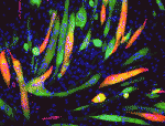
4. A rendered image of a primary neuronal stem cell culture in which cells were labeled with different fluorescently labeled proteins that differentiate between stem cells (orange/yellow) and their neuronal ‘offspring’ (blue/ green/ purple).

5. Confocal image of an adult mouse hippocampus, the area of the brain where new memories are formed. Astrocytes (green) were observed around the granule cell layer of the dentate gyrus, as indicated by cell nuclei (red). Some astrocytes were derived from neural precursor cell population (blue).


Thumbs up (10 votes)
Loading...

Tags: ,
Categories: Images, News