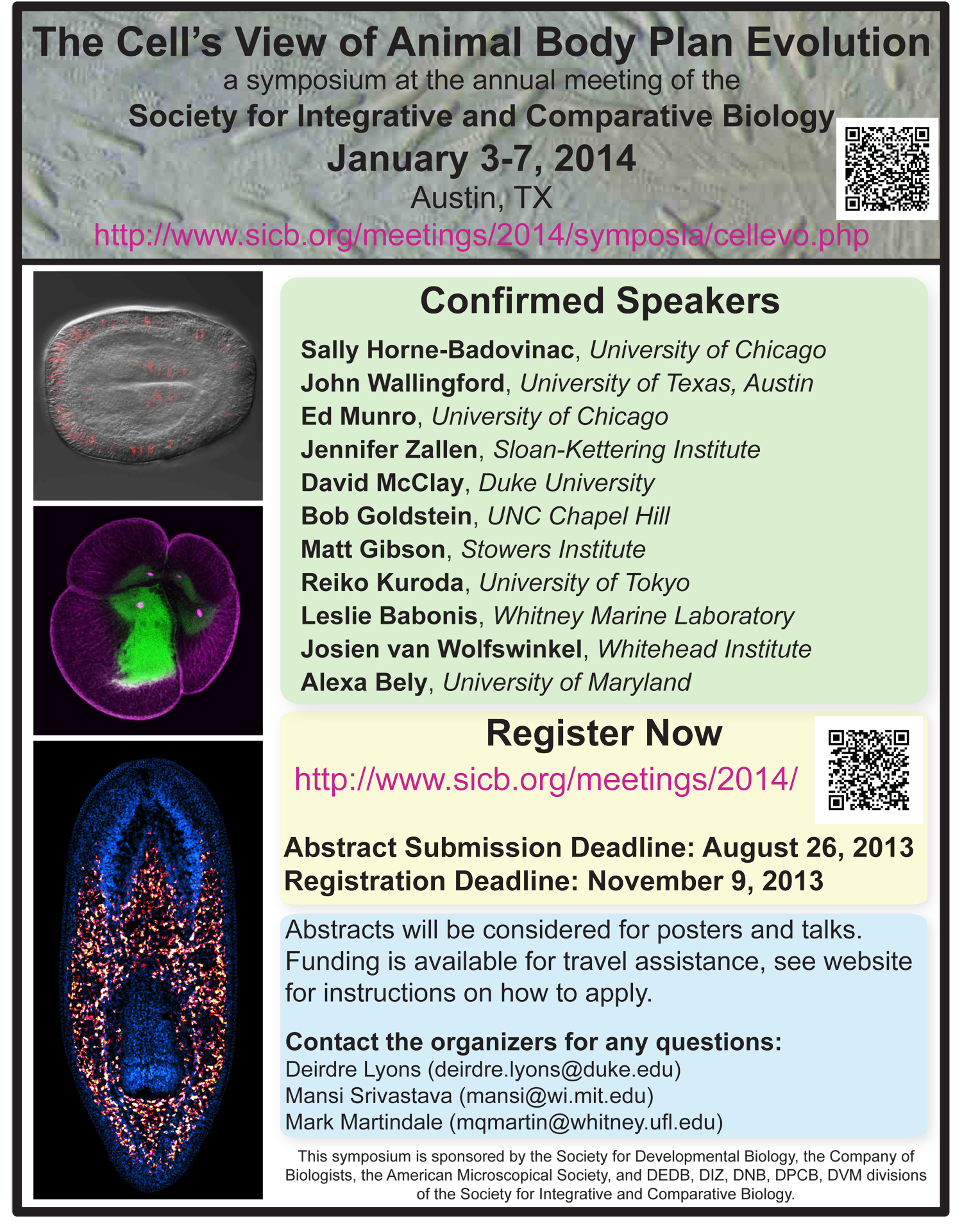
Symposium: The cell’s view of animal body plan evolution
Society for Integrative and Comparative Biology (SICB) Annual Meeting
January 3-7, 2014 Austin, TX
http://www.sicb.org/meetings/2014/symposia/cellevo.php
Abstract submission deadline: August 26 2013
http://www.sicb.org/meetings/2014/abstracts/
Registration deadline: November 9, 2013
Understanding how diverse animal body plans evolved remains one of the most exciting and challenging goals for evolutionary and developmental biologists alike. Over the past few decades, genomic and molecular genetic approaches have provided insights into which gene networks regulate cell fate specification. It is less well understood how specification states launch specific cell biological properties, such as polarity, migration, and adhesion. Yet, cells are the fundamental unit of all biological structures and phenomena – evolution shapes phenotypes by ultimately tinkering with cellular characteristics. With recent advances in applying modern molecular, live-imaging, and modeling techniques to a broader range of experimental systems, can we now compare cell types across animal species to understand how they have mediated organismal evolution? This symposium will bring together researchers who use varied approaches to test hypotheses at multiple levels of biological organization, ranging from systems-level studies of gene regulatory networks for cell behaviors to modeling cytoskeletal dynamics that drive tissue morphogenesis.
“Cellular Evolutionary Developmental Biology” does not exist as a codified field. Because of recent breakthroughs in research methods, this is the ideal time to discuss what it will look like in the near future. The diversity of expertise and perspectives present at the annual SICB meeting makes it an ideal venue to consider such an integrative topic. We hope that this symposium will stimulate a synthesis that can inform new directions in the field in the future. The invited speaker symposium covers topics including cytoskeletal dynamics underlying patterning and morphogenesis of tissues, specification and gene regulatory networks leading to cellular behaviors and comparative cell biology of regeneration.
A poster session will follow this day long symposium. A complementary session of shorter contributed talks will be held on a different day. We encourage researchers to submit abstracts on a broad range of research topics pertaining to the evolution of development and developmental cell biology. We will select 10-15 short (15 minute) talks from submitted abstracts. We have a limited number of travel scholarships available to support the participation of students, postdoctoral researchers, and earl-career professors.
Make sure to select our symposium from the pull down menu when you submit your abstract (http://www.sicb.org/meetings/2014/). Feel free to contact the organizers if you have any questions.
Organizers:
Deirdre Lyons, Postdoctoral Researcher, Duke University (deirdre.lyons@duke.edu)
Mansi Srivastava, Postdoctoral Fellow, Whitehead Institute (mansi@wi.mit.edu)
Mark Martindale, Directory, Whitney Marine Laboratory (mqmartin@whitney.ufl.edu)
Funding Opportunities:
Some funding is available for those who present a poster or talk as part of this symposium to help defray the cost of attending the meeting. Preference will be given on a primarily need basis to junior scientists (students, post-docs and junior faculty), current members of the Society for Developmental Biology (SDB), and those from under-represented minorities and those with disabilities. To apply, send an email by September 1, 2013 to Dede Lyons (deirdre.lyons@duke.edu) with the following information:
1) Name, institution, lab and position (student, post-doc, etc.)
2) The title and abstract of your poster or talk. Please indicate if you requested a talk or poster
3) Indicate whether you are a current SDB member and/or belong to an under-represented minority or have a disability
3) Cost of attending the meeting, broken down by travel, lodging, and registration costs
4) Alternative funding sources and amounts available to you
5) The amount you are requesting for funding from the symposium
Other sources of funding are available through SICB:
http://www.sicb.org/students/awards.php3#support
http://www.sicb.org/students/skinner.php3
Confirmed Speakers:
Sally Horne-Badovinac
Assistant Professor, Molecular Genetics and Cell Biology University of Chicago)
(http://shb-lab.org/)
Title: “Mechanisms of egg chamber elongation in Drosophila”
Ed Munro
Professor, Molecular Genetics and Cell Biology University of Chicago
(http://munrolab.bsd.uchicago.edu/index.html)
Title: “Dynamics of tissue morphogenesis in ascidians”
Jennifer Zallen
SICB_CellEvoDevoAssociate Member, Sloan-Kettering Institute
(http://www.mskcc.org/research/lab/jennifer-zallen)
Title: “Co-ordination of cell movements to shape tissue morphology in Drosophila”
Dave McClay
Professor, Department of Biology Duke University
(http://fds.duke.edu/db/aas/Biology/david.mcclay)
Title: “Sequential control of morphogenesis and pattern formation in the sea urchin embryo”
Bob Goldstein
Professor, Biology Department UNC Chapel Hill
(http://labs.bio.unc.edu/Goldstein/index.html)
Title: “Using C. elegans and other organisms to understand conserved mechanisms of morphogenesis”
Matt Gibson
Associate Investigator, Stowers Institute for Medical Research
Assistant Professor, Anatomy and Cell Biology University of Kansas School of Medicine
(http://research.stowers.org/gibsonlab/)
Title: “Cellular and molecular mechanisms of tentacle morphogenesis in the sea anemone Nematostella vectensis”
Reiko Kuroda
Professor, Department of Life Science University of Tokyo
(http://bio.c.u-tokyo.ac.jp/labs/kuroda/englishpage.htm)
Title: “How a single gene twists a snail”
Leslie Babonis
Postdoctoral Researcher, Kewalo Marine Laboratory
(http://www.kewalo.hawaii.edu/martindale/mmpeople.html)
Title: “The influence of the extracellular matrix on differentiation of cnidocytes”
Josien van Wolfswinkel
Postdoctoral Associate, Whitehead Institute for Biomedical Research
(http://jura.wi.mit.edu/reddien/)
Title: “Heterogeneity in planarian neoblasts by single cell analysis”
Alexa Bely
Associate Professor, Department of Biology, University of Maryland
(http://www.life.umd.edu/biology/bely/web/belylab/home.html)
Title: “Using live imaging to probe the cellular basis of annelid regeneration”
John Wallingford
Associate Professor, Molecular Cell and Developmental Biology University of Texas Austin
(http://www.bio.utexas.edu/faculty/wallingford/)
Title: TBD
Supported by:
SICB Divisions: DEDB, DIZ, DNB, DPCB, DVM
American Microscopical Society
Society for Developmental Biology
Company of Biologists
 (1 votes)
 Loading...
 (No Ratings Yet)


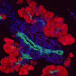 Innervation of the mammalian pancreas is crucial for endocrine and exocrine function. Neural crest cells that give rise to neural progenitor cells are responsible for intrinsic pancreatic innervation, but the specific cues that guide this process in the developing embryo are largely unknown. Now, on
 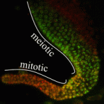 Transcriptional elongation via RNA polymerase II (Pol II) is an essential component of gene expression. It has long been thought that a necessary step of elongation is the serine 2 phosphorylation (Ser2-P) of Pol II, which is catalysed by a CDK-9/cyclin T complex called P-TEFb. Although this dogma holds true in the soma, Elizabeth Bowman, William Kelly and colleagues now present evidence (see
 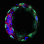 The blastocoel of mammalian embryos is a fluid-filled cavity surrounded by a monolayer of trophectoderm, and its formation marks one of the earliest stages of embryonic development. During this process, the trophectoderm undergoes a metabolic shift toward oxidative phosphorylation, which increases the level of reactive oxygen species (ROS) and places the blastocyst under oxidative stress. In this issue (
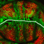 The Drosophila wing primordium is a simple epithelial structure with significant regenerative capacity. As in any regenerative system, it is essential to maintain correct tissue size and shape during the repair process, yet little is known about the mechanisms that enable such control. In this issue (
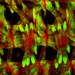 The proper formation of somatic muscle depends on morphological and molecular events that orchestrate the specification, maturation and terminal differentiation of muscle precursor cells during development. Many of the transcriptional regulatory networks that regulate these processes have been defined, but little is known about the possible post-transcriptional mechanisms that might also be involved. In this issue (
 The ubiquitin proteasome system regulates protein expression at the post-translational level by tagging certain proteins with ubiquitin and thereby marking them for degradation by the proteasome complex. This system is highly conserved and is used by almost every eukaryotic cell to degrade and recycle proteins. In this issue (
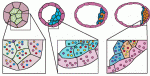 Early mammalian embryos exhibit remarkable plasticity, and recent studies of mouse embryos implicate a network of transcription factors in governing this plasticity and the establishment of embryonic lineages. Here, Alfonso Martinez Arias and colleagues summarise this information, link it to classical embryology and propose a molecular framework for the establishment and regulation of developmental plasticity. See the Hypothesis article on p.
  François Guillemot heads the Division of Molecular Neurobiology at the MRC National Institute of Medical Research in London, where his research focuses on the transcriptional control of neurogenesis and the epigenetic regulation of gene expression in neural development. François recently became an editor for Development, and we asked him about his research and career, as well as his lab’s future move to the Crick Institute. See the Spotlight article on p.