The community site for and by
developmental and stem cell biologists

In Development this week (Vol. 139, Issue 12)

Posted by , on 22 May 2012

Here are the highlights from the current issue of Development:


Mechanical changes in cochlea development

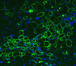
Correct patterning of the mammalian inner ear sensory epithelium, which contains mechanosensory outer hair cells (OHCs) that detect and amplify sound vibrations and non-sensory supporting cells such as pillar cells (PCs), is essential for hearing. The cell surface mechanical properties of both OHCs and PCs are important for their function but how are these properties regulated during development? On p. 2187, Katherine Szarama and colleagues use atomic force microscopy to show that OHCs and PCs have different cell surface mechanical properties that develop over different time courses. By pharmacologically modulating cytoskeletal elements, they show that the increase in OHC stiffness observed during development depends primarily on actin whereas the development of the cell surface mechanical properties of PCs depends on microtubules. In addition, they report that fibroblast growth factor signalling regulates the developing cell surface mechanical properties of OHCs and PCs, in part by altering cytoskeletal dynamics. These new insights into inner ear development may eventually lead to better treatments for hearing loss.


Resetting after quiescence

During development, networks of regulatory genes control precisely timed sequences of developmental events. In C. elegans, heterochronic genes, which encode several transcription factors and microRNAs (miRNAs) that regulate the expression of these transcription factors, control stage-specific cell-fate decisions. Under adverse conditions, however, second larval stage (L2) worms enter a quiescent state called dauer. Intriguingly, when conditions improve, dauer larvae complete development normally. Here (p. 2177), Xantha Karp and Victor Ambros investigate how cell-fate progression is reset after dauer. Progression from L2 to L3 requires downregulation of the transcription factor Hunchback-like-1 (HBL-1), and, during continuous development, HBL-1 downregulation relies mainly on three let-7 family miRNAs. However, after dauer, the researchers report, lin-4 miRNA and an altered set of let-7 family miRNAs downregulate HBL-1. This shift in the programming of HBL-1 downregulation, they propose, involves the enhancement of lin-4 and let-7 miRNA activity by miRNA-induced silencing complex (miRISC) modulators. The employment of alternative genetic regulatory pathways can, therefore, ensure the robust progression of cell-fate specification after temporary developmental quiescence.


ExE progenitors make an eXit

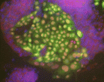
In female mammalian embryos, inactivation of one of the two X chromosomes in each cell regulates X-linked gene expression. X chromosome inactivation (XCI) is dependent on the non-coding RNA Xist, which is expressed from and coats the inactivated X chromosome. Inheritance of a paternally derived Xist mutation causes embryonic lethality because the inactivation of the paternally inherited X chromosome that occurs in the extra-embryonic lineages of female mouse embryos during imprinted XCI fails. Now, Terry Magnuson and colleagues (p. 2130) describe the exact consequences of failed XCI within the extra-embryonic ectoderm (ExE). The ExE of X/XXist– embryos consists mainly of differentiated giant cells and their progenitors, they report, and less differentiated spongiotrophoblast precursors are not maintained. At E6.5, the ExE lacks CDX2, which is required to maintain the ExE’s multipotent state. Moreover, trophoblast stem cell lines derived from X/XXist– blastocysts completely reverse normal imprinted XCI patterns. These results suggest that dosage compensation is indispensable for the maintenance of trophoblast progenitors and that imprinted XCI is probably erased in ExE cells.


Gibberellin regulation of flowering

Several environmental cues, including day length, and endogenous developmental signals regulate the transition from leaf production to flower formation in plants. In Arabidopsis, the growth regulator gibberellin promotes this transition most strongly under short day (SD) conditions. Here (p. 2198), George Coupland and colleagues show how gibberellins also promote flowering in response to long days (LDs). The researchers deplete gibberellins in the vascular tissue or the shoot apical meristem by tissue-specific overexpression of GA2ox7, which catabolises gibberellins. Under LD conditions, gibberellins are needed in the vascular tissue to increase production of a systemic signal that is transported from the leaves to the meristem during floral induction. However, in the meristem, instead of activating the expression of the transcription factor SOC1 (which is needed to induce flowering under SD conditions), in response to LDs, gibberellins regulate the expression of SPL transcription factors, which are needed later during floral induction. Thus, the researchers conclude, gibberellins play spatially distinct roles in promoting flowering under long photoperiods.


Migrating primordial germ cells exploit endoderm remodelling

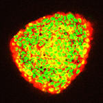
Cell migration through epithelial tissues occurs during development, infection, inflammation, wound healing and cancer metastasis. But how do cells overcome the impermeable junctions between epithelial cells? Leukocytes move out of blood vessels by loosening endothelial cell-cell junctions but do all cells actively remodel tissue barriers during migration? According to Jessica Seifert and Ruth Lehmann, who are studying Drosophila primordial germ cell (PGC) migration through the endodermal epithelium to the gonadal mesoderm, the answer to this question is no (see p. 2101). Although PGC migration requires activation of the G protein-coupled receptor Trapped in endoderm 1 (Tre1) within PGCs, the timing of PGC migration is dictated by the developmental stage of the endoderm. Now, using live imaging and genetic manipulation, the researchers show that PGCs take advantage of developmentally regulated epithelial remodelling, which causes discontinuities in the endoderm, to gain access to the gonadal mesoderm. Thus, Seifert and Lehmann conclude that, rather than actively remodelling tissue barriers, some migrating cells exploit existing tissue permeability.


Tcf21 seals cardiac fibroblast fate

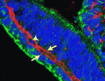
The primary source of cardiac fibroblasts, which are essential for normal heart physiology, is a subpopulation of epicardial cells that has undergone epithelial-to-mesenchymal transition (EMT) and entered the myocardium. But does cardiac fibroblast specification occur early in the formation of the epicardium (which is a multipotent mesothelial layer of cells that spreads over the developing myocardium) or after the epicardial-derived cells have entered the myocardium? Here (p. 2139), Michelle Tallquist and colleagues resolve this puzzle by investigating the role of the transcription factor Tcf21 in cardiac fibroblast specification in mice. The researchers use lineage tracing to show that Tcf21-expressing epicardial cells are committed to the cardiac fibroblast lineage before the initiation of epicardial EMT. Moreover, Tcf21-null embryos fail to develop cardiac fibroblasts and Tcf21-null fibroblast progenitors do not undergo EMT. These results indicate that cardiac fibroblast specification occurs in the epicardium before EMT occurs and, importantly, these findings identify Tcf21 as an essential transcription factor for cardiac fibroblast cell-fate determination.


Plus…


X chromosome inactivation in the cycle of life

Bakarat and Gribnau review new insights into the molecular events occurring during the life cycle of X chromosome inactivation and, in the accompanying poster, provide an overview of the mechanisms regulating X inactivation and reactivation.

See the Development at a Glance poster article on p. 2085


Evolutionary crossroads in developmental biology: cyclostomes

Shimeld and Donoghue summarise the development of cyclostomes (lamprey and hagfish) and discuss how studies of cyclostomes have provided important insight into the evolution of fins, jaws, skeleton and neural crest.

See the Primer article on p. 2091

(note that this article is part of a series of Primer articles on organisms that represent an evolutionary crossroads in the study of evolutionary developmental biology – see the online Featured Topic to view other articles in this series)


Thumbs up (No Ratings Yet)
Loading...

Categories: Research

Navigate the archive

Use our Advanced Search tool to search and filter posts by date, category, tags and authors.

Postdoctoral position in invertebrate stem cell biology

Posted by , on 22 May 2012

Closing Date: 15 March 2021

Postdoc position available at School of Natural Sciences and Regenerative Medicine Institute (REMEDI), National University of Ireland, Galway, to study the molecular mechanisms that control pluripotency and early lineage commitment in stem cells of the cnidarian Hydractinia. This animal is an established cnidarian genetic model organism, amenable to gene expression analysis and manipulation and classical genetics.

The successful candidate will have a PhD in developmental biology, cell biology, molecular biology or related area.

The position is available for 2 years in the first instance. Starting date will be September 2012 or shortly thereafter. Salary will be €38,286 per annum.

For further details please go to http://www.nuigalway.ie/about-us/jobs/ or to the lab webpage at http://www.nuigalway.ie/frank . To apply, email cover letter, CV and the names and contact info for 2-3 references to Dr. Uri Frank at uri.frank@nuigalway.ie .


Thumbs up (No Ratings Yet)
Loading...

Categories: Jobs

Publishing ‘dirty’ data

Posted by , on 22 May 2012

How much does it matter that the images we publish are neat and tidy? It’s a question I’ve been dealing with over the past couple of weeks, and I wanted to share some thoughts. Here at Development, as at many journals, we check all figures before publication to try and identify potentially inappropriate image manipulation. Whenever we do come across a figure that doesn’t comply with our guidelines on image processing, we contact the authors to ask for clarification, request that the author provides us with the original data – so we can check that nothing fraudulent is going on – and often also ask that the final figure be changed to properly represent the original data. I’m happy to say that problems are few and far between, and that those issues I have come across in the short time I’ve been here have been more a case of beautification than of fraud. But is it okay for authors to ‘clean up’ their images with Photoshop paintbrush tools or the like: not touching the data itself, but rather getting rid of specks of dust or extraneous bits of tissue that are there on the slide?

The images shown here don’t come from any paper, but have been kindly provided by a researcher to illustrate what I’m talking about.

This is a Drosophila wing disc, where clones of cells are marked with GFP, and the entire disc stained with phalloidin in red. Very often in preps like this, you get bits of irrelevant tissue associated with the disc on the slide. But this one looks very clean, right? Wrong. Here’s the original version – you can see that there’s a piece of trachea, stained red, off the left side of the wing disc.

So, thinking that this bit of extraneous tissue is problematic, the researchers have taken the simple solution of photoshopping it out: something that’s very clearly revealed by the standard checks we run on our figures: as shown here.

I’ve seen a seen a few of these cases recently, and in each, the aim of the authors was to ensure that the images were easily interpreted, and that readers weren’t diverted from the data by the extraneous bits of stuff. This may seem innocent, but it could be the first step on a dangerous slope, at the bottom of which lie the clearly fraudulent activities of deleting the bits of data that don’t fit our hypothesis, or making up data that do. Journal guidelines are (or at least should be) pretty unambiguous, and the case above falls foul of this statement taken from our Guide to Authors: “Unacceptable manipulations include the addition, alteration or removal of a particular feature of an image, and splicing of multiple images to suggest they represent a single field in a micrograph or gel.” So while it may seem innocuous, it’s not permitted. Nor is it, at least to my mind, in any way necessary: are we really that easily distracted? Does that little bit of trachea really stop us from seeing the clones in the wing disc? It’s been pointed out to me that the image above could have simply been re-cropped to remove the offending tissue, and if it’s okay to do that, why isn’t it okay to selectively black out those parts of the panel? That’s a reasonable point, and selective cropping is an issue to which I’m not convinced there is a straightforward answer. But I’m guided by the basic principle that the presented data should accurately reflect what you saw down the microscope or on the blot or whatever, and that what may seem irrelevant to you (a higher molecular weight ‘background’ band on your Western) might actually be important to someone else (“Oooh look – this might be a post-translational modification of my protein”).

I well remember from my time in the lab the agony of discovering that the perfect picture was ‘ruined’ by a bit of fluff to the side of the embryo, or because the vibrotome knife had left streaks across the section. And then spending hours re-mounting or re-sectioning to avoid these imperfections. But we all know that science can be an inherently messy endeavour: cells don’t grow in neat rows, and Western blots often give us background bands. So why do we need to hide this when it comes to publication? Of course, it’s vital that the data are clearly presented and understood, but what’s most important is that they accurately represent the experiment, and there’s a danger of losing sight of this in the desire for a beautiful image.

Initiatives like publishing all the uncropped blots that have gone into making the figures in a paper (as pioneered by Nature Cell Biology) are aimed at addressing this issue: by all means show only the relevant bit of the blot in the main figure, but for those interested in the (literally) bigger picture, the whole thing – warts and all – is available. But it can be a pain to find and assemble these files, and we don’t want to make publishing harder than it already is – although there’s a school of thought that says if you can’t lay your hands on the original data, you need to be better at archiving it in the first place!

So what do the Node readers think? Have you been tempted to ‘prettify’ your data for publication, or have you actually done it? Are our guidelines clear enough on what you can and can’t do? Do you support initiatives to make the raw data available to the reader, or is it all too much of a hassle? We’d really love your input on what kind of requests or demands a journal should make in terms of data presentation, so please answer the poll below (it’s completely anonymous!) and give us your feedback in the comments section.


Katherine Brown is the Executive Editor of Development

Thumbs up (4 votes)
Loading...

Tags: , ,
Categories: Discussion, Images, Research

Senior Research Assistant

Posted by , on 17 May 2012

Closing Date: 15 March 2021

A research assistant position is available in the laboratory of Dr. Brad Davidson, soon to be located in the Department of Biology at Swarthmore College.  We investigate fundamental questions of Cell Signal Integration and Gene Network Evolution by focusing on the earliest steps of heart cell specification. For more information see http://www.mcrp.med.arizona.edu/html/braddavidson/index.html.

This position is a great match for a recent Ph.D. in biology, biochemistry or a related field who is interested in exploring fundamental questions in developmental cell biology while expanding their technical expertise and lab-management skills.   Employment would begin in July or August of 2012 and continue for one or two years, with the possibility of a longer term dependent on continued funding.

You will initially work directly with me to set up the lab, establish some key assays and initiate pre-defined projects.  You will then be given more flexibility and independence; helping to interpret data and plan future experiments. This effort will require diligence and a conscientious attention to detail.   I will also expect you to participate with an active sense of curiosity and demonstrate an intellectual engagement with relevant ideas and scientific literature.  It will be essential for you to think critically about your results and have the ability to troubleshoot and resolve experimental problems as they arise.

Major research techniques will include generation and manipulation of transgenic invertebrate embryos, molecular cloning and confocal microscopy.  The position will also involve training/supervision of undergraduates and lab management (maintaining seawater aquaria, purchasing lab supplies, reagent preparation, maintaining lab organization and computer/data management).

Requirements:  A Ph.D. in Biology or a related discipline with a solid background in molecular biology is essential. The ideal candidate will have a track record demonstrating their enthusiasm for bench-work as well as their ability to carry out a creative and productive research program. Excellent written and oral communication skills are required. Training in informatics or programming would be a strong asset.

Please apply for the Post Doctoral Research position listed at http://www.swarthmore.edu/hr.xml under the ‘Apply for a job’ link.  Make sure to include your CV, cover letter and two letters of recommendation.

Swarthmore College is a highly selective liberal arts college located in the suburbs of Philadelphia, whose mission combines academic rigor with social responsibility. Swarthmore has a strong institutional commitment to excellence through diversity in its educational program and employment practices and actively seeks and welcomes applications from candidates with exceptional qualifications, particularly those with demonstrable commitments to a more inclusive society and world.


Thumbs up (No Ratings Yet)
Loading...

Categories: Jobs

Postdoctoral Position in Zebrafish Lab at Indiana University School of Medicine, Indianapolis Indiana, USA

Posted by , on 16 May 2012

Closing Date: 15 March 2021

postdoctoral position is available in the zebrafish laboratory of Ryan M. Anderson at the Wells Center for Pediatric Research, Indiana University School of Medicine, Indianapolis, Indiana (http://www.wellscenter.iupui.edu/researchers/ryan-m-anderson). We use zebrafish as a genetic/epigenetic model to study pancreatic endocrine cell differentiation during development and regeneration. The fellow will study cell reprogramming in vivo using newly isolated mutants and transgenic lines.

We seek a motivated scientist who is focused, productive, and possesses exceptional skills in cellular and molecular biology. Candidate must hold a doctorate in Cell/Developmental Biology, Genetics, or related field. Background in zebrafish and bioinformatics are desirable, not essential. The ideal scientist is a good communicator of ideas and data, and a team player who enjoys working collaboratively within our group.

Please forward applications to Ryan M. Anderson ryanande@iupui.edu

Include a letter describing research experience and future interests, contact information for three references, and CV. Potential start date is immediate.


Thumbs up (No Ratings Yet)
Loading...

Tags: , , , , , , ,
Categories: Jobs

Funding for international postdocs

Posted by , on 11 May 2012

Rupinder emailed the Node recently to ask if we knew any sources for postdoc funding for international postdocs.

Many PhD graduates travel to another country for their postdoc, but a lot of government grants in host countries are unavailable to international applicants – even if they’re already in the country on a study or work permit. That means you have to rely on your supervisor’s lab grant, which limits your choice of lab to those that can afford you. An external grant increases your chances of finding a postdoc lab abroad, so it’s worth trying to get one.

I had a look, and it seems that there are three main sources of external funding for international postdocs:

1.) Grants set up to promote international exchanges and collaborations.

There are several sources of funding that support the general cross-border exchange of information. Here are a few that are relevant to postdocs in the life sciences.

The Wellcome Trust supports fellows who want to work in particular countries.

The Human Frontier Science Program has postdoc grants specifically for postdocs moving to a new country to broaden their research.

EMBO has fellowships available for postdocs moving either to or from a member state of the EMBC

The American Association of University Women offers funding to female international graduate students and postdocs who want to work in the US:

For international postdocs wanting to work in Europe, the European Commission has funds available.

2.) Charities that support disease-specific biomedical research

Unlike tax-funded government agencies, donor-supported medical charities often don’t restrict grant eligibity to citizens or visitors of a particular country. They just want the best researchers to find a cure or treatment for the disease or disorder they represent. These charities are too numerous to list, but the National Postdoc Association (of the US) has a page where you can download a spreadsheet with many of the US-based charities. They also have other information for international postdocs in the US. If you’re in another country, you may want to search for charities in that country that relate to the work you’re interested in.

3.) Grants from your host country

Some countries encourage young scientists to work abroad for a few years, and then return with their new-found knowledge. If this funding is available to you, these are likely to be government grants from your country of citizenship or country of (permanent) residence, so check with your home country.
A list of some of these funds – as well as other sources of funding for international postdocs – can be found on the EMBL website, under “other potential sources…”.

That was all I could find, but if you know of any other sources of funding for international postdocs, please leave a comment below.

Thumbs up (6 votes)
Loading...

Categories: Funding, Resources

Smart signaling in the developing brain

Posted by , on 10 May 2012

The WNT pathway functions in so many processes during development that it is easy to be jealous of its multi-tasking abilities.  A recent paper in Development describes the role of WNT signaling in neural stem cell proliferation.

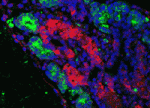
WNT signaling plays an important role in neural development, axon guidance, cell polarity, and stem cell biology.  WNT pathway mutations are linked to several different cancers, including medulloblastomas.  Medulloblastomas are malignant tumors found in the cerebellum of the brain and are more commonly found in children.  Recently, Pei and colleagues asked which cells in the developing cerebellum were responsive to canonical WNT signaling and found that WNT signaling promotes proliferation of neural stem cells (NSCs), the major source of neurons on the cerebellum.  WNT signaling, however, did not induce proliferation in granule neuron precursors, the other major class of progenitors in the cerebellum.  In addition, Pei and colleagues used transgenic mice with an inducible allele of β-catenin to find that constitutive activation of WNT signaling induced NSC proliferation in vivo.  This increase in proliferation, however, caused NSCs to lose the ability to undergo self-renewal or differentiation.  The images above show cerebellum tissue from a control mouse (left) and a transgenic mouse with activated β-catenin (right).  Constitutively active WNT signaling caused an increase in the population of NSCs (G-FAP, Sox1), which were also actively proliferating (BrdU, bottom).

For a more general description of this image, see my imaging blog within EuroStemCell, the European stem cell portal.

ResearchBlogging.orgPei, Y., Brun, S., Markant, S., Lento, W., Gibson, P., Taketo, M., Giovannini, M., Gilbertson, R., & Wechsler-Reya, R. (2012). WNT signaling increases proliferation and impairs differentiation of stem cells in the developing cerebellum Development, 139 (10), 1724-1733 DOI: 10.1242/dev.050104

Thumbs up (1 votes)
Loading...

Tags: , ,
Categories: Images, Research

Homeward bound

Posted by , on 10 May 2012

It’s been longer than I’d hoped since I last wrote, but a whole heck of a lot has been going on. Monday last week, the professor in charge of international cooperation at the Chinese Academy in Urumqi came out to the field station with a couple of colleagues to host lunch with us. I knew they were coming, but I didn’t know that they had called ahead to arrange a dumpling-making party for us. So while we waited for their arrival, the cook had prepared all of the fillings and bought the wrappers, and all of the students working at the field station came to join us in making our own lunch. Great fun! After the bigwigs arrived, plates and plates and plates of food hit the table, and the three of us Americans decided that we should eat family style at one long table rather than dispersed among the 5 tables around the room. So we moved furniture around to join up three of the long tables into one feast table. The cool thing about that is that as far as I know those tables are still arranged in that manner. The atmosphere at meal times went from groups of students that work together sitting together and not interacting much to one big happy family laughing and chatting throughout dinner. And I noticed they all started playing pool more and even moved the pingpong table down to where it could be used. I like to think we had a little something to do with that.
Around the celebratory mood, we still had to get a lot of work done, so we’ve been going out to the desert each night for Talia’s data collection. I don’t think I gave a clear description of her work before now, so here it goes. The jerboas are bipedal animals (see previous reference to my motivation for studying this animal here.) Talia works in a lab that studies biomechanics, and her interest is in the nature of that bipedality. In most animals, there is a rule that is followed where an animal predictably switches gaits as it increases in speed (e.g. horses). In the jerboas, it seems that they can use multiple gaits at any speed and perhaps switch from two feet hitting the ground at once to one foot at a time depending on how maneuverable they need to be. So we’ve been setting up enclosures on different natural substrates and filming the animals at night when they’re most active. Unfortunately this requires the use of headlamps which draws giant moths to our faces and bats encircling our heads. If it’s not one troublesome species, it’s another.
So upon completing this part of the grand adventure, we made it back to Urumqi, survived yet another banquet, said our goodbyes and thank yous,  and boarded a 46 hour train to Shanghai. The Hangover Express (see banquet reference above). It was wonderful to be on a train for two days with no one to be held accountable to and no one trying to pour 50% or greater alcohol down our throats to show our happiness! Talia and I slept and slept and slept and read two books each and watched the Chinese countryside whiz past. Our cabin companions were friendly. One was a man who designs jade jewelry and has a fondness for good tea. He shared some tea with us and showed us photos of his work. Talia showed him some of her high speed video of the jerboas which he seemed to find pretty cool. I’m just glad we made it with all of our belongings and the box of embryos was still cool on arrival to the lab in Shanghai. That was the purpose of the two day train – to get the embryos from Xinjiang to Shanghai. Since 2009, it is difficult to do much out there. Shipping or flying with anything remotely questionable is nearly impossible. I can’t tell you how many times I’ve heard the phrase “Xinjiang is special.” But we now have 415 jerboa embryos in a freezer in Shanghai, the paperwork has been initiated, and in about 2 months they ought to arrive in my lab in Boston. That’s when the real fun of RNA sequencing and in situ validation starts, and it’s a little terrifying to think that the fate of my career for the next several years sits awaiting the approval of the Chinese government!
Thumbs up (2 votes)
Loading...

Categories: Lab Life

What does a cell know and how does it know it (Just a thought on Dennis Bray’s Wetware, Yale University Press) Alfonso Martinez Arias (Dpt Genetics, University of Cambridge, Cambridge UK. ama11@hermes.cam.ac.uk)

Posted by , on 10 May 2012

It is a time of gene counting, mapping, function guessing in a narrow way: a gene for this or a gene for that. If one reads the indexes of journals one might believe that by adding genes one gets an organism. It is for this reason that reading a book like “Wetware” by Dennis Bray is a stimulating exercise and a gift to the mind. Dennis Bray is a pioneer of what some of us call Systems Biology, not the one related to genomics and proteomics but ‘the other one’, the one that aims to understand Biology from the perspective of organized assemblies of molecules that perform specific tasks. For a long time he has been interested in the idea that proteins perform computations and this notion lies at the heart of his view of life. In Wetware, distilling thoughts gathered over a career probing the engineering of cells, Dennis Bray presents a personal view of a cell as a biomolecular device that senses its environment, whether a specific milieu or other cells, and through a computation of sorts, responds by changing itself and the environment. Sure, one can read his primary papers on many of the topics developed here but there is beauty and insight in the way the argument is put together in the book, its accessibility and the inspired way in which notions of what is life flow- from the reactions of Stentor to human intervention to the information processing capacities of biochemical networks.

The book can be construed as a popular work but this will betray what I perceive as an ambition to open ways of thinking which, if taken seriously, should lead many cell and developmental biologists to pose questions about calculations and dynamics rather than about genes and their function as revealed by more or less complex genetic experiments.

Dennis Bray has written before an excellent book on cell movement (Cell movement: from molecules to motility) where he explores in detail some of the issue raised in Wetware and which has guided many of us on our trips into the structure and function of the cytoskeleton. But Wetware has a lot more because here he develops a panoramic view of Life from the inside of the cell. The pervasive metaphor of the computer will irritate some computational buffs but this will only reveal that they miss the point: a cell is not a piece of hardware in the human sense and the book makes this clear that any analogy one might draw will have to bear in mind the peculiar characteristics of the molecular make up of a cell. One example: the notion that Diffusion of molecules within a cell might be a correlate of the cables that bring together the pieces of a machine, is a far reaching notion that deserves to be explored, as is the implicit notion that a signature of life is the molecular make up of sensory experiences.

I shall not go on about the book because the main object of this posting is to encourage you to read it. I believe that it should be required reading for graduate students in cell and developmental biology. Towards the end there is a quotation from Andre Gide which is an enticement to act: “one doesn’t discover new lands without consenting to lose sight of the shore for a very long time”. Wetware is a gust of wind that should encourage you to sail into the current of the unknown, without fear, with the imagination that is denied by the current interest in publications rather than Discovery.

Thumbs up (9 votes)
Loading...

Tags: , , ,
Categories: Book Reviews

Drosophila Research Technician

Posted by , on 9 May 2012

Closing Date: 15 March 2021

University College London – UCL Cancer Institute

Full Time : The appointment will be on UCL Grade 6. The salary range will be £25,927 – £30,384 per annum., inclusive of London Allowance.

The successful applicant will use molecular biology, Drosophila genetics and immunohistochemistry to study the molecular mechanisms regulating gene expression during development. They will also be responsible for maintenance of the laboratory’s collection of Drosophila stocks and for supporting the day-to-day running of the laboratory.

The post is intially funded till September 2013.

The candidate must hold a BSc (Hon) in biological sciences or equilvelant. Experience in basic molecular biology techniques along with good organisational and communication skills are essential for this post. While experience working with Drosophila would be an advantage, training will be provided as required.

For further details about the vacancy and how to apply on line please go to http://www.ucl.ac.uk/hr/jobs/ and search on Reference Number 1250187.

If you have any queries regarding the vacancy or application process please contact Cancer Vacancies Team e-mail: vacancies@cancer.ucl.ac.uk

Please quote the Reference Number with all correspondence.

Closing Date: 23/5/2012

We particularly welcome applications from black and minority ethnic candidates as they are under-represented within UCL at this level.

Thumbs up (No Ratings Yet)
Loading...

Categories: Jobs