Submitted by Tohru Yano:
“This paper tell us the way of the formation of sharp morphogen gradient in development by using computer simulation and experimental data.”
From December 1 to 24 we are featuring Node readers’ favourite papers of the past year. Click the calendar in the side bar each day to see a new paper. To see all papers submitted so far, see the calendar archive.
If you got into the habit of reading the Node on your tea breaks, we’ve got just the thing for you! We now have Node tea bags, custom made for us. They’ll be at our stand at the ASCB meeting next week (stand 1303) and at several other conferences. Below are some photos of the tea bags in production, showing the custom tags and pouches.
Submitted by Heather Buschman:
“To determine if microRNAs influence germ layer formation in early embryonic development, the authors sequenced the entire mouse genome’s worth of microRNAs in embryonic stem cells. In doing so, they discovered that two microRNA families in particular—let-7 and miR-18—block endoderm formation, but permit mesoderm and ectoderm formation. The authors also went on to pinpoint a mechanism—these microRNAs direct germ layer formation by dampening TGFβ signaling.”
From December 1 to 24 we are featuring Node readers’ favourite papers of the past year. Click the calendar in the side bar each day to see a new paper. To see all papers submitted so far, see the calendar archive.
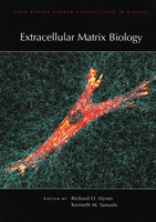
This book review originally appeared in Development. Charles Streuli reviews “Extracellular Matrix Biology ” (Edited by Richard O. Hynes and Kenneth M. Yamada).
Book info:
Extracellular Matrix Biology. Edited by Richard O. Hynes, Kenneth M. Yamada Cold Spring Harbor Laboratory Press (2012) 387 pages ISBN 978-1-936113-38-5 $67.50 (hardcover)
To be metazoan is to have an extracellular matrix (ECM). ECM components evolved simultaneously with animals and provided the architectural scaffold onto which multicellularity could emerge several hundred million years ago. From a primitive basement membrane toolkit and the primordial fibrillin and thrombospondin genes that allowed the evolution of multilayered animals, successive gene duplications and crises permitted the appearance of protostomes and deuterostomes, leading to vertebrates and eventually to us. Along the way, the evolutionary shuffling of pre-existing and novel protein domains to form tenascins, fibronectin and other matrix proteins coincided with the emergence of neural crest, vasculature and the nervous systems that characterise chordates and advanced vertebrates. Coinciding with this, metazoans evolved complex plasma membrane machines containing adhesome proteins that link the ECM to the cytoskeleton and intracellular signalling pathways.
Core ECM proteins make up ∼1.5% of the mammalian proteome. This enormous parts-list of around 300 matrix proteins, newly christened the ‘matrisome’, highlights the diverse and extraordinary roles of the matrix in metazoan biology. ECMs range from pericellular matrices, such as basement membranes, to the grander structures of connective tissue that give organs their shape, and the tendons, cartilage and bones that bestow humans and animals with their form. It is therefore not surprising that the ECM pervades every aspect of metazoan life. The matrix is essential for early embryonic development and it regulates nearly every facet of cell behaviour. Moreover, altered ECM and ECM-cell interactions lie at the root of many diseases, including cancer, diabetes, inflammatory disorders and osteoarthritis.
The ancient history of the association between the ECM and cells effectively means that the two are inseparable. Although the prosaic view of cells is that they end at the plasma membrane, the cell and its pericellular matrix are actually a continuum in all complex animals. A cell’s ECM coat provides the architectural hardware for cell positioning and migration, which is meshed together with intracellular cytoskeletal networks by myriad components of the adhesome. The matrix enables a first-pass checkpoint for growth and developmental signals, it is the vehicle for morphogenetic gradients in development, and it is the spatial rheostat for intracellular signalling pathways. ECM receptors thus convert both chemical and physical extracellular inputs to determine cell survival, proliferation, migration and tissue-specific gene expression.
A book on such a profoundly important topic requires a clear vision of the breadth of the ECM and its roles in development, cell and tissue function and disease. Extracellular Matrix Biology has been organised by two of the most eminent scientists in the ECM research field, Richard Hynes and Kenneth Yamada, and, predictably, the book hits the mark. This volume, one in a series from Cold Spring Harbor Laboratory Press, contains a superb collection of review articles. Well-known opinion-makers in the field have written the chapters, which are detailed, authoritative and up to date. The organisation of the book is sensible, beginning with an overview of the matrisome and its evolution, and then working through the key ECM components, basement membranes, collagens, proteoglycans, tenascins, thrombospondins and fibronectin. This is followed by chapters on integrin activation, genetics, and links with TGFβ, as well as adhesion complexes, mechanotransduction, and matrix remodelling. Towards the end, links between ECM and tissues are considered, with chapters on cell migration, embryonic development, angiogenesis, skin, and the nervous and haemostatic systems. I am slightly disappointed that mucins have been omitted, as they are crucial components of the pericellular matrix, but nevertheless the coverage in the book is excellent overall.
Looking through my (rather dusty) volume of Betty Hay’s Cell Biology of Extracellular Matrix (2nd edition, 1991), which was one of the last comprehensive books on ECM, it is startling to see how many advances have been made since then. Prior to our era of genomics, knockouts and dynamic imaging, much of the knowledge about ECM was based on biochemistry and cytology. Disease associations with altered ECM were known, but many of the molecules involved had not been identified and mechanisms were largely unexplored. Little was understood about the structures of matrix molecules or their receptors, and there was only an indication that integrins could transmit signals from the ECM to the cell interior. It is also interesting that the focus of illustrations has largely switched from electron microscopy images and some relatively sketchy diagrams to detailed lists of ECM components, domain organisations, interaction networks, disease associations, and mouse and human mutations. This new book is therefore a timely addition, and many of the chapters also consider where the field is going now and what unknowns might be unravelled in the cell-matrix field over the next 20 years. Overwhelmingly, the shift in focus from the organisation of the ECM in tissues, to cataloguing its vast array of constituents and interactions with cells, indicates that a central challenge is to understand how all the matrix and adhesion components fit together and how they control cell fate and function.
Although crystal ball gazing is inevitably difficult, there are certainly some exciting opportunities that are alluded to in the book. The emerging awareness that biology occurs in four dimensions and is completely dependent on the ECM (incidentally, this was well understood by Hay) will undoubtedly be a major focus for cell-matrix research at least for the next few years. For example, knowledge of how matrices are assembled in 3D and remodelled in 4D is beginning to appear. Most cells function within communities, often of different cell types, and we are just beginning to understand how the matrix influences cell function and dynamics in 4D and how different cell types use the ECM to communicate, e.g. via stromal-epithelial interactions. The notion that the physics of the ECM profoundly influences cell and tissue function is fuelling new research on how forces are built into ECMs, how they are sensed by cells, and how they are converted to chemical signals that control transcription and cell fate. The structures of ECM proteins and some adhesion signalling proteins have been solved (some only at low resolution), leading to a new focus on atomic interactions at the protein-protein interface. ECM and adhesion receptors are crucial regulators of the immune system, and the dawn of immunomatrix biology is changing the thinking about immunological control. And, most profoundly, the basic discoveries of cell-matrix research are yielding novel small-molecule, bioengineered and genetic therapies for the vast array of diseases caused by altered ECM and interactions between the cell and the matrix. For example, there are no effective cures for osteoarthritis beyond surgery; advances in this (and other) areas using genetic analysis based on genome-wide association studies and systems biology are needed to dissect all of the component parts that cause debilitating diseases of injury and old age, and thereby to develop new treatments.
One of the challenges posed by reviewing this book was to ask who it is for. The original chapters appeared over several months in the online publication Cold Spring Harbor Perspectives in Biology, and they have now been collated for the book. Although some of the articles are available online (depending on your library’s subscription), by combining them in a single volume the editors have created a very nice resource that will provide a valuable reference work for several years to come. I would recommend those entering the field of cell-matrix biology, as well as those already in it, to read the book from cover to cover. The ECM is now understood as being so much more than a tissue scaffold and a precursor for building bones, and this new book definitively indicates where the field is now and where it is going. The intimate connections between the ECM and most cellular processes in animal development are gradually being recognised by mainstream biologists, and this could be accelerated when the key advances in this book are used to update current undergraduate texts on cell biology. At a broader level, once some of these concepts are incorporated within school biology education, then we’ll know that ECM biology has truly come of age.
As you may have spotted, we’ve just posted a job ad for community manager for the Node. That is currently my job, but I will be leaving at the end of February, so there will be a vacancy.
I’ve had this job since the Node launched, and even before then! I’ve had a great time setting up the site, meeting so many scientists around the world, and seeing the Node grow from nothing to a well-visited resource for the developmental biology community. You’ve all been great to work with, but now it’s time for me to move on, and for someone else to take over. Have a look at the job ad and apply if you’re interested. The office is based in Cambridge, UK. We… erm, I mean THEY are actually moving to a nice new office in a few months, too. The company publishes five journals, so you’ll learn a lot about publishing while you’re here as well.
For everyone else, to make it easier on the next community manager and in the transition phase, please remember that you do not need to ask for permission to post on the Node. If your account is approved, you can post. I’ve personally been okay with reading over some posts for grammar and spelling if you’ve asked me to, but this is really not necessary. The concept of the Node is such that you can share news without editorial input. Take advantage of your freedom to post!
My new job will be in London, and still in the scientific publishing-adjacent field, so I’m not going far, but I won’t be involved with the Node anymore. If you’d like to stay in touch with me, and find out what I’m doing next, your best bet is to find me on Twitter as @easternblot.
The Company of Biologists and its journal Development are seeking to appoint a new Community Manager to run its successful community website the Node and the journal’s social media activities.
Launched in 2010, the Node is the place for the developmental biology community to share news, discuss issues relevant to the field and read about the latest research and events. We are now looking for an enthusiastic and motivated person to develop and maintain the site.
Core responsibilities of the position include:
– Creating and commissioning content for the Node, including writing posts and soliciting content from the academic community, societies and other organisations
– Providing creative and practical input into the development of the site
– Maintaining and developing Development’s and the Node’s presence on social networking sites such as Facebook and Twitter
– Contributing non peer-reviewed content to the journal
– Representing Development and the Node at international conferences
– Contributing to the Company of Biologists’ outreach activities
The successful applicant will have:
– Research experience in the biological sciences, ideally a PhD in developmental or stem cell biology
– Proven blogging and social media skills, ideally including experience with WordPress
– A clear understanding of the online environment as it applies to scientists
– Excellent writing and communication skills
– Excellent interpersonal and networking abilities – both online and in person
This is an exciting opportunity to develop an already successful and well-known site, engaging with the academic, publishing and online communities. The Community Manager will work alongside an experienced and growing team, including Development’s Executive Editor, as well as with the journal’s international team of academic editors. Additional responsibilities may be provided for the right candidate. The position will be based in our office in Cambridge, UK.
The Company of Biologists (www.biologists.com) is a not-for-profit organisation, publishing five journals in the biological sciences: the three established journals Development, Journal of Cell Science and The Journal of Experimental Biology, as well as two newer Open Access journals, Disease Models & Mechanisms and Biology Open. The organisation has an active programme of charitable giving for the further advancement of biological research, including travelling fellowships for junior scientists and contributions to academic societies and conferences.
Applicants should send a CV along with a covering letter that summarises their relevant experience (including, if possible, links to online activities), salary expectations, and why they are enthusiastic about this opportunity.
Applications should be sent by email no later than January 20th 2013 to miriam@thecob.co.uk
Informal queries to Miriam Ganczkowski on +44 (0)1223 426 164
Applicants should be eligible to work in the UK.
Submitted by Eva Amsen:
“I saw Mitinori Saitou present this work at the EMBO meeting and it made me realize how far this fast-moving field has already come.”
From December 1 to 24 we are featuring Node readers’ favourite papers of the past year. Click the calendar in the side bar each day to see a new paper. To see all papers submitted so far, see the calendar archive.
There are a lot of situations in life where the “middleman” is unnecessary and costly. In cells, that middleman is necessary and fascinating at the same time. The sequence of DNA to middleman mRNA to protein provides our cells with countless ways to regulate complex events, including those surrounding stem cell divisions.
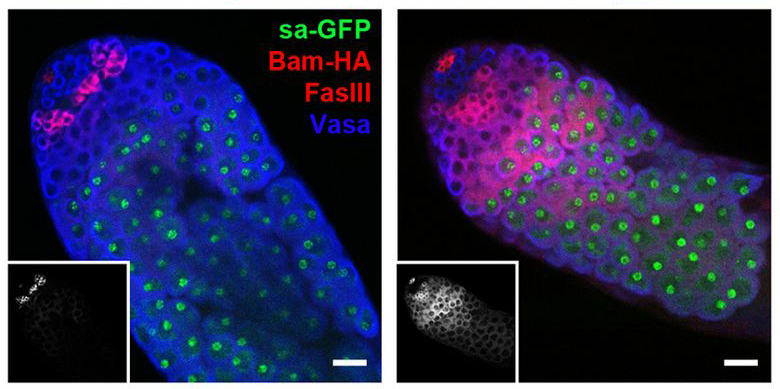
When stem cells divide, one daughter cell maintains stem cell characteristics while the other daughter cell follows a path towards differentiation. Before differentiation, this cell can divide several times during a stage called transit-amplification. In the fruit fly testes, the division of the germline stem cell (GSC) produces another GSC and transit-amplifying cells called spermatogonial cells. Spermatogonial cells begin differentiating as they pass through the spermatocyte stage on their way to becoming sperm. A recent paper in the journal Development investigates the regulation of a key differentiation factor, Bam (Bag of marbles), during this transition. Bam protein is found in spermatogonial cells, but is not found in the later spermatocyte stage (yet bam mRNA is present). According to Eun and colleagues, post-transcriptional regulation of Bam levels occurs through microRNA binding at the bam 3’UTR. Overexpression of the two microRNAs involved delayed the proliferation-to-differentiation transition, while failure of Bam down-regulation caused differentiation problems leading to male sterility. The images above show fruit fly testes stained to show the GSC hub (small red hub), spermatocytes (green) and Bam protein (red, white in inset images). In a control testis (left), Bam protein is found in spermatogonial cells near the GSCs. When the bam 3’UTR sequence was replaced with the 3’UTR of a constitutively expressed tubulin gene, Bam protein is found throughout spermatocytes as well.
For a more general description of this image, see my imaging blog within EuroStemCell, the European stem cell portal.
Eun, S., Stoiber, P., Wright, H., McMurdie, K., Choi, C., Gan, Q., Lim, C., & Chen, X. (2012). MicroRNAs downregulate Bag of marbles to ensure proper terminal differentiation in the Drosophila male germline Development, 140 (1), 23-30 DOI: 10.1242/dev.086397
“This beautiful paper uses cadherin-coated magnetic beads applied to single Xenopus gastrula cells in culture to demonstrate that tension polarises the cell and determines the direction of migration, and to investigate the mechanosensory pathway involved. The work exemplifies our increasing appreciation of the role that physical forces have in determining cell and tissue behaviour during development.”
From December 1 to 24 we are featuring Node readers’ favourite papers of the past year. Click the calendar in the side bar each day to see a new paper. To see all papers submitted so far, see the calendar archive.
Today, at the Company of Biologists Christmas lunch, we held a small inter-journal cover competition. Over the past few weeks, all our colleagues from Development, Journal of Cell Science, Biology Open, Disease Models and Mechanisms and the Journal of Experimental Biology voted for their favourite (and least favourite) covers submitted by editors of each journal. Development had submitted the skate cover – and won! It was a close race with JEB’s chameleon cover.
Of course this was all YOUR doing. Thanks David, Lynn, and Meghan for taking the image, and thanks to everyone else for voting it onto the cover. You helped us win chocolate for the office!



 To be metazoan is to have an extracellular matrix (ECM). ECM components evolved simultaneously with animals and provided the architectural scaffold onto which multicellularity could emerge several hundred million years ago. From a primitive basement membrane toolkit and the primordial fibrillin and thrombospondin genes that allowed the evolution of multilayered animals, successive gene duplications and crises permitted the appearance of protostomes and deuterostomes, leading to vertebrates and eventually to us. Along the way, the evolutionary shuffling of pre-existing and novel protein domains to form tenascins, fibronectin and other matrix proteins coincided with the emergence of neural crest, vasculature and the nervous systems that characterise chordates and advanced vertebrates. Coinciding with this, metazoans evolved complex plasma membrane machines containing adhesome proteins that link the ECM to the cytoskeleton and intracellular signalling pathways.