The community site for and by
developmental and stem cell biologists

A career as editor

Posted by , on 19 May 2011

I was a PhD student with Peter Walter, studying protein translocation across the endoplasmic reticulum. I did very well as a student, publishing six research papers during that time. After that, I was a postdoc with Christiane Nüsslein-Volhard and then with Yuh-Nung Jan, studying oogenesis and pattern formation in Drosophila. Throughout my training, people handed me their papers to read and asked me to attend their practice qualifying exams because I was always looking for the big picture, always needing to know why they were doing the experiments in the first place. Time and again, friends suggested I become an editor. I mostly laughed at them, in part because I wasn’t really sure what an editor did, and in part because there seemed to be so few jobs for editors that I never thought I’d get one. Anyway, I was good at the bench and I couldn’t imagine disappointing my father by not taking a job in academia.

In 1993-4, I went on the job market, looking at standard faculty positions. I received some offers, including one from Vanderbilt University, where I am now. But I was resisting accepting a position, and some friends – who were also on the job market at the time – sent me to a career counselor. The counselor’s husband was a bench scientist, so she had some sense of my career until that point, and asked me a very simple question, one that I had never asked myself: “If you didn’t have to worry about how much money you made, or what anyone else thought of you, what would you do?” What surprised me was that I knew the answer to that question: I’d be a student for the rest of my life.

When I said that, I realized that what I loved about being in science was knowing something today that no one knew yesterday, and that it didn’t matter so much if I learned it by my own hands, or over coffee with a friend. This calmed me down a lot about the idea of starting up a lab, as I had been worrying about going around claiming other people’s work as my own.

When I got back to the lab that afternoon, I went into our lunch room and opened an issue of Cell. Near the front cover, there was an ad for an editor, and they were looking for someone with expertise either in cell biology (my graduate training) or developmental biology (my postdoctoral training) – and I thought to myself that it seemed an awful lot like being a student, so I applied for the position. The short version of this story is that I got the job, and I was a full time professional editor for about a dozen years, including a few very exciting years as the Executive Director of Public Library of Science, before returning to academia to my current position at Vanderbilt University. I still spend most of my time as an editor, most notably as the Editor-in-Chief of Development’s newest sister, Disease Models & Mechanisms.

Being an editor is really very much like being a student. You encounter lots of interesting and new science every day in a broad range of fields. But, at least for those of us who decide which research papers to publish in high profile journals such as Cell, it is also about being able to judge science. As an editor, you will have to turn away most of the papers you receive, and explain your reasoning. I think that editorial decisions need to be timely, constructive, transparent, and fair – or at least as much as they can be, given the need to turn complicated issues and shades of gray into stochastic decisions, and the need to keep confidential information confidential.

Editors and journals can be important partners to science. Through editorial policy they can help scientists do the right thing, such as sharing information and reagents; and through publishing policies, such as leaving copyright with the author or providing free access to published work, they can contribute to accelerating science itself. Editors are both gatekeepers and guardians of our treasury of scientific information, and editors need to behave responsibly and ethically. Thankfully, the Committee on Publication Ethics, of which Company of Biologists is a member, helps editors know and do the right thing.

For any trainees interested in being an editor at a journal like Cell, I encourage that you participate in journal clubs. A lot of the work of an editor is to assume the best of all possible worlds – that the conclusions are justified by the data and the interpretations are reasonable – and then assess how important the conclusions are to and beyond the field. Journal clubs are great ways to practice this – but be careful not to miss the forest for the trees, and to get too focused on the weakest part of the paper, which may be tangential to the overall conclusions. Also, take the time to go to seminars and meetings and talk to scientists in other fields and at other institutions. A scientist too buried in his own experiments to pay attention to the exciting discoveries around him is unlikely to enjoy or succeed at an editorial career.

Oh, and when I told my father, he was delighted. You see, he’d wanted me to be a writer, and considered this an enlightened combination of my twinned loves of science and language. And he was right.

Thumbs up (11 votes)
Loading...

Tags: ,
Categories: Careers

Navigate the archive

Use our Advanced Search tool to search and filter posts by date, category, tags and authors.

Help me select some Node highlights

Posted by , on 17 May 2011

The Node’s birthday is coming up on June 22nd and we’re currently preparing some things for that behind the scenes.

One thing that I’m working on are a few slideshows to present this summer at departments and conferences. In these presentations, I want to include some highlights from the Node’s first year, and I could use some help finding those. I could include my own favourites, but I’d rather hear what you liked, so tell me, please: what was the best thing you’ve seen on the Node all year? Favourite posts, favourite topics, favourite parts of the site, or any other comments are all welcome.

Have a look at the archives in the sidebar or the intro page for inspiration and reminders of what was on the site a few months ago. So many memories already…

Thanks for helping out!

Thumbs up (1 votes)
Loading...

Tags:
Categories: Discussion, News

Imaging stem cells in the Drosophila ovary

Posted by , on 12 May 2011

To accompany our paper “Long-term live imaging provides new insight into stem cell regulation and germline-soma coordination in the Drosophila ovary” I have been asked by staff at the Node to discuss the path we took when developing a successful imaging protocol.

Germline follicle formation in the Drosophila ovary is a very dynamic process – involving the coordinated migration and division of many different cells and cell types. I was trying to study how two of these cell populations – somatic escort cells and germline stem cells together build germline cysts, the 1st stage of follicle development. Whilst lineage data had suggested that germline cysts are generated by the coordinated division of one germline stem cell and two escort cells, actually finding examples of escort cells dividing in vivo was proving difficult, whereas dividing germline stem cells are found frequently. Escort cells are quite extraordinary cells; their nuclei are jammed in little cracks between large germ cells yet they have incredibly long, thin cytoplasmic processes that wrap the germ cells. These characteristics make escort cells pretty hard to see and therefore to study. I decided that the only way I was going to be able to make any progress in unraveling the escort cell/germline stem cell coordination mystery was to be able to watch both cell populations live in their native environment whilst they built a germline cyst. Given that cyst formation involves highly dynamic cellular behaviours and takes around 12 hours this was going to be quite a challenge.

Upon trying to image live germaria (the structure within the ovary that builds germline follicles) I immediately encountered a big problem – movement. The ovaries of Drosophila are subdivided into strings of developing follicles (called ovarioles) with the germaria at one end. Each of these ovariolar strings is surrounded by a sheath of muscle. Upon dissection the muscle contracts so that regular, peristaltic movements pass down the ovarioles causing them to flap around in the culture dish. The only method I found to prevent these contractions was to manually remove the muscle layer. Even then, I still had a movement problem; given that germaria are attached to strings of follicles some of which are an order of magnitude larger than a germarium, any little rocking or rolling movements of the large follicle would result in the attached germarium flying out of the field of view, and this appeared to happen frequently. I partially fixed this problem by dissecting flies that have just emerged from the pupal case, whose ovaries do not contain the more mature, larger follicles. Additionally, programming the microscope stage to move slowly and smoothly between each position during imaging helped. However, some movement remains despite trying many different methods to immobilize the tissue (including coating the dish with extra-cellular matrix components, draping the tissue with membranes and placing it in gels). Given that follicle formation is such a dynamic process, this is not entirely surprising. By re-focusing the microscope at regular intervals during imaging up to half of the germaria can be kept in focus for 12 hours.

Although overcoming tissue movement difficulties was a lengthy process, once achieved the path towards imaging success went quickly. This was thanks to the previous development by the Montell lab of culture medium designed to nurture more mature follicle stages that I found also supported follicle formation. Germaria could be cultured and imaged for greater than one entire cycle of follicle formation (14 hours). Once the method was developed and the first movies made the answer to the escort cell/germline stem cell coordination problem was almost immediately evident: Escort cells do not divide with gemline stem cells to generate a germline cyst which then migrates as one unit down the germarium, instead escort cells remain in one place, dividing rarely, and simply ‘hand-over’ the germ cells from one escort cell to the next. Demonstrating the ease with which dynamic processes can be studied when viewed unfolding in living tissue.

ResearchBlogging.orgMorris, L., & Spradling, A. (2011). Long-term live imaging provides new insight into stem cell regulation and germline-soma coordination in the Drosophila ovary Development, 138 (11), 2207-2215 DOI: 10.1242/dev.065508

Thumbs up (5 votes)
Loading...

Categories: Research

When F-actin dynamics and Hippo signalling activity meet to regulate tissue growth.

Posted by , on 11 May 2011

Genetic screens in flies brought me by chance to have a look at one of the basic apparatus of the cell: the actin cytoskeleton. At that time, I remembered my cell biology courses at University and since the actin cytoskeleton was not one of the hot spot, I though it was just a machinery required for basic cellular functions, from which, we knew almost everything. I quickly realized that in multicellular organisms, it was definitively worth to have a closer look at it.

Given the crucial role for F-actin in numerous cellular processes, it came to us as a surprise that triggering excess F-actin polymerization, by disrupting the activity of the actin-Capping Protein (CP) heterodimer, did not automatically lead to cell lethality, but could trigger tissue growth. We therefore focused our efforts on investigating how the control of F-actin could be involved in preventing cell proliferation. This gave rise to our recent story published in Development ” Actin-Capping Protein and the Hippo pathway regulate F-actin and tissue growth in Drosophila”.

We quickly realized that many of the targets genes controlled by the Hippo (Hpo) tumor suppressor pathway were upregulated in cells lacking CP. This conserved signal transduction pathway, has emerged as a critical regulator of tissue size both in Drosophila and mammals. Central to the Hpo pathway is a kinase cascade, which involves the Hpo and Warts (Wts) kinases and their adaptor proteins Salvador (Sav) and Mats. Phosphorylation of Wts by Hpo prevents nuclear translocation of the transcriptional co-activator Yorkie (Yki) through phosphorylation, leading to transcriptional downregulation of target genes that positively regulate cell growth, survival and proliferation. Multiple upstream inputs are known to regulate the core Hpo kinase cassette at various levels. However, how they do so is poorly understood (http://dev.biologists.org/content/138/1/9.abstract). We observed that in cells lacking CP, upregulation of Yki target genes was associated to a decrease in phosphorylated Yki and its relocalization to the nucleus. These behaviours resulted from defects in F-actin since knocking down the Cyclase associated protein Capulet (Capt), which sequesters actin monomers, also caused abnormal F-actin accumulation and ectopic Yki activity.

At around this time, we were happy to hear that the laboratory of Georg Halder had also identified CP and Capt in a S2 cells genome-wide RNAi screen for genes that inhibit expression of a Yki-dependent luciferase reporter gene. Their story, just published in EMBO Journal (Sansores-Garcia et al.), shows a sticking correlation between the F-actin levels and Hpo signalling output. While knocking down actin modulators that prevent F-actin accumulation triggers Yki activity, depleting actin regulators that promote F-actin accumulation has the opposite effect. Their work nicely shows that this link is conserved in mammalian cells. They also confirmed that extra F-actin polymerization of cells knocked down for CP or overexpressing an activated version of the actin nucleator Diaphanous (DiaCA), causes tissue growth in vivo through Yki activity and not through disruption of apical-basal cell polarity or signalling in general. Interestingly, dsRNAi targeting the Cofilin twinstar (tsr) was also tested positive as a modifier of Yki activity in their assays. However, in Drosophila epithelia, we found that loss of tsr had no effect on Yki target genes. Although, epithelial and S2 cells require both a proper F-actin network to regulate Hpo signalling activity, the different effects of tsr loss on Hpo signalling output suggest that Hpo signalling activity uses different F-actin networks in tissues and in single cells. In epithelia, CP, Capt and DiaCA control F-actin formation near the apical surface, while Tsr acts around the entire cell cortex. Because the integrity of the apical domain of epithelial cells seems critical for the maintenance of Hpo signalling, in epithelia, but not in S2 cells, a specialized population of polarized F-actin,regulated by CP, Capt and DiaCA at the apical cell membrane, may promote Hpo signalling activity.

One interesting issue was then to determine at what level of the pathway, CP or F-actin dynamics in general intersect with Hpo signalling activity. We observed that overexpressing Hpo or the upstream regulator Expanded (Ex) suppressed growth of CP-depleted cells, suggesting a role for F-actin upstream or in parallel to Ex. However, overexpressed, Ex and possibly Hpo, also suppressed F-actin accumulation of Cpa-depleted cells. Moreover, we found that Hpo signalling activity prevented F-actin accumulation, independently of Yki activity. We were therefore confronted to the problem: what is first, the egg or the chicken. Nevertheless, our results indicated an interdependency between Hpo signalling activity and F-actin dynamics in which CP and Hpo pathway activities inhibit F-actin accumulation, and the reduction in F-actin in turn sustains Hpo pathway activity, preventing Yki nuclear translocation and upregulation of proliferation and survival genes.

In contrast, Sansores et al. observed that the DiaCA-induced overgrowth was not suppressed by Hpo or Ex overexpression, whereas Wts could do so. Thus, they conclude that DiaCA and therefore F-actin affect the Hpo pathway upstream of Wts but in parallel to Ex and Hpo. Interestingly, they also show that, unlike loss of CP, the accumulation of F-actin caused by DiaCA overexpression was not suppressed by Wts, Ex, or Hpo overexpression. The different effects of DiaCA and CP loss on Hpo signalling activity, when Ex and Hpo are overexpressed, argue that the control of F-actin by Hpo pathway activity is required to sustain its activity. By preventing F-actin accumulation of CP-depleted cells, increased Ex or Hpo may sustain Hpo pathway activity. In contrast, because overexpressed Ex or Hpo cannot prevent excess F-actin resulting from DiaCA overexpression, F-actin accumulation can still inhibits Hpo pathway activity. Alternatively, the different outcome of cells depleted of CP or overexpressing DiaCA, when Ex or Hpo are overexpressed, could result from different strengths of the CP loss of function and DiaCA phenotypes on growth. The effect of expressing DiaCA on growth were stronger than those caused by loss of CP, suggesting that Hpo signalling activity is only partially affected by the loss of CP. Overexpressed Ex or Hpo might therefore counteract the mild effect of CP loss on Hpo signalling activity but not the one of DiaCA overexpression. Finally, it is possible that F-actin acts at several levels to regulate Hpo signalling activity. Consistent with this possibility, F-actin has also been shown to control the activity of the MST1/2 Hpo orthologs in mouse fibroblasts. Moreover, we noticed that clones of cells mutant for CP affected Hpo pathway activity cell autonomously but also non-autonomously. Thus, the control of F-actin by CP activity may have a dual function in controlling Hpo signalling activity. Sansores et al. did not observe any non-autonomous effect on Hpo signalling activity in cells knocked down for CP using RNAi, suggesting that the non-autonomous disruption of Hpo signalling activity is less sensitive to F-actin accumulation.

All these data took us to bring one more piece to the complex puzzle of how Hpo pathway activity is regulated and convinced us, more than ever, that different populations of F-actin filaments exist in the cell that have specialized functions. The challenge will now be to identify these populations, understand how they are regulated and characterize their role in controlling specific cellular events.

ResearchBlogging.orgFernandez, B., Gaspar, P., Bras-Pereira, C., Jezowska, B., Rebelo, S., & Janody, F. (2011). Actin-Capping Protein and the Hippo pathway regulate F-actin and tissue growth in Drosophila Development DOI: 10.1242/dev.063545

Sansores-Garcia, L., Bossuyt, W., Wada, K., Yonemura, S., Tao, C., Sasaki, H., & Halder, G. (2011). Modulating F-actin organization induces organ growth by affecting the Hippo pathway The EMBO Journal DOI: 10.1038/emboj.2011.157

Thumbs up (6 votes)
Loading...

Tags: ,
Categories: Research

Interview with Beddington Medal winner Carlos Carmona-Fontaine

Posted by , on 11 May 2011

Each year, the British Society for Developmental Biology awards the Beddington Medal for the best PhD thesis in developmental biology. At the 2011 BSDB meeting, this award went to Carlos Carmona-Fontaine, who completed his PhD in Roberto Mayor’s lab at UCL. Now a postdoc at Sloan-Kettering Institute in New York, Carlos returned to the UK to present his thesis work at the BSDB meeting. He gave a brilliant talk that included comparisons between insect migration and neural crest cell movement, as well as an auditory interpretation of the effect of chemo-attractants on harmonic collective cellular movement. Everyone in the audience (including Carlos’ parents!) enjoyed the talk immensely. I caught up with Carlos at the end of the conference to talk a bit more about locusts, music, and the trick to a successful PhD thesis.

Congratulations on the Beddington Medal. What was your thesis about?

Thank you. My thesis was about neural crest migration. Neural crest cells are a cell population in the embryo that can differentiate into different kinds of cells. But before differentiating, they have to migrate, and the way they do that is as a group of cells. This is called collective migration. What was interesting to me was that neural crest cells seem to be able to self-organise in order to get this coherent, co-ordinated movement, so I studied which kinds of cell-cell interaction allow for these coherent group movements.

Your Beddington talk yesterday covered a lot of ground. You were even talking about locust migration. What do locusts have to do with cells?

I had found a paper in Current Biology, by the group of Iain Couzin, about locust collective movement. I noticed that the kind of interactions they described were so similar to the interactions I was finding in neural crest cells, that I thought similar rules may apply, and we started collaborating.
Obviously locusts are very different to cells, but in mathematical terms the interactions between locusts or between migrating cells are very, very similar. If you look at attraction and repulsion, for example, “attraction” in insects could be a visual cue, whereas in cells it could be a chemo-attractant, but at the end of the day, mathematically speaking, they are the same thing: simple interactions that allow collective movement.

Besides locusts and cells, you also included some music in your talk. Can you explain what that was about?

One of the striking things of collective movement is not only that the cells remain together, but that they move in a coordinated way. I considered this to be harmonic movements, and was thinking of a way to represent that.
Then I heard a concert by Steve Reich. He’s a 21st century composer – one of my favourites. He has this piece called “Drumming”, which is only percussion. There’s a specific moment with a lot of xylophones and some drums, and they all seem to coordinate in this very random pattern. I was just amazed by this piece.
This gave me the idea of representing cell coordination in a similar way. It ended up as something a lot less musically pleasing than what Steve Reich does, but the idea came from there.

What are you doing now? Are you still working on neural crest cells?

No. I started a postdoc in New York about a month ago, and there I will eventually work on computer models of tumour growth. But the project is not entirely defined yet, and the idea is to explore a little bit at this point.

Do you have any advice for new PhD students?

Don’t stress, have fun – and pay attention to mathematical, and more quantitative means of biology. Even if you’re not an expert in mathematics, you can still try to get some inspiration from more physical and mathematical points of view. And have fun.

Read more:
ResearchBlogging.orgCarmona-Fontaine, C., Matthews, H., Kuriyama, S., Moreno, M., Dunn, G., Parsons, M., Stern, C., & Mayor, R. (2008). Contact inhibition of locomotion in vivo controls neural crest directional migration Nature, 456 (7224), 957-961 DOI: 10.1038/nature07441

Bazazi, S., Buhl, J., Hale, J., Anstey, M., Sword, G., Simpson, S., & Couzin, I. (2008). Collective Motion and Cannibalism in Locust Migratory Bands Current Biology, 18 (10), 735-739 DOI: 10.1016/j.cub.2008.04.035

Thumbs up (12 votes)
Loading...

Tags: , , , ,
Categories: Interview, News

In Development this week (Vol. 138, Issue 11)

Posted by , on 10 May 2011

Here are the highlights from the current issue of Development:

Dronc regulates Numb and neuroblast formation

The ability of stem cells to maintain a balance between self-renewal and differentiation is crucial for development and tissue homeostasis. In Drosophila neuroblasts, the tumour suppressor Numb restricts proliferation and self-renewal in differentiating daughter cells but how its activity is regulated is unclear. Here, Bingwei Lu and colleagues reveal a novel mechanism for the control of Numb activity (see p. 2185). They show that phosphorylation of Numb at conserved sites modulates its tumour suppressor activity and that the antagonist actions of Polo kinase and protein phosphatase 2A control Numb phosphorylation. Expression of Numb-TS4D (a phosphomimetic form of Numb) abolishes Numb activity, they report, and leads to the formation of ectopic neuroblasts. They also identify the Dronc caspase as a Numb binding partner and show that Dronc overexpression suppresses the effects of Numb-TS4D in an apoptosis-independent and possibly non-catalytic manner. By contrast, reduction of Dronc activity enhances phospho-Numb-induced ectopic neuroblast formation. Together, these results provide new insights into neural stem cell homeostasis in Drosophila.

Stem cell development goes live

Stem cells are maintained by signals from their local microenvironment but it has been hard to study exactly how stem cell behaviour is controlled. Now, Lucy Morris and Allan Spradling describe a culture method for live imaging Drosophila ovarian development within the germarium and use it to test some long-held beliefs about ovarian follicle development (see p. 2207). The germarium is a structure at the anterior tip of ovarioles that produces new ovarian follicles by controlling follicle and germline stem cell (GSC) division and nurturing their developing daughters. The researchers confirm, for example, that GSC divisions are oriented with respect to the germarium’s anteroposterior axis. They also show that somatic escort cells (the glial-like partners of early germ cells) do not adhere to and migrate with GSC daughters as previously proposed, but pass the GSC daughters from one escort cell to the next using dynamic membrane activity. These and other results establish the live imaging system as a valuable tool for the study of stem cell biology.

p45NF-E2 controls intrauterine growth

In mice, the absence of the leucine zipper transcription factor p45NF-E2 results in thrombocytopenia (reduced platelet numbers), impaired placental vascularisation and intrauterine growth restriction (IUGR). It is generally assumed that the lack of embryonic platelets causes the growth problems seen in p45NF-E2-deficient embryos, but now, Berend Isermann and colleagues report that the placental defect and IUGR of p45NF-E2 null mouse embryos is unrelated to thrombocytopenia (see p. 2235). Instead, they show that p45NF-E2 is expressed in trophoblast cells where it is required for normal syncytiotrophoblast formation, placental vascularisation and embryonic growth. Expression of p45NF-E2 in labyrinthine trophoblast cells, they report, colocalises with the expression of Gcm1, a zinc-finger transcription factor crucial for syncytiotrophoblast formation. Finally, they show that p45NF-E2 cell-autonomously represses Gcm1-dependent syncytiotrophoblast formation by inhibiting acetylation in vitro and in vivo. The identification of this novel function for p45NF-E2 during placental development provides new insights into the mechanisms underlying IUGR, a poorly understood but common complication of human pregnancies.

miR165: a plant dose-dependent positional cue

Cell fate determination by positional cues occurs during both plant and animal development. Although some positional cues have dose-dependent effects in animals, this type of cue has not been identified in plants. Here, however, Keiji Nakajima and colleagues show that microRNA165 (miR165) non-cell-autonomously regulates the differentiation of multiple cell types in the Arabidopsis root in a dose-dependent manner (see p. 2303). The Arabidopsis root consists of a central stele (which contains the pericycle layer and the xylem) surrounded by layers of endodermis, cortex and epidermis. Endodermis-derived miR165/166 is known to specify xylem differentiation in the root meristem by suppressing the expression of the transcription factor PHABULOSA (PHB) in the stele. Using an inducible miR165 expression line, the researchers now reveal that endodermis-derived miR165 acts in a dose-dependent manner to establish a PHB expression gradient across the stele that controls the differentiation of two xylem cell types and the pericycle. Thus, these studies reveal that plant development requires at least one dose-dependent positional cue.

Extracellular Engrailed signals get direct

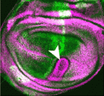
Although homeoprotein transcription factors are best known as cell-autonomous developmental regulators, several homeoproteins have direct non-cell-autonomous activities in the developing vertebrate nervous system. But do homeoproteins also act as signalling molecules during invertebrate development? On p. 2315, Alain Joliot, Florence Maschat and co-workers present the first in vivo evidence for homeoprotein signalling in Drosophila. They use detergent-free immunostaining to reveal an extracellular pool of the homeoprotein Engrailed (En) in the fly wing disc. They then use a secreted single-chain anti-En antibody to show that En is a short-range signalling molecule that participates in the development of the wing’s anterior crossvein. Finally, they report that, in contrast to the repressive effect of En on decapentaplegic (dpp) expression, where it acts intracellularly as a transcription factor, extracellular En activity helps to form the anterior crossvein by enhancing Dpp signalling. The researchers propose, therefore, that direct signalling by homeoproteins is an evolutionarily conserved mechanism that is involved in the development of multiple tissue types.

Blood vessels guide 3D lung branching

Traditionally, blood vessels are regarded as an inert network of tubes that supply tissues with nutrients and oxygen, but recent studies suggest that blood vessels play perfusion-independent roles in early development. Now, on p. 2359, Eli Keshet and co-workers report that blood vessels also determine the reproducible branch pattern of lung airways in mice. During lung development, the coordinated branching of epithelial and vascular tubes culminates in their co-alignment in the mature organ. By ablating the lung vasculature in vivo and in lung explants, the researchers show that, although the first two-dimensional round of epithelial branching proceeds at a nearly normal rate, branching events that require rotation to change the branching plane into the third dimension are selectively affected. This role of the vasculature is independent of perfusion, flow or blood-borne substances but can be partly explained by perturbation of the expression of stereospecific branching regulators such as FGF10. Together, these results reveal a novel perfusion-independent role for the vasculature in directing three-dimensional organogenes.

Thumbs up (No Ratings Yet)
Loading...

Categories: Research

Zinc Finger Nucleases targeting genes in a frog near you!

Posted by , on 9 May 2011

Loss-of-function studies in Xenopus have been, until recently, limited to transient knockdowns by injection of morpholino antisense oligonucleotides.  In part because of X. laevis’ complex allotetraploid genome, the system lacked techniques for targeted gene disruption.  In recent years the use of the closely related Xenopus tropicalis, a true diploid with one of the smallest tetrapod genomes, has allowed the addition of genetics to the suite of molecular and embryonic techniques enjoyed by frog researchers, and in a recent PNAS paper from Richard Harland’s lab, John Young and colleagues provide the first example of targeted gene knockout in amphibians using zinc finger nucleases.

The C2H2 zinc finger, the most common DNA binding domain, was first discovered, appropriately enough in Xenopus.  Each zinc finger recognizes a specific trinucleotide sequence, and several fingers can be linked in tandem to bind a longer DNA sequence, with a high degree of specificity.  In addition it has been possible to engineer zinc finger protein domains with novel specificities and to couple them to the DNA cleavage domain of the restriction enzyme Fok1, giving the possibility of engineering zinc finger nuclease (ZFN) that targets a unique genomic locus and produces a double strand break (DSB).  In the absence of a homologous template, cells repair DSBs by non-homologous end joining which sometimes introduces small insertions or deletions. If these result in a frame-shift, and the ZFN is targeted to the coding sequence of a gene, a null or hypomorph allele may be produced.  So far ZFNs have been used to target genes in drosophila, zebrafish and rat.  The Young et. al. paper has now extended the use of the technique to Xenopus.

The authors first tested whether somatic ZFN genome editing was possible in amphibian embryos by injecting mRNA encoding a nuclease targeted against GFP into eggs heterozygous  for a single-copy ubiquitous GFP transgene.  Lower doses of mRNA resulted in a mosaic loss of fluorescence and when the mRNA concentration was increased most cells in the resulting embryos were not fluorescent.  Sequencing of the GFP locus in injected embryos showed insertions and deletions of 5-20 bp in the targeted sequence.

A panel of ZFNs targeting noggin was then designed and 6 constructs that were active in a yeast-based single stranded annealing assay were tested in Xenopus embryos.  All ZFNs were tolerated well by the embryos and produced mutant amplicons at a frequency of 10-47% with insertions/deletions ranging from 5-195 bp.  To test heritability, injected embryos carrying mutated loci were raised to sexual maturity and crossed to wild type frogs.  3 mutated noggin alleles were recovered in the next generation, including a 4bp insertion resulting in a premature stop codon, probably a null allele, and a 12 bp deletion which might be a hypomorph, showing that ZFNs can be used to produce an allelic series of any gene of interest.

An exciting future possible application of ZFNs is the ability of DSBs to increase the rates of homologous recombination, in the presence of template DNA with homologous ends, by many orders of magnitude.   Although this has not been tested in embryos yet, it appears to work well in cell-lines, raising the possibility of using ZFNs to generate knockins as well as knockouts in genes of interest.  For the time being, however, the ability to disrupt specific genes in Xenopus is an exciting development for loss-of-function studies in this model system.

ResearchBlogging.orgYoung, J., Cherone, J., Doyon, Y., Ankoudinova, I., Faraji, F., Lee, A., Ngo, C., Guschin, D., Paschon, D., Miller, J., Zhang, L., Rebar, E., Gregory, P., Urnov, F., Harland, R., & Zeitler, B. (2011). Efficient targeted gene disruption in the soma and germ line of the frog Xenopus tropicalis using engineered zinc-finger nucleases Proceedings of the National Academy of Sciences, 108 (17), 7052-7057 DOI: 10.1073/pnas.1102030108

Thumbs up (8 votes)
Loading...

Tags: , ,
Categories: Research

The story behind the screen – flashbacks from the first RNAi screen in a whole vertebrate

Posted by , on 9 May 2011

The story of our recently released Development paper ‘FatJ acts via the Hippo mediator Yap1 to restrict the size of neural progenitor cell pools’  involves hundreds of dozens of fresh free-range eggs and not trivial amounts of time spent peering down a microscope.  I have written this with Nick van Hateren, who is the joint first author of this paper along with me.

We had recently developed a short hairpin based system for carrying out RNA interference in the chicken called the pRFPRNAi system. This was an exciting time in the lab, as there had previously been no such system to carry out functional genetics in our favourite model system, and we were looking forward to make full use of it. This was also a great chance for us to demonstrate to the community that the chicken really is an excellent model system to do RNAi screens.

During this time, RNA interference screens were all the rage, and several screens had already been carried out in Drosophila cell lines and the worm, but never in a whole vertebrate. Even though shRNA was possible in the mouse, introducing constructs into mouse embryonic tissue was not a trivial matter, and carrying out a screen even on a small scale would present significant challenges, and indeed still would. The main obstacle here was the inaccessibility of the mouse embryos as they developed within the mother. The chick embryo, on the other hand came conveniently packaged inside an egg, and transfecting tissues by electroporation is a well-established and efficient technique. The spinal cord, in particular, was ideal for our studies. It is shaped like a tube, making it easy to inject it with a DNA solution. The DNA can then be transfected to only one side of the spinal cord by electroporation, while the other side would remain as a convenient internal control.

Armed with these reassuring facts, we began to search for suitable candidates for an RNAi screen. We had previously carried out a microarray analysis of the chick spinal cord, and amongst the thousands of genes expressed there, there were 40 genes that contained cadherin domains. These appeared to be the perfect choice, since they were a reasonable number and also because the large size of the cadherins makes it difficult to carry out overexpression studies.

We decided it would be prudent to target three different regions of each gene, which meant that we would need to sub-clone 120 shRNA sequences. Even though this sounded like a daunting task, the reality was far from it. Our cloning strategy was already well optimised, and we were done sooner than we expected. It was time to get down to the interesting work…

The screen itself was carried out very systematically. We had planned out the whole week so we could get in two rounds of electroporations and end up with a batch of fixed and frozen embryos ready for sectioning and analysis. The longest part was actually sectioning the vast numbers of embryos we generated, but we were lucky that our friendly lab technician Vicky was happy to give us a hand with this.

The screen was a rollercoaster ride of emotions – ranging from euphoria to dejection when a whole batch of our antibodies went bad. By the end of it we had many more positive hits than we had expected, and the range of phenotypes was also reassuringly diverse (Examples of two are in the image below). We were very excited that this would be a wonderful showcase for the feasibility of RNAi screens in the chick embryo.

Two examples of phenotypes from the screen

One of our most intriguing hits was FatJ, a cadherin that appeared to be important for controlling the number of a small sub-population of interneurons. Loss of FatJ caused a small but robustly reproducible increase in the number of these interneurons, and we were intrigued to understand more about this phenotype.

We found that FatJ expression is restricted to the intermediate region of the neural tube,  and we were very encouraged to find that this domain corresponded to the progenitor pools for the interneurons whose numbers were increased following FatJ knockdown. We then examined the number of cells in different progenitor pools within the FatJ expression domain. After a great many cell counts and many hours of confocal microscope time, we determined there was a corresponding increase in the number of progenitor cells within the FatJ expression domain. This gave us a valuable clue to the mechanism of FatJ action: the loss of FatJ causes an increase in the number of progenitors which then differentiate normally to produce a corresponding increase in the number of interneurons. We confirmed this by double labelling with progenitor and differentiated interneuron markers and ensuring no cells expressed both markers simultaneously.

At that time, there were relatively few studies of FatJ reported in the literature; however we noticed that FatJ was the closest vertebrate orthologue of Drosophila Fat (dFat) which was known to be involved in planar cell polarity and was upstream of the newly-discovered Hippo pathway that controls tissue size in Drosophila. Many components of this pathway are highly conserved in vertebrates so we reasoned that FatJ might act through the Hippo pathway to regulate proliferation of neural progenitors. The Hippo pathway is a MAP Kinase cascade that phosphorylates the transcriptional regulator Yorkie (Yap in vertebrates) and this prevents the expression of proliferative and anti-apoptosis genes. Our hypothesis was that, in the absence of FatJ, there was no signalling through the Hippo pathway so Yap1 was not phosphorylated and proliferative genes continued to be expressed. This would lead to an increase in the number of cells within the progenitor pool. To test this theory, we designed shRNAs to target Yap1 and Tead4 (the transcription factor partner of Yap1) and electroporated these at the same time as FatJ shRNAs. We found that loss of Yap1 or Tead4 at the same time as loss of FatJ produced a normal number of interneurons and therefore rescued the FatJ phenotype.

Around this time, a paper by Cao et al (Genes Dev. 2008 Dec 1;22(23):3320-34) was published reporting the regulation of neural progenitor pools by the Hippo pathway and that dominant repressor forms of Yap1 and Tead produce an increase in the number of Lim1/2 positive cells – the same phenotype we observed after FatJ knockdown! Crucially, the authors did not focus on the upstream signal controlling the hippo pathway, which we believed to be FatJ. To address this, we attempted to determine more directly if loss of FatJ caused a change in the phosphorylation state of Hippo pathway components. This was a time-consuming process involving many electroporations followed by sub-dissection of transfected cells and then western blot analysis with phospho-specific antibodies. Unfortunately, the anti-phosphoMst antibody (the Hippo orthologue) did not work well enough to detect a change in activity of the Hippo pathway. However, we did detect a decrease in the level of phospho-Yap1 after FatJ knockdown and this decrease was also evident by immunohistochemistry of neural tube sections. Therefore, we had confirmation that loss of FatJ causes a decrease in phosphorylation of a downstream hippo component.

This gave us a mechanism for the observed phenotype; FatJ normally acts via downstream Hippo pathway components to limit the size of specific progenitor pools in the neural tube. In the absence of FatJ, these progenitors continue to proliferate resulting in a corresponding increase in the number of the interneurons. Intriguingly, the FatJ-/- mutant mouse phenotype displays a wider neural tube than wild-type littermates suggesting that longer-term loss of FatJ expression could lead to significant tissue overgrowth.

This brought us to the end of a long journey; starting from an RNAi screen and ending with a mechanism. Even though our screen focused on a specific group of genes, we ended with a range of phenotypes – this really highlights the usefulness of the chick as a model system and has proven that RNAi screens are indeed feasible in this system, opening up new possibilities for functional genomics in higher vertebrates.

Van Hateren, N., Das, R., Hautbergue, G., Borycki, A., Placzek, M., & Wilson, S. (2011). FatJ acts via the Hippo mediator Yap1 to restrict the size of neural progenitor cell pools Development, 138 (10), 1893-1902 DOI: 10.1242/dev.064204

Thumbs up (5 votes)
Loading...

Tags: , , , , , , ,
Categories: Research

6th International Chick Conference

Posted by , on 6 May 2011

Can I just bring to eveyone’s attention that the 6th International Chick Conference is now to be held at The Roslin Insititute, UK. Sept 17-20, 2011.
This forum often attracts a strong developmental biology contingent and we anticipate the 2011 conference will include many relevant themes (e.g Morphogenesis; Organogenesis; Patterning, Cell Fates and Organizers; Genetic manipulation of chickens;Imaging and Image Analysis). Not only that but this is the first conference to be held at the new and beautiful Roslin Building, in the stunning location of the Pentland hills of Edinburgh.
Please register your interest now at-
http://www.roslin.ed.ac.uk/chick6/

Thanks
Megan

Thumbs up (6 votes)
Loading...

Tags:
Categories: Events, News

Healing an injured heart

Posted by , on 5 May 2011

Regenerative medicine and stem cell research go hand-in-hand when it comes to dreaming up future strategies for treating disease and injury in humans.  Today’s image is from a recent Development paper discussing how damaged heart tissue regenerates in zebrafish, and serves as a great model for devising strategies to help human heart attack patients.

When a person suffers a heart attack, white blood cells move into the injured area of the heart and create scar tissue.  This scar tissue is important to maintain the structural integrity of the heart, but causes long-term changes in the heart’s architecture that may lead to heart failure.   A recent paper in Development looks at this process in zebrafish, and describes how the zebrafish heart can undergo regeneration after injury to cardiac tissue.  In this paper, researchers used cryocauterization to cause localized injury to the heart that appears similar to that seen in humans after a heart attack.  Cryocauterization caused myocardial cell apoptosis within the injured area, followed by formation of scar tissue, followed by complete regeneration.  This regeneration included key cardiac tissue types, including epicardium, myocardium, endocardium and coronary vasculature.  This amazing regeneration ability of the zebrafish heart may provide a framework for how this process may be engineered for human patients after suffering heart attacks.

The images above show zebrafish heart tissue after injury (dpi = days post-injury), with bottom images showing higher magnification views of the boxed regions.  The injured area (IA) lacks tropomyosin staining (red).  Shortly after injury (A), the presence of Mlck (myosin light chain kinase, green) at the border of the injury indicates the presence of activated platelets, which promote scar formation.  After a few days, the Mlck-positive cells in the injured area (B,C) indicates the presence of smooth muscle scar tissue.  Many days after injury (D), the lack of Mlck suggests that the scar tissue has been replaced with new, healthy tissue.

For a more general description of this image, see my imaging blog within EuroStemCell, the European stem cell portal.

ResearchBlogging.orgGonzalez-Rosa, J., Martin, V., Peralta, M., Torres, M., & Mercader, N. (2011). Extensive scar formation and regression during heart regeneration after cryoinjury in zebrafish Development, 138 (9), 1663-1674 DOI: 10.1242/dev.060897

Thumbs up (5 votes)
Loading...

Categories: Images, Research