The 2026 Loke CTR annual meeting theme, “Epigenetics of embryogenesis and placentation” brings together leading clinical and basic scientists to explore topics including the interplay of environment, genetics and epigenetics in DOHaD, mechanisms of cell fate and lineage development, genomic imprinting, X chromosome inactivation, and the role of repetitive elements in trophoblast and early development.
Where: Old Divinity School, St John’s College, All Saints Passage, Cambridge CB2 1TP
Pedro Martinez. Departament de Genètica, Microbiologia i Estadística, Universitat de Barcelona
Gene Regulatory Networks: An Introduction and Their Historical and Conceptual Context.
The concept of a gene regulatory network has, by now, a long history. It was essentially developed in the papers that Britten and Davidson published in 1969 and later. In these works, they proposed the idea that genes—particularly transcription factors—constitute the underlying mechanisms that causally explain development and evolution. This paradigm shifted earlier views of developmental processes as “linear” epistatic relationships, which had incorporated concepts such as “master regulatory genes” positioned at the top of epistatic hierarchies. Although ideas like pleiotropy had long been recognized, implying that linear sequences of gene action were unrealistic representations of how genes build structures, the notion that development could still be depicted as a set of linear processes in which transcription factors act on one another remained attractive and was, in some sense, reinforced by classical developmental genetics.
The advent of genomic technologies and the ability to study interactions between numerous transcription factors and cis-regulatory sequences enabled a more complex view of development. In this framework, many factors act through mutual interactions, in hierarchical architectures, driving processes forward in a largely directional manner. Gene regulatory networks (GRNs, from now on) were instrumental in giving mechanistic form to older concepts such as developmental trajectories in Waddingtonian landscapes, where developmental decisions were visualized as the movement of a system downstream along a valley shaped by both external physical constraints and internal constraints built into the architecture of the network. Moreover, GRNs offered hints about what “emergent properties” may depend on—phenomena in which novel, nonlinear properties arise at different biological scales, even though the proposed underlying explanations were never entirely clear.
However, as with many concepts introduced in science, the use of the term gene regulatory network has gradually lost precision. In a substantial fraction of articles, it is employed in a very loose way. This trend has run parallel to the pressure to use “fashionable” terminology in papers, grant proposals, reports, and other scientific outputs—a problem that is not foreign to many disciplines.
Gene Regulatory Networks: Clarifying What Counts—and What Does Not
A Gene Regulatory Network is a graphical representation of the physical interactions between transcription factors (TFs) and their target cis-regulatory regions that drive a specific developmental process. The representation aims at completeness: it should indicate the full set of transcription factors involved, as well as all the cis-regulatory elements to which they bind (often located in other transcription factors). Needless to say, TFs also regulate other classes of genes, including those coding for signalling molecules and structural proteins. These gene products are, of course, essential for establishing intercellular communication and for generating the specific phenotypes of cells and tissues in which they are expressed. However, TF networks are the most relevant component because they constitute the driving engine of the process. TFs are the true, physical effectors that modulate new gene activities and are ultimately responsible for pushing development forward.
While all of the above may seem almost self-evident, what is striking in many papers is that the links between TFs and their regulatory binding sites are often hardly demonstrated. There is no such thing as a network of factors if the interactions are not experimentally established; otherwise, the implied causality collapses. Once again, we end up with mere correlations between TF activities, without any explanation of how the interactions drive specific parts of the developmental process. A network cannot be based on the assumption that correlations between expression patterns—whether in natural conditions or after gene perturbations—and the presence of putative open chromatin sites are sufficient, even if all these data appear together in correlated datasets (e.g., ATAC-seq profiles or transcriptomic datasets). TF binding relationships must be demonstrated. Moreover, the binding events must not only occur but also be functionally significant. For instance, binding sites may be occupied without being functional.
In this context, only those representations whose nodes and edges have been experimentally verified can truly be called networks. Moreover, we should be cautious about calling a small set of genes a network. There is an obvious impossibility in representing a bona fide network with a very limited number of components. A good network should aspire to completeness, and this requires a substantial number of experimentally demonstrated interconnections.
Alternatively, what we often have is merely a collection of correlated data, which may generate preliminary hypotheses. These hypotheses must then be rigorously tested if they are to become genuine GRNs with any degree of predictive power.
It is the completeness of a GRN that provides a genuinely mechanistic understanding of a developmental process—not merely the description of the actors involved, even when some interactions can be demonstrated. By definition, causal explanatory frameworks (Johansson et al., 2024) should always include:
Linking antecedent causes to subsequent effects;
Describing the pathways from cause to effect (not just establishing a correlation);
A clear temporal sequence, in which the cause must precede the effect; and
An explanation of the “why” of a process, offering reasons for the observed events or states.
In this context, only GRNs reveal the causal and mechanistic relationships needed for a veritable (and verifiable) understanding of development. This does not mean that GRNs are the only explanatory tool—physical forces and complex cell behaviors also account for complementary aspects of developmental processes. However, these should be integrated into a holistic framework in which the core explanatory level is provided by the nuclear events that contribute to build a GRN.
REFERENCES
Britten RJ, Davidson EH. Gene regulation for higher cells: a theory. Science. 1969 Jul 25;165(3891):349-57. doi: 10.1126/science.165.3891.349.
Johansson, LG. et al. (2024). Causal Explanations. In: A Primer to Causal Reasoning About a Complex World. Springer Briefs in Philosophy. Springer, Cham. https://doi.org/10.1007/978-3-031-59135-8_8
Peter, I. S., & Davidson, E. H. (2015). Genomic control process : development and evolution / Isabelle S. Peter, Eric H. Davidson. (1st ed.). Elsevier. (for a general review)
1Department of Microbiology and Immunology, University of Rochester Medical Center, Rochester, NY 14642, USA
2Department of Environmental Medicine, University of Rochester Medical Center, Rochester, NY 14626, USA
Introduction
Plastic particles and fibers shed from plastic debris, termed microplastics (MP), have become ubiquitous environmental pollutants found everywhere globally throughout marine and freshwater ecosystems (reviewed in [1]). Improper disposal, accidental loss, and fragmentation of plastic materials have led to an increase in MPs, which range in size from as large as 5 mm to as small as 1 mm. These MPs which can also be airborne, pollute environments as varied as urban landscapes, remote terrestrial regions, aquatic ecosystems. One of the highest waterborne MP concentrations reported in the USA is where Rochester’s Genesee River meets Lake Ontario [2]. In the air, soil and water, these MPs are consumed by a wide variety of organisms from invertebrates including mollusks and crustaceans to vertebrates such as fish, amphibians and ultimately humans. In humans, MPs accumulate in breast milk as well as various organs and tissues including the brain , liver, and placenta [3, 4]. Recently, MPs present in human tissues have increased from 2016 to 2024, especially in the brain [5]. While there is increasing evidence suggesting that MPs pose serious threats to aquatic ecology and human health, many aspects of their potential biological activity remain unclear. Notably, little is known about the potential lasting impacts of exposure to MPs during early development on immunity.
The study of biological effects of MPs has revealed multiple challenges including the wide diversity of plastic types that may or may not induce similar effects and the need for reliable biological models. While it is estimated that there are over 5000 different types of plastics composed of different polymers (e.g., nylon, polyethylene terephthalate, etc.) and chemical additives (e.g., flame retardants, plasticizers, etc.), the large majority of biological studies of MPs have used manufactured sterile polyethylene or polystyrene spherical beads of uniformized size. Moreover, under the actions of UV, temperature and pH in the environment, plastics fragment into a myriad of sizes and shapes, while their physical properties (e.g., porosity, hydrophobicity) are further altered during this process of aging. Finally, plastic debris sinking in water is associated with the formation of biofilms composed of diverse microbial communities, which may include pathogens [6].
To help fill these current gaps in knowledge surrounding MPs, we leveraged the amphibian Xenopus laevis as a robust comparative model. The X. laevis Research Resource for Immunology at the University is specialized in the development and use of Xenopus for immunological research. Fully aquatic tadpoles are ideal experimental organisms for addressing the acute and persistent biological impacts resulting from exposure to MPs, because their post-embryonic development, including the immune system differentiation is external and not protected by the maternal environment, which increases their sensitivity to perturbations by water pollutants. Furthermore, the development and physiology, as well as the immune system of Xenopus, are remarkably similar to that of humans which has led to fundamental discoveries about pathophysiology, development, and medical immunology [7-9].
Lake Ontario MicroPlastics Center
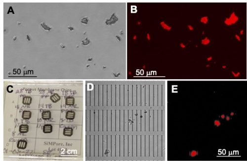
The research program using Xenopus is integrated into the Lake Ontario MicroPlastics Center (LOMP). This is a new collaborative initiative between the University of Rochester (UR) and the Rochester Institute of Technology (RIT). LOMP is a hub for research, translation, and community engagement, interested in investigating how different types of real-life plastics enter and move through the Great Lakes ecosystems and how MPs may affect human health under different environmental conditions. LOMP is one of six Centers for Oceans and Human Health jointly funded by the National Science Foundation and the National Institute of Environmental Health Sciences. As mentioned above, besides marine ecosystems, significant quantities of MPs have been detected in Upstate New York lakes, rivers, and the drinking water of cities such as Rochester, NY [10]. This has led to the establishment of this productive collaborative research program between UR and RIT. An important component of LOMP is its Materials and Metrology Core that develops standardized protocols and produces optimized materials for the different research teams. For example, the Core provides silicon nanomembranes used for water and air filtration to quantify environmental MPs; it also prepares by cryomilling lab-made MP stock solutions in defined size ranges to mimic real-life MPs (Fig. 1A-C).
Figure 1: Polyethylene terephthalate (PET) MPs. (A) Bright field image and (B) RFP image obtained with an epifluorescence microscope of real-life PET-MPs of variable shapes and sizes (1-20 μm) stained with fluorescent Nile red dye. (C) Photograph of silicone nanomembranes (SimPore, Inc.). (D) Bright field image, and (E) RFP image of PET-MPs isolated on a nanomembrane (2 μm × 50 μm microslits) from liver lysates of tadpoles exposed for 7 days.
Biodistribution and biological impact of MP exposure in water on Xenopus
As part of the LOMP program, the objectives of the Xenopus research project are to determine the biodistribution and biological impacts of MP water contaminants using a sensitive and reliable experimental platform in X. laevis. The overarching hypothesis is that the developmental exposure to MPs will induce composition and size dependent long-term perturbations of immune homeostasis, chronic inflammation, decreased resistance to microbial pathogens and poorer antimicrobial immunity.
Using X. laevis tadpoles, we are conducting a rigorous assessment of the biodistribution and biological effects of MP ingestion, and specifically their potential to affect immune homeostasis and weaken the immune response to viral pathogens. In contrast to studies often using unrealistically large amounts of spherical microspheres (greater than 1 g/L), we are using smaller amounts of MPs (from 25 to 0.1 mg/L) that are closer to what is found in the environment. As a point of comparison, it is estimated that mineral water bottle can contain up to 0.6 mg/L of MPs [11], while European drinking water has been reported to contain 4,889 MPs per liter, which would correspond to about 2.6 mg/L [12]. We are focusing on two types of environmentally relevant plastics: polyethylene terephthalate (PET), which is extensively used in the packaging industry, and is a significant contributor to environmental plastic pollution [13] but is under-investigated [14]; and nylon 66 (nylon), which despite being one of the most abundant MPs in the microenvironments and detected in high amount in human tissues [5], there is little research about its biological impact. In collaboration with the other LOMP research teams, we also plan to test MPs isolated from Lake Ontario.
We first defined in detail the biodistribution, accumulation, and persistence of PET-MPs cryomilled to different size ranges: 1–100 μm [15] and 1-20 μm. These MPs were fluorescently labelled with Nile red for their detection by fluorescence microscopy on whole mount tissues and isolated peritoneal macrophages. The biodistribution was also evaluated by enzymatic digestion and silicon nanomembrane filtration (Fig. 1D and E). Even at concentrations as low as 0.1 mg/L, there was a rapid intestinal transit of PET-MPs leading to their accumulation as early as 24 hrs. of exposure in tadpole intestines, liver, kidneys, brain, and peritoneal macrophages, where they persisted for over a week after the initial exposure. We were able to estimate that a 2–3-week-old tadpole weighing approximatively 300 grams could ingest up to 2 mg of PET-MPs during the 24-hr. exposure. When transferring exposed tadpoles into clean MP-free water, a total of 1.7 mg of the MPs were released within 7 days. Thus, we estimated that on average 0.3 grams of MPs were retained in tadpoles one week after exposure, which correspond to 0.5-1 mg of MPs per gram of tadpole tissue.
To determine whether exposure to PET-MP has any effect on tadpole immunity, we took advantage of the ranavirus FV3, a major amphibian pathogen, which is a large double-stranded DNA virus. We have extensively characterized the pathogenesis and immune response against FV3 in X. laevis (reviewed in [16]). Notably, exposure to PET-MPs at a concentration of 10 mg/L for 1 month significantly increased tadpoles susceptibility to viral infection and altered innate antiviral immunity without inducing overt inflammation [15]. Further analysis of gene expression by qPCR revealed an altered expression of several genes critical for macrophage function (e.g., IL-34, MHC-II), which suggest that exposure to PET-MPs induces some macrophage dysfunction. Regarding the effect on development, we also noted that 1 month of exposure to PET-MP significantly delayed metamorphosis completion.
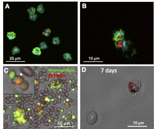
We are now focusing our investigation on the effects of MPs on macrophage function using both in vitro and in vivoapproaches. Interestingly, our preliminary results indicate that, compared to polystyrene or polyethylene manufactured spherical beads that are phagocytosed by a majority (>90%) of peritoneal macrophages from adult frogs at the same concentration, PET, and nylon 1-20 μm MPs fragments are internalized by only a minor fraction (~10%) of peritoneal macrophages. In addition, fewer PET and nylon MPs are internalized by individual peritoneal macrophage compared to manufactured spherical beads (Fig. 2A and B). We are in the process of developing a controlled aging process by UV treatment to determine if aged MPs might be phagocyted at a different rate than pristine MPs. To assess MP effect on macrophage function in vivo, we are taking advantage of the X. laevis transgenic line mpeg::GFP, where macrophages express the fluorescent GFP reporter. Intraperitoneal injection of a small amount of Nile red-stained PET-MPs in adult frogs allows the detection of GFP+ macrophages that have engulfed red fluorescent PET-MPs (Fig. 2C). Similarly, tadpole peritoneal macrophages can phagocytose red fluorescent PET-MPs following intraperitoneal injection. Moreover, we can detect macrophages with internalized PET-MPs up to 7 days after exposure (Fig. 2D), which suggests that, as also observed in vitro, MP engulfment does not induce marked cell death. This in vivo system will now allow us to follow the fate of macrophages with internalized MPs and determine whether their function in homeostasis, regeneration and immune response is altered.
Figure 2. In vitro and in vivo MP internalization assay. (A) Confocal microscopy images of adult X. laevis peritoneal macrophages (Mø) incubated with 10 μg/mL of Nile red fluorescently stained PET-MPs (1-20 μm) for 24 hours in vitro. (B) Similar peritoneal macrophages incubated with 10 μg/mL of Nile red fluorescently stained nylon-MPs (1-20 μm) for 24 hours in vitro. (C) Three days following the elicitation of Mø in the peritoneum, mpeg::gfp transgenic frogs with green fluorescent Mø were intraperitoneally injected with 10 μg of Nile red fluorescently stained PET-MPs. PLs were harvested 24 hrs. later by lavage and Mø with internalized MPs (orange) were visualized under a fluorescent microscope. (*) Magnification of a Mø with ingested PET-MPs. (D) Peritoneal macrophage from a tadpole (3 weeks of age) 7 days post-intraperitoneal injection with 10 μg Nile red fluorescently stained PET-MPs.
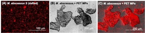
Regarding association of microorganisms with MPs, mycobacteria spp. were found in biofilms generated on plastic debris in a field mesocosm study in Lake Ontario and in the laboratory setting by our LOMP collaborators at RIT [6]. To further investigate the potential of MPs to interact with pathogens, we incubated PET-MPs with different mycobacteria species. M. marinum, a non-tuberculosis mycobacterium found in water around the world, can tightly bind to PET-MPs in vitro, as well as M. abscessus, which is a notable emerging human pathogen (Fig. 3). We plan to determine whether association of mycobacteria with MPs promotes their colonization in tadpoles and whether it affect infection and survival in macrophages.
Fig. 3: Tight association of non-tuberculous Mycobacterium abscessus with PET-MPs. M. abscessus expressing dsRED fluorescent reporter incubated overnight at 1×10⁵ cfu in (A) 1 ml of amphibian PBS or (B, C) 10 µg of PET-MPs in 1 ml of amphibian PBS. The mixture was gently resuspended before examination under a fluorescent microscope.
In summary, our study using Xenopus carries substantial significance, raising developmental immunotoxicity (DIT) concerns not only for aquatic vertebrates but also for human health. The demonstrated impact on immune function underscores the broader ramifications of MP pollution, highlighting the need for comprehensive strategies to mitigate its pervasive DIT effects on both aquatic ecosystems and human populations.
Acknowledgements
The expert animal husbandry provided by Tina Martin is gratefully appreciated. We would like to thank Rachel F. Lombardo, Francisco De Jesus Andino and Hannah Turner for their significant experiment contribution as well as Drs. Lisa De Louise and Alison Elder for their critical review of this manuscript. The Lake Ontario MicroPlastics Center (LOMP) is jointly funded by NIEHS (P01 ES035526) and NSF (OCE-2418255). JR is also funded by NAID (R24-AI059830) and R.L. by the Toxicology Training Grant (T32-ES07026).
This video is the culmination of several years attempting to: (1) Figure out best practices for modeling protein-protein interactions; (2) Understand the outputs of programs like AlphaFold and adjacent software including quantitative metrics; and (3) Communicate my thoughts to unwitting victims through workshops. Hopefully, others like me (molecular biologists, geneticists, cell and developmental biologists) may find some value in the content.
My lab group is interested in understanding the genetic and developmental mechanisms behind the evolution of novel phenotypes (evo-devo). I am seeking a curious student interested in biomineralization, evolution, development, and/or molluscs to join my team. We are aiming to understand how the shell of molluscs has evolved into so many different forms by understanding the developmental mechanisms regulating biomineralization via the mantle.
We are a new lab helping to develop a slipper snail model (Crepidula atrasolea) to test fundamental evolution and development questions. As a member of our lab, you will have the freedom to tailor your project to your interests, ideally relating to the genetics, development, or role of the nervous system in shell formation in gastropods. Depending on your project and research goals, you will have the opportunity to learn microinjection, CRISPR, hybridization chain reaction in situ hybridization, immunohistochemistry, confocal microscopy, animal husbandry, electron microscopy, and more.
We are a welcoming group that celebrates diverse backgrounds and strives for an inclusive environment for biologists of any background, age, race/ethnicity, sexual orientation, gender, or dis/ability.
In mid-January, we will hear from three of Development’s Pathway to Independence (PI) fellows studying development, evolution and the environment. Chaired by one of Development’s first PI fellows, Priti Agarwal, Principal Investigator of the Organ Mechanobiology Lab at The National Centre for Biological Sciences (NCBS), India. Priti’s group studies the role of physical forces and their interaction with biochemical signals in regulating robust organ morphogenesis and homeostasis using C. elegans.
Wednesday 14 January – 15:00 GMT/UTC
Chee Kiang (Ethan) Ewe (Tel Aviv University) ‘Neuronal oversight of germline small RNAs prevents heat-induced sterility in Caenorhabditis elegans’
Max Farnworth (University of Bristol) ‘How the evolution of spatial foraging shaped centres of cognition in Heliconiini butterflies’
Sonya Widen (IMBA, Vienna BioCenter) ‘“Unpacking” eukaryotic horizontal gene transfer: Mavericks transfer novel “cargo” genes across the globe’
At the speakers’ discretion, the webinar will be recorded to view on demand. To see the other webinars scheduled in our series, and to catch up on previous talks, please visit: thenode.biologists.com/devpres
Earlier this year, The Company of Biologists celebrated its 100-year anniversary with the Biologists @ 100 conference in Liverpool – bringing together researchers across a wide range of disciplines. To capture the spirit of the meeting, our three community sites recruited dedicated conference reporters. For the Node, this was Jen Annoh, who set the stage with an excellent beginner’s guide to scientific conferences.
Jen’s interviews
During the Biologists @ 100 conference, Jen spoke with researchers working on a range of different topics. Here we highlight Jen’s interview with Saroj Saurya, who is working to minimise the environmental impact of her biological research at the University of Oxford.
Saroj Saurya
At Biologists @ 100, The Company of Biologists invited attendees to submit essays on how they made they journey to Liverpool more sustainable, you can read the winning essays below:
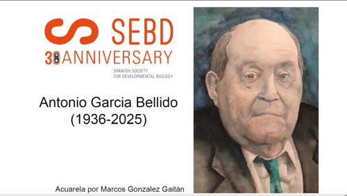
On the past 10th of November, Antonio García-Bellido passed away. Widely regarded as the father of the Spanish school of Developmental Biology and one of the most influential figures in this field worldwide, his work focused on uncovering the genetic, cellular, and molecular foundations of animal development, using the fruit fly Drosophila melanogaster as a model organism.
Among his landmark contributions were the discovery of the concept of “compartments” in the Drosophila wing, transforming our understanding of segmentation and tissue development, the formulation of the selector gene theory, clonal analysis of developing systems, and studies on the genetic control of organ size and shape. These achievements not only revolutionized Developmental Biology in Spain but also marked a turning point internationally, shaping contemporary research and inspiring generations of scientists across the globe.
Antonio served as Research Professor at the Spanish National Research Council (CSIC) and led the Developmental Genetics Laboratory at the Centre for Molecular Biology (CBM) for decades. He received numerous honours, including the Prince of Asturias Award for Scientific and Technical Research, and was elected to prestigious institutions such as the Royal Society of London and the U.S. National Academy of Sciences, in addition to several honorary doctorates. Still his legacy goes far beyond these honours: Antonio inspired generations of scientists, many of whom became part of what was known as the “Madrid school” of developmental genetics. His teaching, visionary ideas, and commitment to rigorous experimental approaches defined an era and continue to shape science today.
He leaves behind a path guided by curiosity, scientific rigor, and the power of ideas. We find comfort in knowing that his work lives on in every laboratory, every publication, and every young researcher who, following his example, chooses to dedicate their career to Developmental Biology.
For SEBD, his passing represents the loss of one of our founding members and first president, a mentor who decisively contributed to giving our discipline international visibility and prestige. His figure will remain an ethical, scientific, and human reference. In his honour, the SEBD has asked his closest collaborators and friends to share a few words about Antonio García-Bellido. You can watch their testimonies in this video in Spanish (subtitles can be added if you wish).
The interviewees are identified in the main part of the video, but at the end there are short testimonies where names are not shown. For this reason, we provide a list of these contributors in order of appearance:
Earlier this year, The Company of Biologists celebrated its 100-year anniversary with the Biologists @ 100 conference in Liverpool – bringing together researchers across a wide range of disciplines. To capture the spirit of the meeting, our three community sites recruited dedicated conference reporters. For the Node, this was Jen Annoh, who set the stage with an excellent beginner’s guide to scientific conferences.
Jen’s interviews
During the Biologists @ 100 conference, Jen spoke with researchers working on a range of different topics. Here we highlight three such conversations, where each researcher tells us more about the work they presented at the conference:
Clare Benson (King’s College London, UK) – exploring how lipids behave in skin cells under mechanical stress, revealing how fat molecules adapt to changes in their environment.
Luke Simpson (University of Nottingham, UK) – investigating early embryogenesis and gastrulation, shedding light on the fundamental processes that shape the embryo.
Matthew Stower (University of Oxford, UK) – studying how the embryo is organised, focusing on how cells find themselves in the right place at the right time.
Clare Benson
Luke Simpson
Matthew Stower
These snapshots offer a glimpse into the research (and the people!) celebrated at the meeting.
More Biologists @ 100 content
For further perspectives, check out the excellent coverage from our sister sites and contributors:
 (No Ratings Yet)
 (9 votes)