A postdoctoral research position is available in the laboratory of Dr. Michael Smutny at the Centre for Mechanochemical Cell Biology at Warwick Medical School, UK. The lab is focused on exploring biochemical and biophysical processes driving cell and tissue morphogenesis during early embryonic development. For a brief overview of the research in the lab, please visit https://mechanochemistry.org/Smutny/research/.
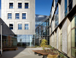
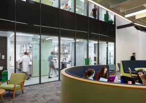
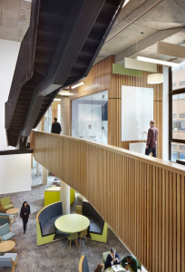
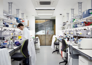
The successful candidate will work in a new state-of-the-art Interdisciplinary Biomedical Research Building (IBRB), which is part of a thriving research community at Gibbet Hill Campus including the School of Life Science, Warwick Medical School and the Centre for Mechanochemical Cell Biology.
The focus of the project will be on identifying biophysical and biochemical determinants controlling collective cell migration of embryonic progenitor cells in the zebrafish embryo. The project will investigate molecular and physical mechanisms underlying polarisation and directed cell migration combining a range of in vivo and in vitro approaches. The suitable candidate will use state-of-the-art microscopy, computational image analysis tools, latest techniques in cell/developmental biology (gene/protein perturbations, optogenetics), biophysical methods and have the possibility to establish interdisciplinary collaborations. Full training in new techniques and career development opportunities will be provided.
Highly motivated candidates with a strong background in advanced microscopy, image processing, biophysical approaches and high interest in interdisciplinary and collaborative research are encouraged to apply. Practical experience in working with a model organism (zebrafish, drosophila or similar) is desired, but not essential. Candidates should be able to work independently, have excellent communication and interpersonal skills, and participate in supervision of students.
For more details about the project and how to apply, please visit Warwick jobs (post 103651-0221), or get in touch for enquires and expression of interest directly to michael.smutny@warwick.ac.uk. The application closing date is 8th March 2021.
Bioinformatics scientist positions at the postdoctoral level are available in the lab of Dr. Sabine Dietmann at Washington University School of Medicine in St. Louis, Missouri, USA. Our research program is dedicated the development of multi-omics and machine learning approaches to the data sets generated in stem cell-based model systems for embryonic development and organoids. (https://informatics.wustl.edu/research-lab-sabine-dietmann). The candidate’s independent research will benefit from our lab’s extensive collaborative network in the Department of Developmental Biology and the Division of Nephrology at Washington University in St Louis and at international institutions. Projects are available in the following areas: (1) comparing developmental trajectories across species (2) machine learning models for cell fate decisions and gene regulatory networks (3) epitranscriptomics in single cells (https://profiles.wustl.edu/en/persons/sabine-dietmann).
We are seeking enthusiastic and talented candidates with high proficiency in scientific programming languages, such as R and Python/PERL. A good understanding of machine learning frameworks in Python (Keras/Tensorflow), experience with creating packages in R or some experience with single cell data analysis and genomics would be very beneficial.
Applicants should have a Ph.D. or master’s degree in Biology, Computer Science, Bioinformatics, Physics or related field plus 2 years of demonstrated relevant research experience.
Consistently ranked among the top 10 US medical schools, Washington University School of Medicine offers a highly interactive and stimulating academic environment for scientists in training, a place where you can be an individual and achieve exceptional things. Washington University in St. Louis is an equal opportunity employer and committed to providing a comprehensive and competitive benefits package. Our lab is located in the Central West End of St. Louis, a vibrant neighborhood adjacent to major cultural institutions.
To apply for this position please submit a CV, a cover letter describing your research interests and contact information for two references no later than March 14 to sdietmann@wustl.edu.
The Tavosanis lab welcomes applications for a post-doc position to study the cellular mechanisms of neuronal dendrite differentiation in Drosophila.
For reference, please, see: Stuerner et al., Development 2019 https://doi.org/10.1242/dev.171397; Castro et al. Elife 2020 doi: 10.7554/eLife.60920; Stuerner et al., Biorxiv https://www.biorxiv.org/content/10.1101/2020.10.01.322750v1; Baltruschat et al., Biorxiv https://www.biorxiv.org/content/10.1101/2020.07.07.191064v1).
This project will combine the generation of molecular tools for acute manipulation of protein activity with high-resolution in vivo microscopy.
For more details, please, contact Gaia Tavosanis directly at: gaia.tavosanis@dzne.de
To apply, you can follow the link: https://jobs.dzne.de/en/jobs/50653/postdoctoral-researcher-fmd-1871202011
Position Summary:
The Paul Lab at the MBL seeks a highly motivated individual to join the Josephine Bay Paul Center for Comparative Molecular Biology and Evolution as a Research Assistant (Level I). The successful candidate will be responsible for carrying out routine laboratory work as outlined below. Our research group is looking at the processes that diversify microbial genes, to better understand the functional significance of protein variation in cells and viruses from a variety of biomes.
Additional Information:
The primary aim of the position is to maintain the molecular lab facilities and to assist in developing genetic experiments with bacteria/archaea primarily derived from marine and freshwater ecosystems. Responsibilities will include establishing and monitoring cell cultures, maintaining lab equipment, ordering lab supplies, and conducting basic molecular experiments.
Basic Qualifications:
A Bachelor’s degree in biology, molecular biology or a related discipline is required. This position requires an independent, organized, and self-motivated individual with strong problem-solving skills and the ability to multitask. Prior experience in a research lab and applying basic molecular biology techniques is required. Excellent written, verbal, and interpersonal skills; attention to detail; and a strong work ethic are essential. Position level and salary will depend upon education and experience.
Preferred Qualifications:
The ideal candidate will have prior experience with nucleic acid purification, PCR, and maintaining (bacterial/archaeal) cell cultures. An understanding of basic molecular biology concepts is important.
Physical Requirements:
Minimal exposure to biohazardous chemicals. Occasional lifting of heavy objects.
Required documents:
Apply on the MBL website and include the following documents in your application package: cover letter, resume/CV, copies of most recent transcripts (unofficial is acceptable), and contact details of 3 references.
By Oriana Q. H. Zinani, Kemal Keseroğlu, Ahmet Ay & Ertuğrul M. Özbudak:
Gene expression is an inevitably stochastic process (Ozbudak et al., 2002). In contrast, embryonic development and homeostasis require cells to coordinate the spatiotemporal expression of large sets of genes. Many mechanisms are known to orchestrate this coordination, such as operons, bidirectional promoters, enhancer sharing, 3-D DNA looping, topologically associated domains, transcription factories or hubs, and shared upstream regulators.
In metazoans, pairs of co-expressed genes often reside in the same chromosomal neighborhood, with gene pairs representing 10 to 50% of all genes, depending on the species (Adachi and Lieber, 2002; Arnone et al., 2012). Although many paired genes encode for essential housekeeping proteins, some encode for signaling regulators and transcription factors (such as CYP26A1–CYP26C1, ETS1–FLI1, MRF4–MYF5, MESP1–MESP2, SIX1–SIX4–SIX6, and STAT1–STAT4), which have transcription start sites that are 9–516 kb apart. Because various other mechanisms can ensure correlated gene expression, the selective advantage of maintaining adjacent gene pairs remains unknown.
To address this question, in Zinani et al, we used two linked zebrafish segmentation clock genes, her1–her7, as the testbed. The subdivision of the anterior–posterior axis into a fixed number of somites is a landmark example of how coordinated gene expression patterns the vertebrate embryo. During somitogenesis, groups of cells synchronously commit to segmentation in a notably short time frame. The pace of segmentation is set by the period of an oscillator, the segmentation clock, in cells of the unsegmented presomitic mesoderm (PSM). Oscillatory expression of the Hes or her clock genes is conserved in vertebrates; disruptions of their oscillations lead to vertebral segmentation defects (i.e., congenital scoliosis in humans). Approximately every 30 min in zebrafish, around 200 cells bud from the PSM to form a new somite. Segmentation is carried out for a species-specific number of cycles (33 in zebrafish). Her1 and Her7 are basic helix-loop-helix (bHLH) proteins that dimerize to bind DNA. The zebrafish segmentation clock relies on a transcriptional negative-feedback loop in which Her7-Hes6 hetero- or Her1-Her1 homodimers repress transcription of her1 and her7 (Ay et al., 2013; Giudicelli et al., 2007; Schroter et al., 2012). In zebrafish, two paired clock genes (her1 and her7) are separated by a 12-kb regulatory intervening sequence. her1 and her7 have similar transcriptional time delays (Hanisch et al., 2013) and RNA half-lives (Giudicelli et al., 2007); therefore, the transcription of her1 and her7 is mainly concomitant in the tissue. To achieve the rapid tempo and reproducible precision of segmentation, the transcription of her1 and her7 should be tightly coordinated.
Chromosomal adjacency was previously shown to cause correlated expression of synthetic reporters (Becskei et al., 2005; Fukaya et al., 2016; Raj et al., 2006). To test the role of gene pairing on transcriptional co-firing, we detected nascent transcription loci in the nucleus of single cells with single-molecule fluorescence in situ hybridization (smFISH) (Keskin et al., 2018; Zinani et al., 2021). We found that the probability of transcriptional co-firing of paired her1 and her7 genes on the same chromosome is significantly higher than the two unpaired her1 genes on homolog chromosomes. This finding demonstrated that gene pairing augments correlated transcription of the two clock genes by triggering transcriptional co-firing.
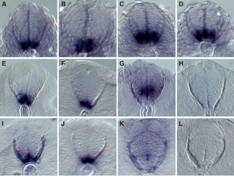
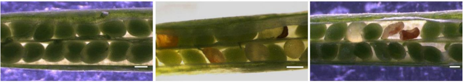
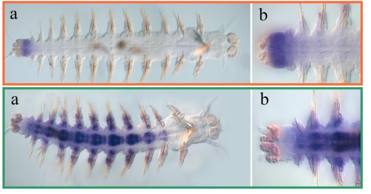
Co-firing of two paired clock genes could be advantageous for somite segmentation as it would coordinate transcript levels. To test this hypothesis, we generated cis and trans double heterozygous embryos by using CRISPR/Cas9 (Figure). The cis heterozygous mutants carried two mutant genes in one chromosome and two wild-type genes on the homologous chromosome (Figure, top row). In contrast, the trans mutants carried a mutant her1 gene adjacent to a wild-type her7 gene on one chromosome, and a mutant her7 gene adjacent to a wild-type her1 gene on the other chromosome (Figure, bottom row). Hence, fish that are compound heterozygous for these alleles will have the same functional gene dose as the previously described double heterozygous embryos with paired her1 and her7 genes (Figure, left column). We next raised the gene-paired and gene-unpaired embryos at 21.5 °C, where wild-type embryos successfully form somites. Gene-unpaired embryos had reduced success in somite segmentation as compared to gene-paired embryos (Figure, right column). These results revealed that maintaining paired genes in the genome is beneficial for successful pattern formation during embryonic development.
We next investigated the mechanism by which gene pairing is beneficial for clock oscillations. Although negative-feedback loops are widespread in gene regulatory networks, they usually do not give rise to oscillations, but instead act as a rheostat to tightly maintain gene expression around a steady-state. For a negative-feedback loop to generate oscillations, several important criteria need to be satisfied. Previous studies revealed the importance of time delays and short RNA or protein half-lives to generate sustained oscillations. However, another important criterion needed to generate oscillations is that the rate of transcription needs to be high enough to push the system into an unstable steady-state, establishing a limit cycle (Lewis, 2003; Novak and Tyson, 2008). We found that when genes are paired on the same chromosome, co-transcription happens more frequently. We hypothesized that co-firing of transcription results in a high rate of RNA production, and thereby overshoots the limit-cycle threshold. Simulations of our stochastic model agreed with our hypothesis.
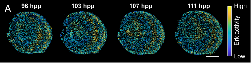
To assess the function of gene pairing in the segmentation clock in real-time, we imaged transgenic Tg(her1:her1-Venus) (Delaune et al., 2012) zebrafish embryos along the entire PSM. We quantified the amplitude of oscillations in the next presumptive somites for 11 somite cycles. We found that the average amplitude was decreased in gene-unpaired embryos compared to gene-paired embryos. The amplitudes of oscillations preceding disrupted boundaries were significantly lower than the ones preceding the successful ones in a given genetic background.
Our results demonstrate that the prevention of gene pairing disrupts oscillations and segmentation in zebrafish embryos. We predict that gene pairing is similarly advantageous in other biological systems, and our findings could inspire engineering of precise synthetic clocks in embryos and organoids.
In our third SciArt Profile we meet Sydney Wyatt, a PhD student based at the University of California, Davis.
Sydney
Where are you originally from, where do you work now, and what do you work on?
I am originally from a small Northern California town, Paradise. I went out of state to get my BS at University of Utah in Salt Lake City, and while I enjoyed my time there, I’m happy to be pursuing my PhD in Integrative Genetics and Genomics at University of California, Davis; it gives me the opportunity to be closer to my parents. My thesis research in Dr. Bruce Draper’s lab currently revolves around interrogating sex determination and early gonad development in zebrafish. Specifically, I am investigating the female sex determination pathway, as well as the role of gap junctions in early germ cell development.
Has science always been an important part of your life?
I remember loving dinosaurs growing up: my favorite is still Parasaurolophus. Understandably, I wanted to be a paleontologist for a long time, although it changed to forensic pathologist by the time I went to college. After my first anatomy lab I quickly changed my mind! Instead, I found I loved genetics and developmental biology through my classes and undergraduate research. Despite working on specific projects now, I still enjoy learning about other fields (and still love learning about dinosaurs!).
I was fortunate that STEM was always encouraged by my family, despite limited exposure growing up. I didn’t have access to the opportunities and mentorship I volunteer for today, namely mentoring high school senior research projects and various public science outreach events like the North Bay Science Discovery Day. I think if I had had those opportunities, I might have learned what I wanted out of my education more quickly.
And what about art – have you always enjoyed drawing/painting/etc?
I was a ballet dancer for many, many years, so performing arts were important to me. I didn’t start drawing until high school when I needed a new creative outlet after an injury. I took classes from a local oil painter, and he mentored my senior community project. I eventually moved into digital art; it allows me to do my art in short bursts in any location without hauling around a pad of paper and a case of pencils. It’s nice to have that outlet to help me avoid burnout.
“Digital art allows me to do my art in short bursts in any location”
What or who are your artistic influences?
I have been drawing a lot of fish lately thanks to my pandemic discovery of #SundayFishSketch created by Rene Martin on Twitter.
How do you make your art?
I pick a subject based either on suggestions from friends and family or a color palette. I try to vary the main color to avoid too much repetition. Varying the subject helps too: I rotate between fish, flowers and birds. Once I have my subject and references, I get sketching. For physical pieces, I use a grid technique to help me capture the relative positions of all the details. For digital pieces, I start with basic shapes (and use the eraser and undo button a lot). Color swatching is important. I’ll do a couple practice swatches to warm up and remind myself of what I was thinking when designing the piece. The main colors get lightly blocked first with shadows and highlights, and then I build up from there. I have a tendency to do entire sections in one sitting, like one entire wing of a bird, to keep my technique consistent.
Does your art influence your science at all, or are they separate worlds?
I work on fish, so maybe a little influence. There are a couple of zebrafish-themed Sunday fish sketches floating around Twitter. Science I read about influences my art, too. I learned about really interesting killifish genomics research and was inspired to create a whole series of non-traditional model organisms, like killifish or gar or delta smelt. These organisms are so important but not widely heard of outside their niches.
What are you thinking of working on next?
I’m excited to keep working on my research projects, as well as continue to build my art and writing portfolios. As far as art goes, I’ve recently been commissioned by friend to help make label for a home-brewed beer called Axolotl Ale. I also manage a sticker shop (mostly fish) based on previous interest in purchasing pieces. My long-term goal is continue to create non-traditional model organism pieces to highlight the diversity in research subjects for the public as part of my science communication efforts.
Zebrafish: This quick painting was 100% inspired by my research—I work with zebrafish as my model organism.
Killifish: I learned about killifish as a model organism in toxicology and aging research only recently. Specifically, the African turquoise killifish is up-and-coming in aging research. For this piece I wanted to play up the deep blues and purples I saw in my reference images.
Three-spined Stickleback: My college evolution and development professor had done research on these fish, and working on this piece inspired me to start my collection of model organisms less well known to the public.
Pacific Spiny Lumpsucker: This is the piece that started my sticker shop. A friend commissioned me to make this for stickers. People were interested in where they could get their own PSL stickers and I opened my shop.
Medaka: This was another sticker commission for a friend who uses these as her model organism in toxicology research.
Mandarin Fish: This was one of my earlier #SundayFishSketch pieces. It took me two weeks to get all the details right! Since then, I do quicker pieces for the prompts and more elaborate ones for myself or commissions.
Fish Gallery (click for full size image & caption)
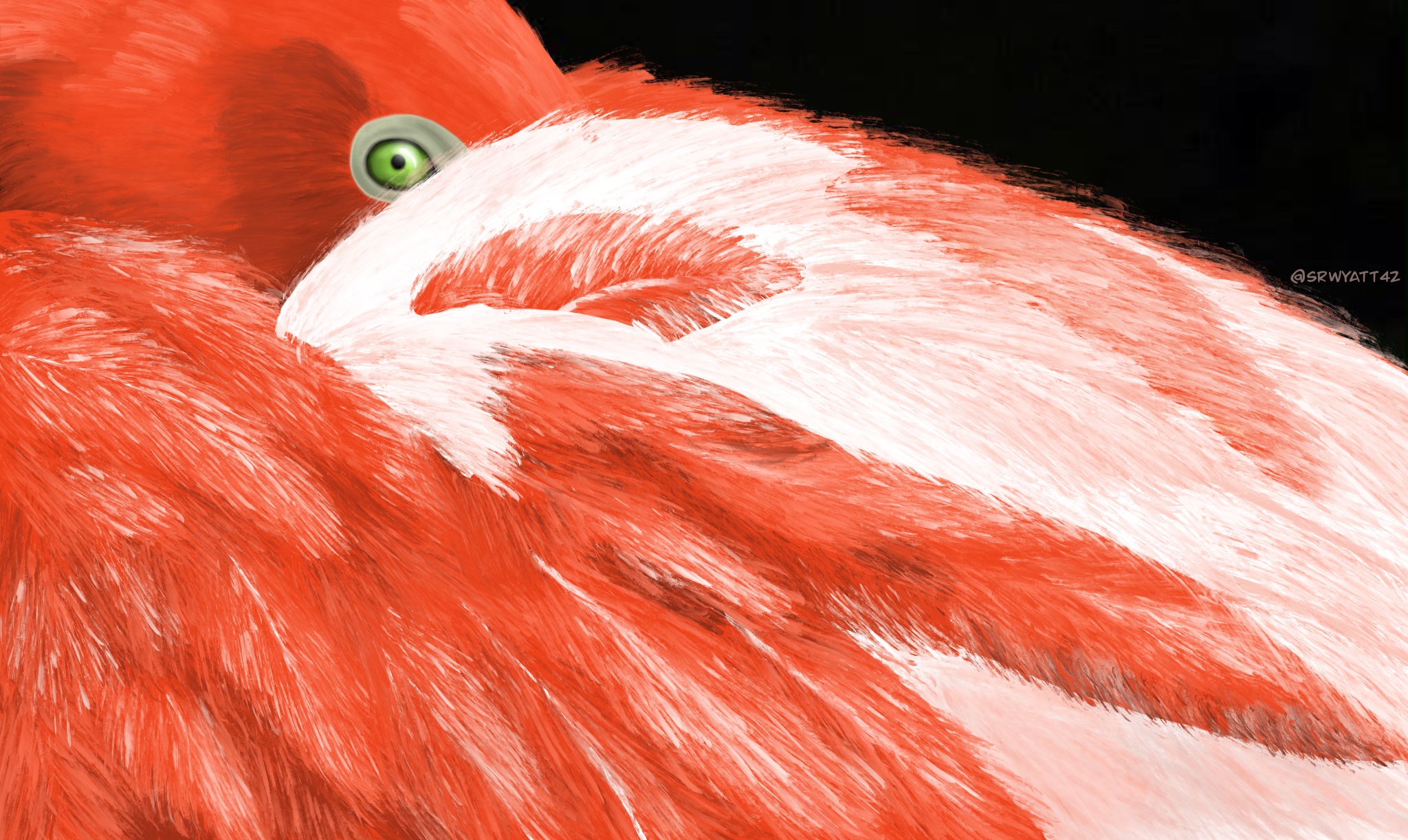
Flamingo: I wanted to depict something other than what one might think of as a flamingo: standing up with their head held high or their beaks down in the water to feed. I think this image is a bit more playful than that.
Yellow Roses: This is an old piece, but still one of my favorites. I love how soft and rich the petals look. It’s a quality I try to capture with all my flower pieces.
We’re looking for new people to feature in this series throughout the year – whatever kind of art you do, from sculpture to embroidery to music to drawing, if you want to share it with the community just email thenode@biologists.com (nominations are also welcome!).
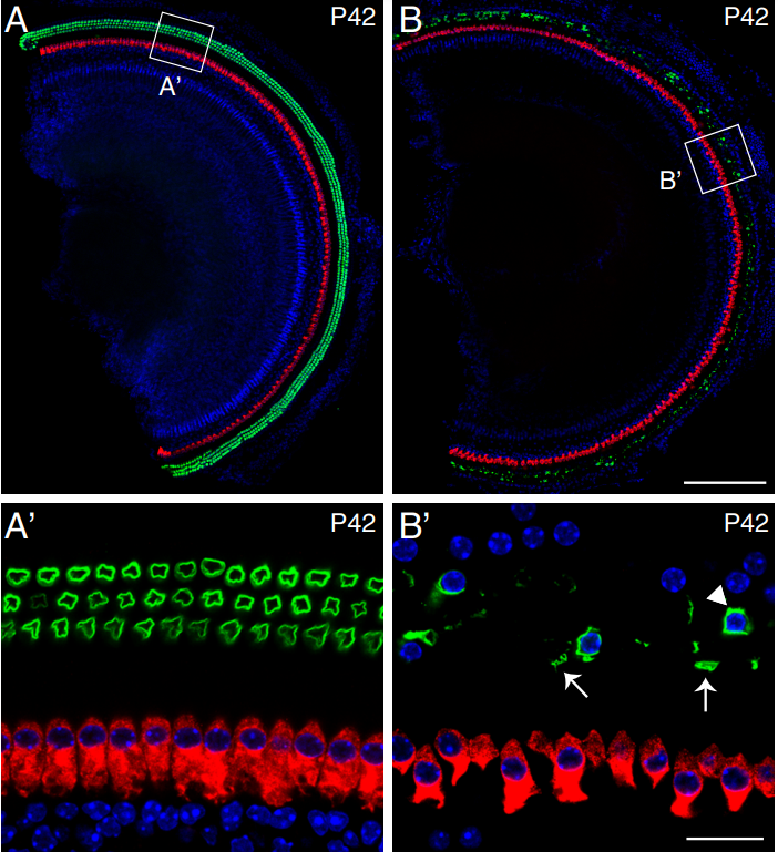
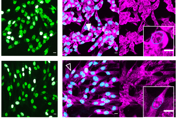
A postdoctoral position is available in the Scholpp lab at the Living Systems Institute (LSI) at the University of Exeter (https://www.exeter.ac.uk/livingsystems/). The mission of the institute is to study the function of living systems by multi-scale and interdisciplinary approaches. A primary focus of our lab is understanding how cells communicate in a tissue. We focus mainly on signals from the WNT protein family, which regulate fundamental processes including cell proliferation and differentiation, cell polarity and migration. Misregulation of this morphogenetic network causes severe diseases such as neurodegenerative diseases. We have demonstrated that WNT proteins can be loaded on signalling filopodia known as cytonemes (yellow arrows) to allow paracrine signalling in vertebrates.
In this pilot project, we will study the interplay of WNT and β-amyloid signalling during the formation and maintenance of synapses. We will use hiPSC-derived cortical neurons to study the underlying molecular mechanism controlling WNT cytoneme function and synaptogenesis by in vivo high-resolution imaging.
What we offer:
A fully-funded position for 12 months (with the possibility of extension, including consumables and travel money) by the Alzheimer’s BRACE Charity ( https://www.alzheimers-brace.org/ )
A highly collaborative team and a collegial environment in the LSI
A stimulating environment with freedom to develop new research directions.
Supportive mentorship for multi-faceted career development and opportunities tailored towards individual career goals.
Regular interactions with world-class colleagues through Biosciences and Medical School.
An institute located in a dynamic city in the South West of England with the affordable cost of living.
What we are looking for:
Applicants with experience in developmental neuroscience and imaging, preferable in hiPSC culture (incl. maintenance and differentiation, and functionality assessment through cellular and molecular biological assays) and high-resolution imaging (incl. confocal microscopy, SIM, 2-photon)
Enthusiastic and ambitious individuals with a keen interest in our research and a collaborative and collegial laboratory environment.
Fearlessness in learning new techniques and designing projects independently.
Willingness to apply for applicable fellowships and grants
Interest in working with junior lab members and project students.
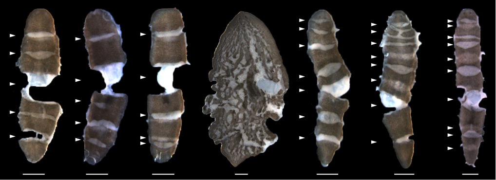
A joint research team co-led by Prof. Chao Tang (Peking University, PKU), Prof. Zhongying Zhao (Hong Kong Baptist University, HKBU) and Prof. Hong Yan (City University of Hong Kong, CityU) has developed a novel computational tool that can reconstruct and visualize three-dimensional shapes and temporal changes of cells, speeding up the analytical process from thousands of hours by hand to a few hours by computer. Furthermore, a total of 17 C. elegans embryos were fully segmented and normalized, generating a time-lapse 3D (i.e., 4D) morphological atlas from the 4- to 350-cell stages, including cell shape, volume, surface area, migration, nucleus position and cell-cell contact with confirmed cell identities. Both the computational tool and standard dataset can advance further studies in developmental biology, cell biology and biomechanics (Figure 1). This interdisciplinary study has been published in Nature Communications, entitled “Establishment of a morphological atlas of the Caenorhabditis elegans embryo using deep-learning-based 4D segmentation” [1].
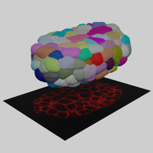
Figure 1. The concept of 3D cell membrane imaging and segmentation (photo source: DOI number: 10.1038/s41467-020-19863-x).
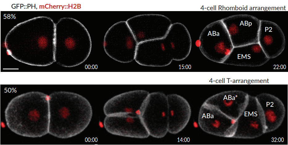
The tool developed by the team is called “CShaper”. “It is a powerful computational tool that can segment and analyze cell images systematically at the single-cell level, which is much needed for the study of cell division (Figure 2), and cell and gene functions,” described Prof. Hong Yan, Chair Professor of Computer Engineering and Wong Chung Hong Professor of Data Engineering in the Department of Electrical Engineering (EE) at CityU.
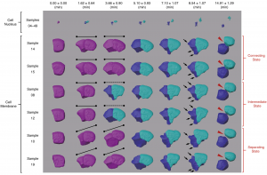
Figure 2. Morphological dynamics of cell division, deformation and migration during C. elegans embryogenesis at single-cell resolution (photo source: DOI number: 10.1038/s41467-020-19863-x).
Breakthrough in segmenting cell images automatically
Cell images are usually obtained by laser beam scanning. The existing image analysis systems can only detect cell nucleus well with a poor cell membrane image quality, hampering reconstruction of cell shapes. Also, there is a lack of reliable algorithm for the segmentation of time-lapse 3D images (i.e. 4D images) of proliferating cells. Image segmentation is a critical process in computer vision that involves dividing a visual input into segments to simplify image analysis. Often, researchers have to spend hundreds of hours labeling many cell images manually.
The breakthrough in CShaper is that it can detect cell membranes, build up cell shapes in 3D, and more importantly, automatically segment the cell images at the cell level. “Using CShaper, biologists can decipher the contents of these images within a few hours. It can characterize cell shapes and surface structures, and provide 3D views of cells at different time points (Figure 3),” said Mr Jianfeng Cao, a PhD student in Prof. Hong Yan’s lab and a co-first author of the paper.
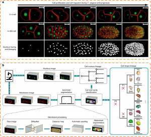
To achieve this, the deep-learning-based model DMapNet developed by the team plays a key role in the CShaper system. “By learning to capture multiple discrete distances between image pixels, DMapNet extracts the membrane contour while considering shape information, rather than just intensity features. Therefore CShaper achieved a 95.95% accuracy of identifying the cells, which outperformed other methods substantially,” he explained. CShaper was also tested on plant tissue cells, showing promising results. The team believe the computer tool can be adopted to other biological studies.
Figure 3. Fluorescent imaging (cell membrane and cell nucleus) and segmentation pipeline (CShaper) (photo source: DOI number: 10.1038/s41467-020-19863-x).
Generating a standard morphological atlas
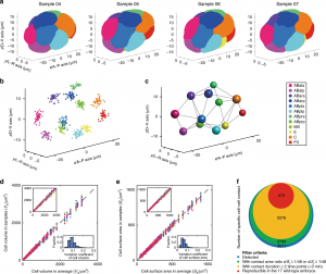
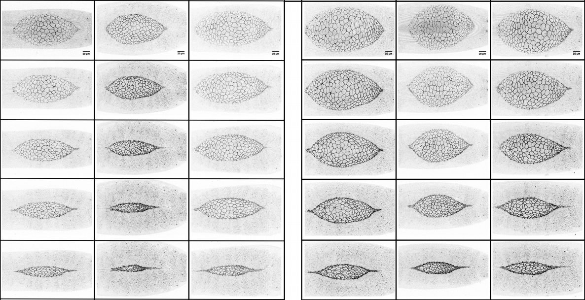
With CShaper, the team collected 17 C. elegans embryos with fluorescence on both cell membrane and cell nucleus. The segmented regions were subjected to spatial and temporal normalization, producing a set of standardized embryo structures placed in a cuboid framework. The 4D morphological atlas lasts from the 4- to 350-cell stages during C. elegans embryogenesis, and includes quantitative information of cell shape, volume, surface area, migration, nucleus position and cell-cell contact with confirmed cell identities (Figure 4). “The morphological atlas at cellular level is information-rich and allows systematic analysis with cell identity, and is needed by the field of developmental biology,” said Prof. Zhongying Zhao.
“With such accurate data, we can not only develop mathematical model to ‘compute’ embryogenesis, but also search the underlying principles which govern the nematode development,” said Prof. Chao Tang. “Using the standard morphological data, recently we built a phase field model to reconstruct the C. elegans embryonic morphogenetic procedure [2], and uncovered a minimal time principle optimizing the C. elegans embryonic cell lineage [3],” said Mr Guoye Guan, a PhD student in Prof. Chao Tang’s lab and a co-first author of the paper.
Figure 4. Standard morphological atlas and multidimensional developmental properties (photo source: DOI number: 10.1038/s41467-020-19863-x).
Prof. Chao Tang (PKU), Prof. Zhongying Zhao (HKBU) and Prof. Hong Yan (CityU) are the corresponding authors of the paper; Ph.D students Jianfeng Cao (CityU) and Guoye Guan (PKU) and Dr. Vincy Wing Sze Ho (HKBU) are the co-first authors; Ming-Kin Wong (HKBU) and Lu-Yan Chan (HKBU) provided experimental assistance.
The funding support for this study included the Hong Kong Research Grants Council, the HKBU Interdisciplinary Research Cluster Fund, the Hong Kong Innovation and Technology Commission, the Hong Kong Institute for Data Science, the Ministry of Science and Technology of China, and the National Natural Science Foundation of China.
Part of this article is from the news release in PKU [4] and CityU [5].
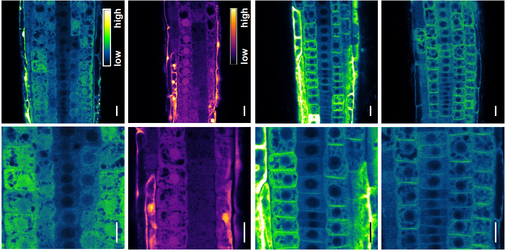
The Schnorrer team is welcoming applications for a PhD position funded by the ERC synergy grant StuDySarcomere to decipher how sarcomere components self-organize at the molecular scale to build a functional contractile muscle.
Project
Background:
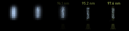
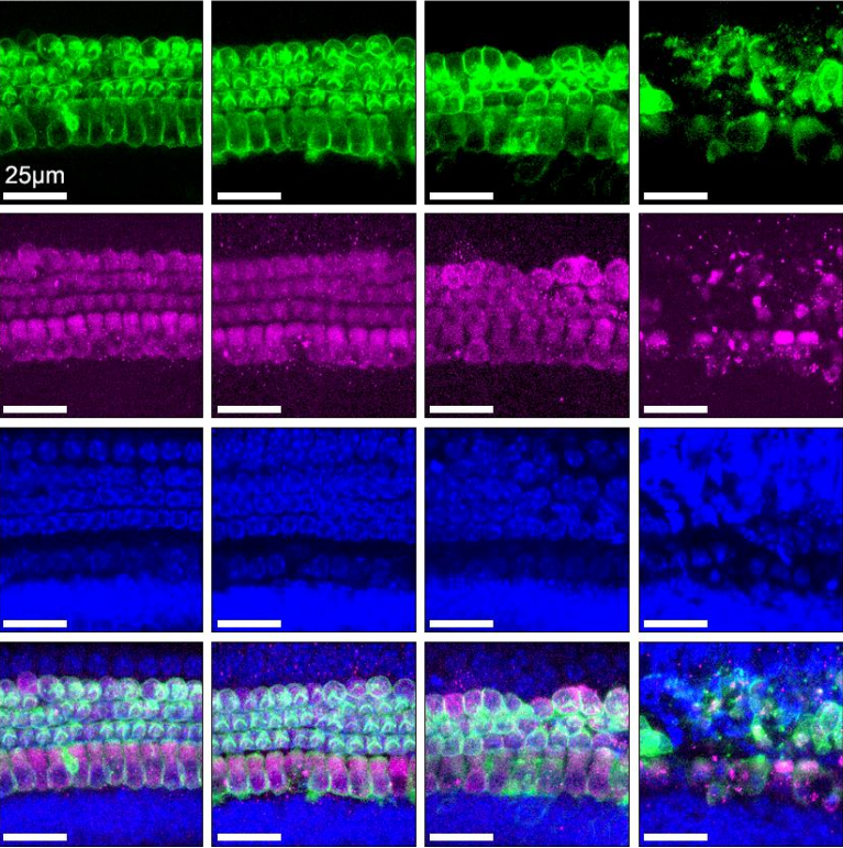
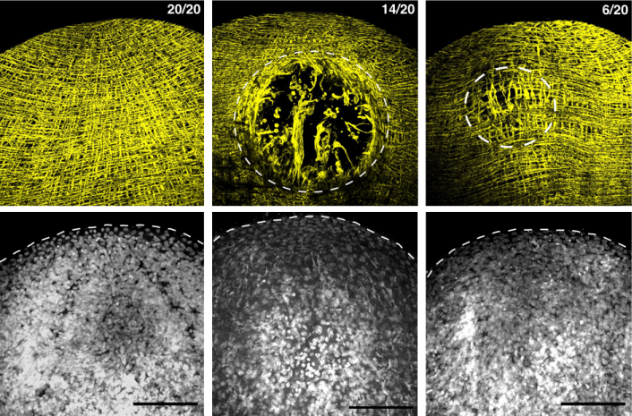
Muscles assemble tens of different proteins into periodic contractile structures called sarcomeres. Although the sarcomere components in the mature sarcomere are known, how these proteins assemble to functional sarcomeric machines is still a mystery. Sarcomeric proteins are large and locate at defined positions of the sarcomere, seen in stripes (see image above). However, with classical imaging techniques, it is nearly impossible to follow how proteins come together at the molecular scale.
Goal:
We apply DNA-PAINT super-resolution imaging, together with a novel labelling method using nanobodies, to decipher how sarcomeric components assemble with nearly two-order of magnitude better resolution than classical confocal microscopy. State of the art image analysis, including deep learning methods, will be instrumental to automate analysis of the large data obtained. The in vivo model Drosophila as well as stem cell derived human muscle fibers will be used in the project.
The team and the environment
This ambitious project will be hosted in a team of biologists and physicists, experts in muscle at the Developmental Biology Institute of Marseille (IBDM), France. The PhD student will benefit from state- of-the-art biological tools and instruments, including a high-end DNA-PAINT dedicated microscope of the team. This project will be supported by several team members and will benefit from collaborations through the European ERC Synergy network StuDySarcomere.
The team is part of the very active multidisciplinary program, The Turing Center of Living Systems, Centuri, which brings together biologists, physicists, and computational scientists. The student will benefit from this environment, including collaborations, courses, seminars, and meetings.
Your profile
You are a Physicist with strong interest in Biology or a Biologist with strong background in computational analysis. You are ambitious, enjoy doing experiments and analyze your data with tools you can develop.
Welcome to our monthly trawl for developmental biology (and related) preprints.
The preprints this month are hosted on bioRxiv, arXiv and, for the first time, preprints.org (check out our new ‘Reviews’ section!) – use these links to get to the section you want.
Amnion signals are essential for mesoderm formation in primates
Ran Yang, Alexander Goedel, Yu Kang, Chenyang Si, Chu Chu, Yi Zheng, Zhenzhen Chen, Peter J. Gruber, Yao Xiao, Chikai Zhou, Nevin Witman, Chuen-Yan Leung, Yongchang Chen, Jianping Fu, Weizhi Ji, Fredrik Lanner, Yuyu Niu, Kenneth Chien
BMP pathway antagonism by Grem1 regulates epithelial cell fate in intestinal regeneration
Martijn AJ Koppens, Hayley Davis, Gabriel N Valbuena, Eoghan J Mulholland, Nadia Nasreddin, Mathilde Colombe, Agne Antanaviciute, Sujata Biswas, Matthias Friedrich, Lennard Lee, Oxford IBD cohort investigators, Lai Mun Wang, Viktor H Koelzer, James E East, Alison Simmons, Douglas J Winton, Simon J Leedham
FOXO1 and FOXO3 cooperatively regulate innate lymphoid cell development
Thuy T. Luu, Jonas Nørskov Søndergaard, Lucía Peña-Pérez, Shabnam Kharazi, Aleksandra Krstic, Stephan Meinke, Laurent Schmied, Nicolai Frengen, Yaser Heshmati, Marcin Kierczak, Thibault Bouderlique, Arnika Kathleen Wagner, Charlotte Gustafsson, Benedict J. Chambers, Adnane Achour, Claudia Kutter, Petter Höglund, Robert Månsson, Nadir Kadri
Intrinsic network activity in human brain organoids
Tal Sharf, Tjitse van der Molen, Elmer Guzman, Stella M.K. Glasauer, Gabriel Luna, Zhouwei Cheng, Morgane Audouard, Kamalini G. Ranasinghe, Kiwamu Kudo, Srikantan S. Nagarajan, Kenneth R. Tovar, Linda R. Petzold, Paul K. Hansma, Kenneth S. Kosik
Muscle-specific Cavin4 interacts with Bin1 to promote T-tubule formation and stability in developing skeletal muscle
Harriet P. Lo, Ye-Wheen Lim, Zherui Xiong, Nick Martel, Charles Ferguson, Nicholas R. Ariotti, Jean Giacomotto, James A. Rae, Matthias Floetenmeyer, Shayli Varasteh Moradi, Ya Gao, Vikas A. Tillu, Di Xia, Huang Wang, Samira Rahnama, Susan J. Nixon, Michele Bastiani, Ryan D. Day, Kelly A. Smith, Nathan J. Palpant, Wayne A. Johnston, Kirill Alexandrov, Brett M. Collins, Thomas E. Hall, Robert G. Parton
Universal DNA methylation age across mammalian tissues
MAMMALIAN METHYLATION CONSORTIUM, Ake T. Lu, Zhe Fei, Amin Haghani, Todd R. Robeck, Joseph A. Zoller, Caesar Z. Li, Joshua Zhang, Julia Ablaeva, Danielle M. Adams, Javier Almunia, Reza Ardehali, Adriana Arneson, C. Scott Baker, Katherine Belov, Pete Black, Daniel T. Blumstein, Eleanor K. Bors, Charles E. Breeze, Robert T. Brooke, Janine L. Brown, Alex Caulton, Julie M. Cavin, Ioulia Chatzistamou, Hao Chen, Priscila Chiavellini, Oi-Wa Choi, Shannon Clarke, Joseph DeYoung, Christopher Dold, Candice K. Emmons, Stephan Emmrich, Chris G. Faulkes, Steven H. Ferguson, Carrie J. Finno, Jean-Michel Gaillard, Eva Garde, Vadim N. Gladyshev, Vera Gorbunova, Rodolfo G. Goya, Matthew J Grant, Erin N. Hales, M. Bradley Hanson, Martin Haulena, Andrew N. Hogan, Carolyn J. Hogg, Timothy A. Hore, Anna J. Jasinska, Gareth Jones, Eve Jourdain, Olga Kashpur, Harold Katcher, Etsuko Katsumata, Vimala Kaza, Hippokratis Kiaris, Michael S. Kobor, Pawel Kordowitzki, William R. Koski, Brenda Larison, Sang-Goo Lee, Ye C. Lee, Marianne Lehmann, Jean-Francois Lemaitre, Andrew J. Levine, Cun Li, Xinmin Li, David TS Lin, Nicholas Macoretta, Dewey Maddox, Craig O. Matkin, Julie A. Mattison, June Mergl, Jennifer J. Meudt, Khyobeni Mozhui, Asieh Naderi, Martina Nagy, Pritika Narayan, Peter W. Nathanielsz, Ngoc B. Nguyen, Christof Niehrs, Alexander G. Ophir, Elaine A. Ostrander, Perrie O’Tierney Ginn, Kim M. Parsons, Kimberly C. Paul, Matteo Pellegrini, Gabriela M. Pinho, Jocelyn Plassais, Natalia A. Prado, Benjamin Rey, Beate R. Ritz, Jooke Robbins, Magdalena Rodriguez, Jennifer Russell, Elena Rydkina, Lindsay L. Sailer, Adam B. Salmon, Akshay Sanghavi, Kyle M. Schachtschneider, Dennis Schmitt, Todd Schmitt, Lars Schomacher, Lawrence B. Schook, Karen E. Sears, Andrei Seluanov, Dhanansayan Shanmuganayagam, Anastasia Shindyapina, Kavita Singh, Ishani Sinha, Russel G. Snell, Elham Soltanmaohammadi, Matthew L. Spangler, Lydia Staggs, Karen J. Steinman, Victoria J. Sugrue, Balazs Szladovits, Masaki Takasugi, Emma C. Teeling, Michael J. Thompson, Bill Van Bonn, Sonja C. Vernes, Diego Villar, Harry V. Vinters, Mary C. Wallingford, Nan Wang, Robert K. Wayne, Gerald S. Wilkinson, Christopher K. Williams, Robert W. Williams, X. William Yang, Brent G. Young, Bohan Zhang, Zhihui Zhang, Peng Zhao, Yang Zhao, Joerg Zimmermann, Wanding Zhou, Jason Ernst, Ken Raj, Steve Horvath
Single Cell Enhancer Activity Maps Neuronal Lineages in Embryonic Mouse Basal Ganglia
Linda Su-Feher, Anna N. Rubin, Shanni N. Silberberg, Rinaldo Catta-Preta, Kenneth J. Lim, Iva Zdilar, Christopher S. McGinnis, Gabriel L. McKinsey, Thomas E. Rubino Jr., Michael Hawrylycz, Carol Thompson, Zev J. Gartner, Luis Puelles, Hongkui Zeng, John L. R. Rubenstein, Alex S. Nord
A Single Cell Atlas of Lung Development
Nicholas M Negretti, Erin J Plosa, John T Benjamin, Bryce A Schuler, A. Christian Habermann, Christopher S Jetter, Chase J Taylor, Peter Gulleman, David Nichols, Brittany K Matlock, Susan H Guttentag, Timothy S Blackwell, Nicholas E Banovich, Jonathan A Kropski, Jennifer MS Sucre
Robust integrated intracellular organization of the human iPS cell: where, how much, and how variable
Matheus P. Viana, Jianxu Chen, Theo A. Knijnenburg, Ritvik Vasan, Calysta Yan, Joy E. Arakaki, Matte Bailey, Ben Berry, Antoine Borensztejn, Jackson M. Brown, Sara Carlson, Julie A. Cass, Basudev Chaudhuri, Kimberly R. Cordes Metzler, Mackenzie E. Coston, Zach J. Crabtree, Steve Davidson, Colette M. DeLizo, Shailja Dhaka, Stephanie Q. Dinh, Thao P. Do, Justin Domingus, Rory M. Donovan-Maiye, Tyler J. Foster, Christopher L. Frick, Griffin Fujioka, Margaret A. Fuqua, Jamie L. Gehring, Kaytlyn A. Gerbin, Tanya Grancharova, Benjamin W. Gregor, Lisa J. Harrylock, Amanda Haupt, Melissa C. Hendershott, Caroline Hookway, Alan R. Horwitz, Chris Hughes, Eric J. Isaac, Gregory R. Johnson, Brian Kim, Andrew N. Leonard, Winnie W. Leung, Jordan J. Lucas, Susan A. Ludmann, Blair M. Lyons, Haseeb Malik, Ryan McGregor, Gabe E. Medrash, Sean L. Meharry, Kevin Mitcham, Irina A. Mueller, Timothy L. Murphy-Stevens, Aditya Nath, Angelique M. Nelson, Luana Paleologu, T. Alexander Popiel, Megan M. Riel-Mehan, Brock Roberts, Lisa M. Schaefbauer, Magdalena Schwarzl, Jamie Sherman, Sylvain Slaton, M. Filip Sluzewski, Jacqueline E. Smith, Youngmee Sul, Madison J. Swain-Bowden, W. Joyce Tang, Derek J. Thirstrup, Daniel M. Toloudis, Andrew P. Tucker, Veronica Valencia, Winfried Wiegraebe, Thushara Wijeratna, Ruian Yang, Rebecca J. Zaunbrecher, Allen Institute for Cell Science, Graham T. Johnson, Ruwanthi N. Gunawardane, Nathalie Gaudreault, Julie A. Theriot, Susanne M. Rafelski
Extracellular histones, a new class of inhibitory molecules of CNS axonal regeneration
Mustafa M. Siddiq, Sari S. Hannila, Yana Zorina, Elena Nikulina, Vera Rabinovich, Jianwei Hou, Rumana Huq, Erica L. Richman, Rosa E. Tolentino, Jens Hansen, Adam Velenosi, Brian K. Kwon, Stella E. Tsirka, Ian Maze, Robert Sebra, Ravi Iyengar, Marie T. Filbin
Neutrophil extracellular traps impair regeneration
Eric Wier, Mayumi Asada, Gaofeng Wang, Martin P. Alphonse, Ang Li, Chase Hintelmann, Christine Youn, Brittany Pielstick, Roger Ortines, Lloyd S. Miller, Nathan K. Archer, Luis A. Garza
Extracellular Matrix Dysfunction in Sorsby Patient-Derived Retinal Pigment Epithelium
Abbi L. Engel, YeKai Wang, Thomas H. Khuu, Emily Worrall, Megan A. Manson, Kaitlen Knight, Aya Yanagida, Jian Hua Qi, Aravind Ramakrishnan, Richard G Weleber, Michael L. Klein, David J. Wilson, Bela Anand-Apte, James B. Hurley, Jianhai Du, Jennifer R. Chao
Developmental and behavioral phenotypes in a new mouse model of DDX3X syndrome
Andrea Boitnott, Dévina C Ung, Marta Garcia-Forn, Kristi Niblo, Danielle Mendonca, Michael Flores, Sylvia Maxwell, Jacob Ellegood, Lily R Qiu, Dorothy E Grice, Jason P Lerch, Mladen-Roko Rasin, Joseph D Buxbaum, Elodie Drapeau, Silvia De Rubeis
| Plant development
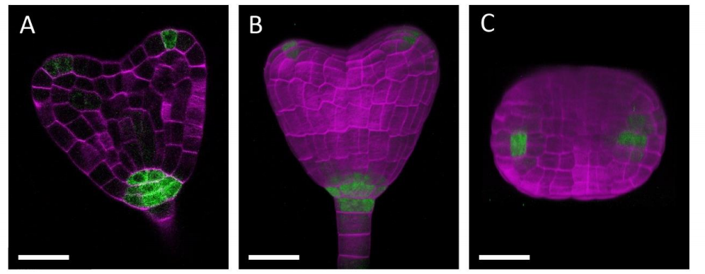
A hetero-oligomeric remorin-receptor complex regulates plant development
Nikolaj B. Abel, Corinna A. Buschle, Casandra Hernandez-Ryes, Sandy S. Burkart, Anne-Flore Deroubaix, Julia Mergner, Julien Gronnier, Iris K. Jarsch, Jessica Folgmann, Karl Heinz Braun, Emmanuelle Bayer, Véronique Germain, Paul Derbyshire, Frank L.H. Menke, Birgit Kemmerling, Cyril Zipfel, Bernhard Küster, Sébastien Mongrand, Macarena Marín, Thomas Ott
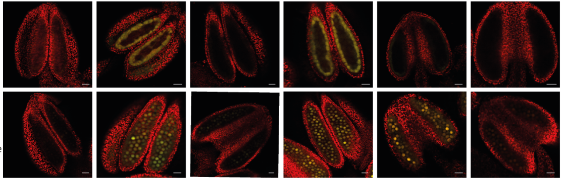
Teosinte introgression modulates phosphatidylcholine levels and induces early maize flowering time
Fausto Rodríguez-Zapata, Allison C Barnes, Karla A Blöcher-Juárez, Dan Gates, Andi Kur, Li Wang, Garrett M Janzen, Sarah Jensen, Juan M Estévez-Palmas, Taylor Crow, Rocío Aguilar-Rangel, Edgar Demesa-Arevalo, Tara Skopelitis, Sergio Pérez-Limón, Whitney L Stutts, Peter Thompson, Yu-Chun Chiu, David Jackson, Oliver Fiehn, Daniel Runcie, Edward S Buckler, Jeffrey Ross-Ibarra, Matthew B. Hufford, Ruairidh JH Sawers, Rubén Rellán-Álvarez
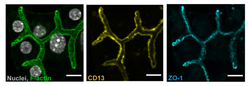
A novel adhesive complex at the base of intestinal microcilli
Klaus T Ebnet, Christian Hartmann, Eva-Maria Thüring, Brigitta E Michels, Denise Pajonczyk, Sophia Leußink, Lilo Greune, Frauke Brinkmann, Mark Glaesner-Ebnet, Eva Wardelmann, Thomas Zobel, M. Alexander Schmidt, Volker Gerke
QUAREP-LiMi: A community-driven initiative to establish guidelines for quality assessment and reproducibility for instruments and images in light microscopy
Glyn Nelson, Ulrike Boehm, Steve Bagley, Peter Bajcsy, Johanna Bischof, Claire M Brown, Aurelien Dauphin, Ian M Dobbie, John E Eriksson, Orestis Faklaris, Julia Fernandez-Rodriguez, Alexia Ferrand, Laurent Gelman, Ali Gheisari, Hella Hartmann, Christian Kukat, Alex Laude, Miso Mitkovski, Sebastian Munck, Alison J North, Tobias M Rasse, Ute Resch-Genger, Lucas C Schuetz, Arne Seitz, Caterina Strambio-De-Castillia, Jason R Swedlow, Ioannis Alexopoulos, Karin Aumayr, Sergiy Avilov, Gert-Jan Bakker, Rodrigo R Bammann, Andrea Bassi, Hannes Beckert, Sebastian Beer, Yury Belyaev, Jakob Bierwagen, Konstantin A Birngruber, Manel Bosch, Juergen Breitlow, Lisa A Cameron, Joe Chalfoun, James J Chambers, Chieh-Li Chen, Eduardo Conde-Sousa, Alexander D Corbett, Fabrice P Cordelieres, Elaine Del Nery, Ralf Dietzel, Frank Eismann, Elnaz Fazeli, Andreas Felscher, Hans Fried, Nathalie Gaudreault, Wah Ing Goh, Thomas Guilbert, Roland Hadleigh, Peter Hemmerich, Gerhard A Holst, Michelle S Itano, Claudia B Jaffe, Helena K Jambor, Stuart C Jarvis, Antje Keppler, David Kirchenbuechler, Marcel Kirchner, Norio Kobayashi, Gabriel Krens, Susanne Kunis, Judith Lacoste, Marco Marcello, Gabriel G Martins, Daniel J Metcalf, Claire A Mitchell, Joshua Moore, Tobias Mueller, Michael S Nelson, Stephen Ogg, Shuichi Onami, Alexandra L Palmer, Perrine Paul-Gilloteaux, Jaime A Pimentel, Laure Plantard, Santosh Podder, Elton Rexhepaj, Arnaud Royon, Markku A Saari, Damien Schapman, Vincent Schoonderwoert, Britta Schroth-Diez, Stanley Schwartz, Michael Shaw, Martin Spitaler, Martin T Stoeckl, Damir Sudar, Jeremie Teillon, Stefan Terjung, Roland Thuenauer, Christian D Wilms, Graham D Wright, Roland Nitschke
Intelligence and academic performance: Is it all in your head?
Katherine L. Bottenhorn, Jessica E. Bartley, Michael C. Riedel, Taylor Salo, Elsa I. Bravo, Rosalie Odean, Alina Nazareth, Robert W. Laird, Erica D. Musser, Shannon M. Pruden, Eric Brewe, Matthew T. Sutherland, Angela R. Laird
 (2 votes)
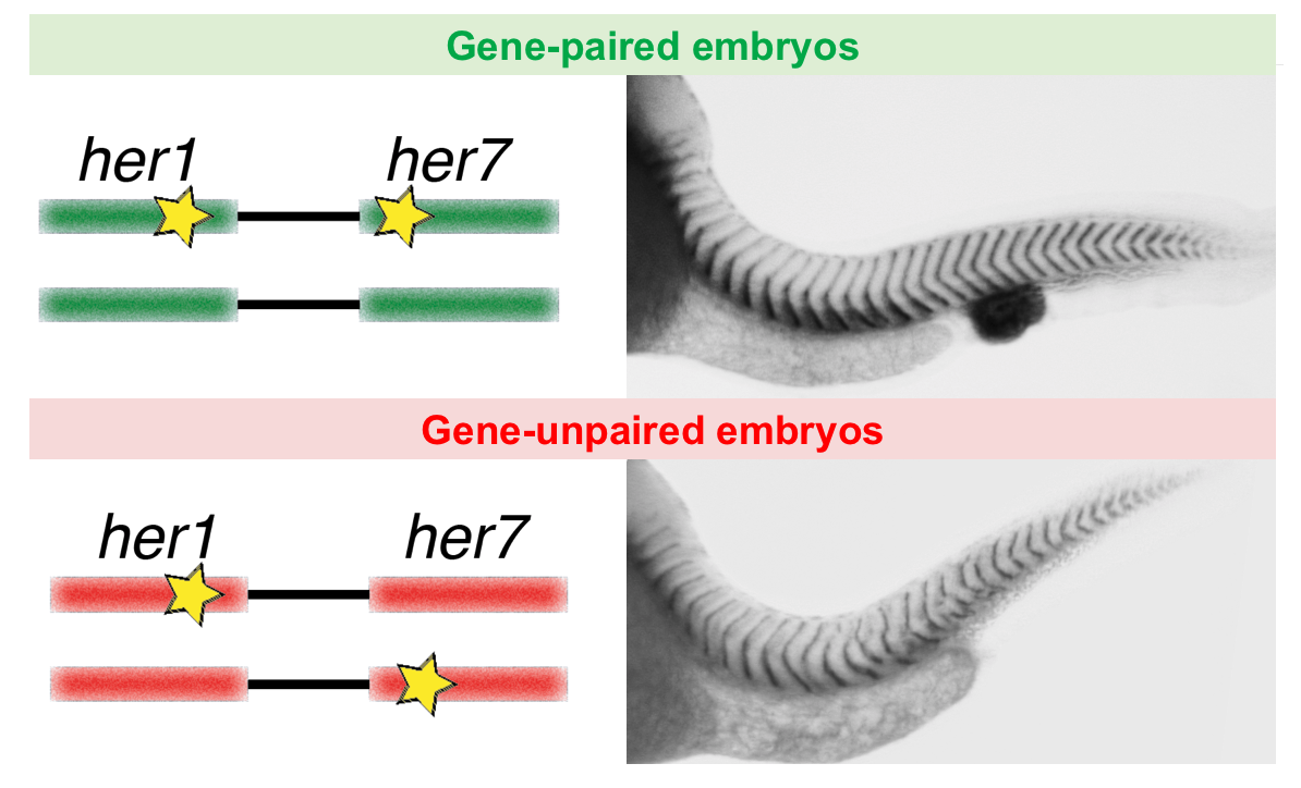
 (6 votes)


 (4 votes)
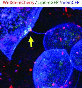 A postdoctoral position is available in the Scholpp lab at the Living Systems Institute (LSI) at the University of Exeter (
 A stimulating environment with freedom to develop new research directions.