We invite applications for an ERC funded postdoctoral research position to study divergence in brains and behaviour in Heliconius butterflies with Dr Richard Merrill’s research group at Ludwig-Maximilians-Universität (LMU) Munich. The project will run in close collaboration with Dr Stephen Montgomery at the University of Bristol, with project partners at Universidad Regional Amazónica (Ecuador) and Universidad del Rosario (Colombia). The position is funded by an ERC starting grant awarded to Dr Merrill, and is initially available for 2 years, with a further 2 years of funding available dependent on progress and interests. The position would be available at the earliest from February 2020.
The postdoc will focus on the evolution and genetic basis of differences in neuroanatomy and associated behaviours between divergent Heliconius taxa. The major aims of the position are to:
a) develop and execute assays of olfactory and visual sensitivity and integration using behavioural experiments,
b) quantify heritable variation in neuroanatomy between populations,
c) determine the behavioural effects of intermediate traits in interspecific hybrids.
The postdoc will also determine whether divergent behavioural and neuroanatomical phenotypes are functionally linked by assaying interspecific hybrids. By combining these data with genomic techniques the researcher will then investigate the genetic basis of shifts in brain and behaviour. The successful candidate will be required to spend substantial periods in the tropics (predominantly Ecuador), which will require excellent project management skills and considerable self-motivation.
Applicants should have a PhD, completed or completion imminent, in evolutionary biology/genetics, sensory biology, neuroethology, animal behaviour, or a related field. Experience of managing animal stocks and conducting behavioural analyses in insects would be desirable. Candidates are expected to work collaboratively, within the group and across the community more generally, and to take an active role in the supervision of students and management of insectaries. Enthusiasm, determination and the capacity to work independently are essential.
LMU is recognized among Europe’s premier academic and research institutions, being consistently ranked among the top Universities worldwide. Within the Division of Evolutionary Biology (http://www.evol.bio.lmu.de), the postdoctoral researcher will be part of vibrant international communities of scientists. In addition, the researcher will join a collaborative and driven community of Heliconius biologists. The working language of the lab and the Division of Evolutionary Biology is English.
Further information can be found at (https://richmerrill.wordpress.com), and questions should be directed to Richard Merrill (merrill@bio.lmu.de). Applications, made up of a single pdf (file name = candidates surname), should include a current CV, letter of motivation and names and contact details of two referees. Please send applications by email (subject: ‘Brain postdoc’) to Richard Merrill (merrill@bio.lmu.de) before the deadline of 30 November 2019.
We are looking for ambitious and motivated postgraduate candidates to join our new computational biology group at the MRC Centre for Regenerative Medicine in Edinburgh. We are using expertise in mathematical modelling of cell interactions to answer biological questions in development and regeneration in close collaboration with experimental colleagues.
There are four projects, competition-funded through the eastbio and Precision Medicine programmes. All projects involve computational as well as experimental or data analysis work. Click through the links below for more information on each project:
Please don’t hesitate to get in touch if you are interested in any of the above projects. Find a list of all opportunities at the Centre for Regenerative Medicine here.
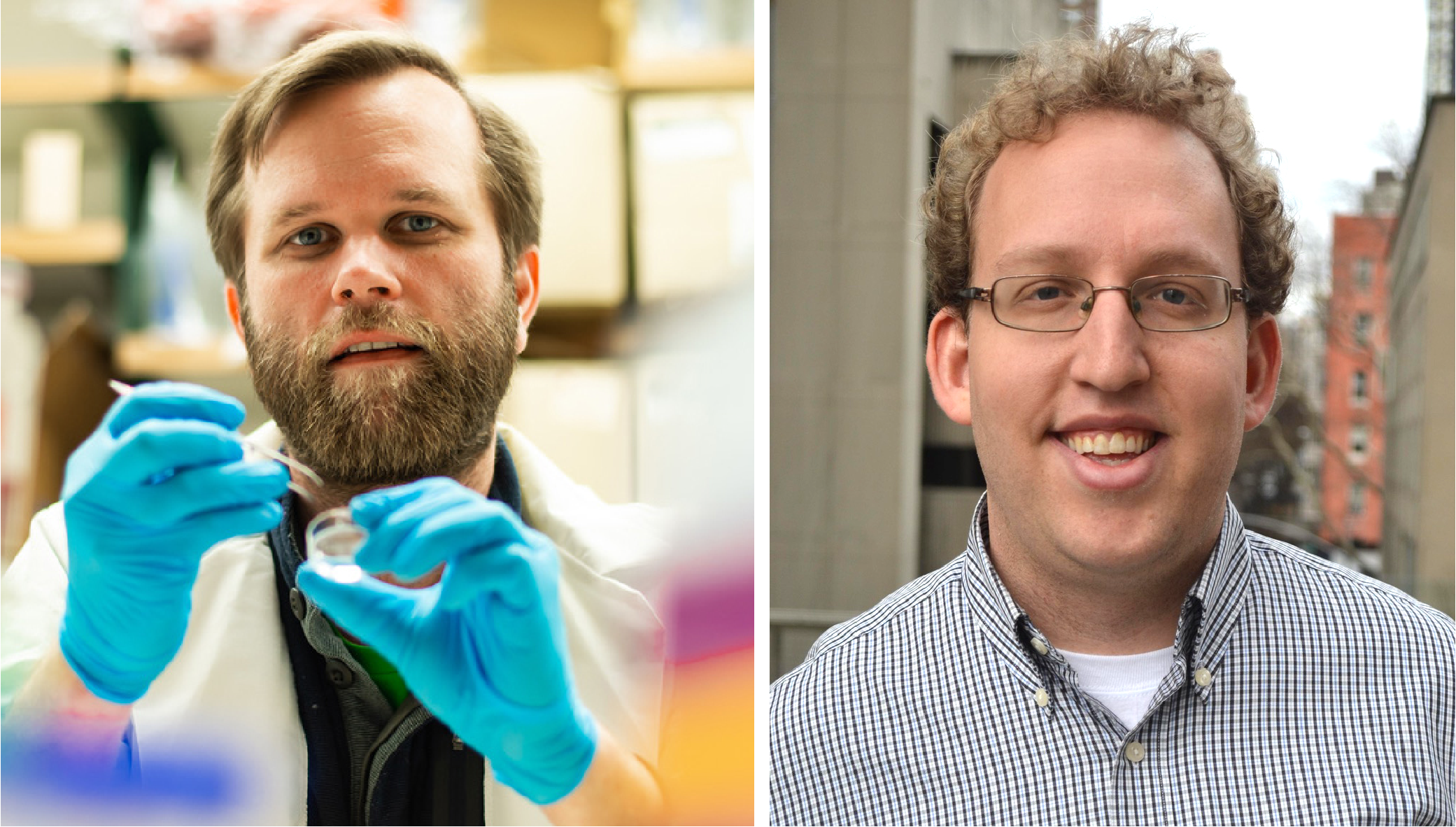
Our understanding of many fundamental aspects of early human development is still in its infancy, but a promising avenue for research uses advanced in vitro culturing techniques. For instance, confining human embryonic stem cells to micropatterned substrates and directing differentiation with signalling molecules has proved a powerful system to mimic (and readily perturb) events usually hidden in the embryo. A paper in Development now applies this technology to the question of how the embryonic ectoderm is patterned into defined domains of progenitor cells. We caught up with first author and graduate student George Britton and his supervisor Aryeh Warmflash, Assistant Professor in the Department of Biosciences at Rice University in Houston, Texas, to find out more about the paper.
George (L) and Aryeh (R)
Aryeh, can you give us your scientific biography and the questions your lab is trying to answer?
AW I was originally trained as a theoretical physicist and spent a good deal of time in graduate school developing techniques for modelling non-equilibrium systems, in Aaron Dinner’s lab at the University of Chicago. I also got involved in creating mathematical models of the development of the immune system and became convinced that the most interesting questions were in developmental biology. I realised that, although we had great collaborators for my graduate work, if I didn’t learn to do experiments, I would always be dependent on others to generate data. It really isn’t possible to make progress in biology from a purely theoretical standpoint. I was interested in working on symmetry breaking and spatial patterning, so, for my postdoc, I moved to Rockefeller University to work with Eric Siggia (a theoretician) and Ali Brivanlou, who studies early development using Xenopus frogs and human embryonic stem cells. I actually started out working with frogs, but found it to be a difficult system for quantitative imaging, and eventually switched to cell culture. During my time there, I developed the two main research directions which I have continued in my own lab – understanding the dynamics of morphogen signalling and how cells interpret them, and understanding how these signals are organised in space to generate patterns. In my lab, we are particularly interested in addressing these questions in early mammalian/human development. This is an attractive area because a lot is known about the identity of the signals that govern development during this time, and the phenotypes associated with their disruption, but much less is known about how these signals operate in space and time. I think that we know enough now to say that mammalian development works quite differently from other systems that have been dissected quantitatively – such as the Bicoid gradient in fly – and so it will be interesting to see what new mechanisms emerge. We have focused a lot of effort on understanding how the germ layers are patterned at gastrulation, and in our new study we extended these methods to a slightly later developmental time point – patterning one of those germ layers, the ectoderm.
George, how did you come to work in the Warmflash lab, and what drives your research today?
GB My previous research training involved the development of ultrasound-guided therapeutic nanoparticles functionalised to reduce necrotic and apoptotic cell death following a traumatic head injury, such as an ischemic stroke. Although our work led to significant improvements in neurological outcomes in animal models, the benefits were limited to early administration. Naturally, it was during this time that I became interested in harnessing the regenerative potential of neural progenitors to overcome the loss of functional tissue due to injury. At that time it was not clear to me what information a collection of transplanted neural progenitors needed and whether the same information could be used to repopulate neural function at any position in the brain. It was then serendipitous that I had the opportunity to join Aryeh’s lab at Rice University as a graduate student. He had developed a reductionist approach to understanding how human embryonic stem cells make fate-based decisions during gastrulation. I thought this was a powerful approach to untangling how a collection of spatiotemporally regulated signals give rise to particular arrangements of fate patterns. Although we aren’t regenerating functional brain tissue, I believe the approach and many of the principles learned can be applied to solve such problems in the future.
Before your study, how much was known about human ectodermal patterning and the extent to which studies in model organisms would translate to it?
GB & AW A great deal has been learned about how ectodermal patterning works in model organisms, but for the most part it wasn’t (and still isn’t) clear how much of this will translate to human. Most of what we knew about specifically human ectodermal development prior to the project was limited to directed differentiation protocols, which were formulated with knowledge gained from decades of experiments in model organisms. For example, the application of BMP and TGFβ inhibitors instructs the formation of neural progenitors from pluripotent stem cells, while appropriately timed BMP or WNT application can generate neural crest or placodes, respectively. So, much of the signalling appears to be conserved but there are also differences – for example, the order in which neural progenitors activate the key transcription factors SOX1 and PAX6 is different. One of the exciting things about micropatterned systems is the ability to compare species while standardising the geometry and culture conditions. However, this also requires the development of analogous systems with pluripotent cells from different species, which is a big job. This has begun to be done for gastrulation micropatterns: a system for mouse embryonic stem cells has been developed in Kat Hadjantonakis’s lab, and it would be interesting to do this for our system as well.
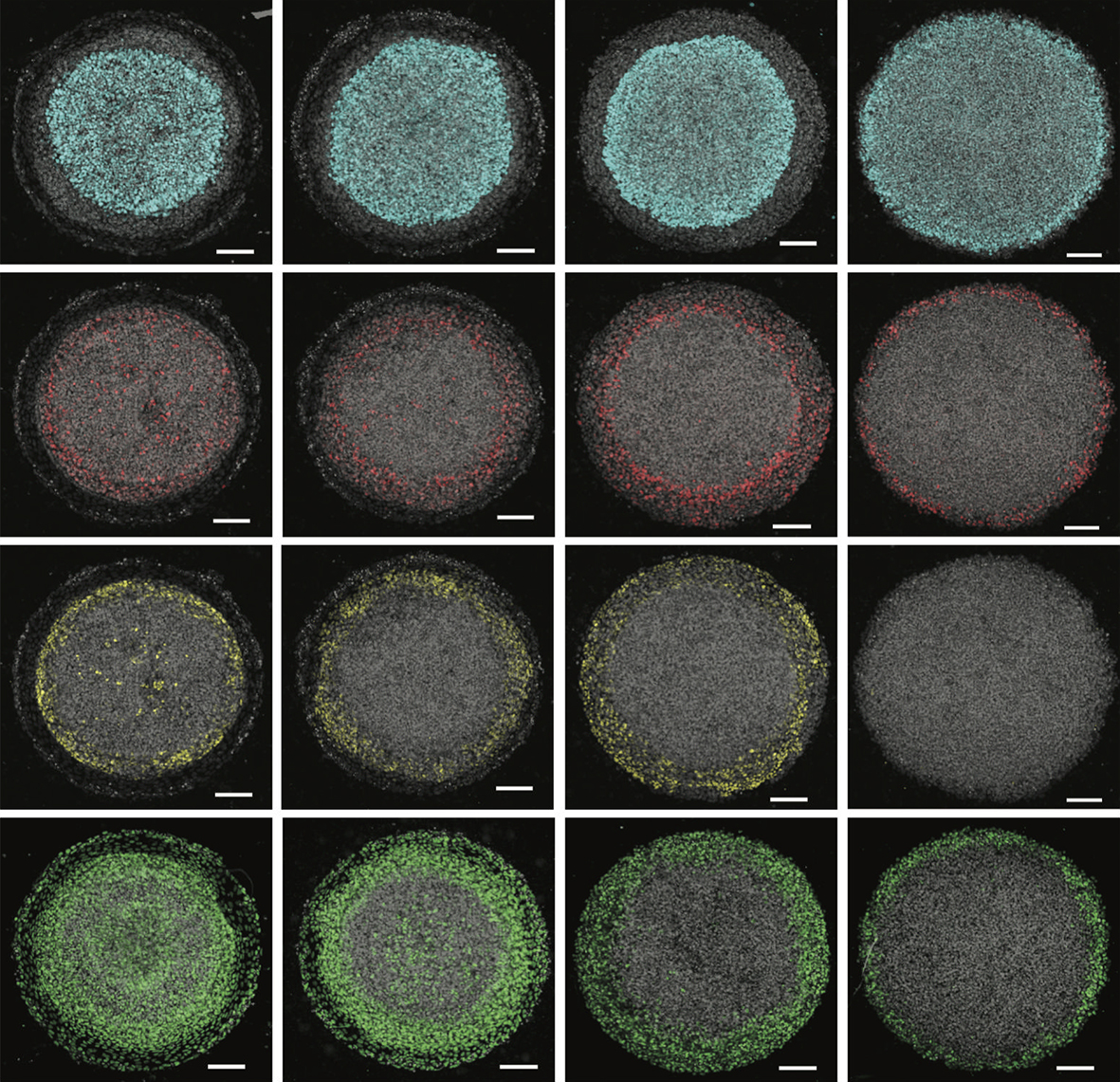
A selection of micropatterned colonies stained for different differentiation markers following a three-step ectoderm induction protocol followed by patterning with different concentrations of BMP4.
Can you give us the key results of the paper in a paragraph?
GB & AW We first mapped the competence of human embryonic stem cells to be diverted to alternative fates during ectodermal differentiation, and identified a time period when cells were primarily restricted to ectoderm but still capable of generating all ectodermal fates. We used this information to develop a platform that generates self-organised patterns of human neural, neural crest, placodal and epidermal progenitors on micropatterned surfaces with a similar organisation as found in vivo. While developing the protocol, we realised that inhibiting endogenous WNT signals was essential for limiting the extent of neural crest differentiation, which led us to identify WNT signalling duration as a crucial parameter controlling the decision between neural crest and placodes. We also showed that both BMP and WNT are involved in this decision, and that it is the relative rather than absolute levels of signalling that determines the patterns. Surprisingly, we also found that manipulating these signals during the initial ectodermal differentiation phase could influence their later competence. For example, early inhibition of BMP signals prevents cells from forming surface ectoderm in response to BMP later during patterning. Finally, we used the ease of manipulating the system to establish the necessity and sufficiency of these signals in activating particular key genes for neural plate border formation and patterning.
You find that the duration of WNT signalling can control ectodermal fates: how do you think this relates to what might be happening in the embryo?
GB & AW This is an interesting question and the answer is likely to be complex. At one level, the requirement to inhibit WNT signalling in our model probably arises because we only model one germ layer, not the interactions between germ layers. WNT inhibition in vivo has been shown to come from the visceral endoderm as well as the head mesoderm. Another role of WNT signalling is to set the coordinate along the anterior-posterior (AP) axis, and what we see is consistent with this – shortest WNT signalling yields anterior patterns with placodes and no neural crest, while the longest WNT signalling gives posterior patterns with neural crest and no placodes. Other studies within the nervous system and neural crest have shown that longer durations of WNT signalling are needed for more posterior fates. However, as WNT seems to induce neural crest specifically, it needs to do so at a particular position along the medial-lateral (ML) axis as well. So at each AP position, WNT signalling must also be controlled to give the correct ML pattern. How the AP and ML patterns are coordinated is an interesting open question.
How do you think cells measure the relative levels of WNT and BMP signalling at the molecular level?
GB & AW Another interesting question, and we are investigating two possibilities. It is possible that cells perform this calculation at the level of signalling through interactions between BMP and WNT, several of which have been described in the past. Alternatively, pathway interaction may not be required, and the computation could be performed by the downstream networks that govern cell fates. In the simplest example, if two mutually repressive transcription factors formed a toggle switch that implemented a binary cell fate decision, and one signal induced each of these transcription factors, this circuit would naturally take ratios between the transcription factor levels in making the cell fate decision. Of course, the real circuit is much more complex, but this serves to illustrate that transcriptional networks could definitely perform these computations on multiple inputs. To sort this out, we will need to measure the signalling activity through both pathways under different conditions as well as how different transcription factors interpret those levels.
When doing the research, did you have any particular result or eureka moment that has stuck with you?
GB There was definitely one instance that I will remember for a long time. In the beginning of the project I had formulated a basic micropatterning protocol (the two-step induction protocol) that generated patterns of neural, neural crest and surface ectoderm. It was nice that patterns emerged at all, but I had grown frustrated with the lack of placodal expression. Thankfully, I had a patient advisor and a great platform to scan a range of signalling perturbations. Then, one late evening in the ‘scope room I observed a beautiful ring of SIX1 expression (a placodal marker) as a result of WNT inhibition. Thinking I was alone, I jumped and danced out of excitement but soon after noticed I wasn’t alone. In my moment of celebration, the janitor had stepped into the room for cleaning but found a grad student dancing alone with no music. It was a bit embarrassing at the time, but she had a great laugh.
I jumped and danced out of excitement but soon after noticed I wasn’t alone
And what about the flipside: any moments of frustration or despair?
GB I don’t think there is a single moment that stands out. It’s natural to run into roadblocks or become frustrated when developing a project from scratch. However, one point of frustration I learned to manage was the longer time required to conduct a single experiment compared with my lab mates, as most of them were studying gastrulation stage patterning with experiments that take 1-2 days. When it was time to present at lab meetings I consistently had fewer results to share, which led to feelings of self-doubt or insufficiency as a member in the lab. Eventually, with more experience, I developed strategies to increase the throughput of my experiments, which has helped push the project in directions I didn’t expect.
So what next for you after this paper?
GB Eventually I need to graduate, but I plan to conduct single cell analysis of signalling and fate in our ectoderm patterns as a follow-up paper. Unfortunately, I am still unsure what direction I should take for a postdoc but I am actively searching.
Where will this work take the Warmflash lab?
AW We are building on this system in a few different ways. We are quite interested in directly measuring the signalling dynamics of BMP and WNT with reporters that we have previously developed and used to study signalling during germ layer patterning. We are also interested in whether we can determine the AP position at which we are achieving this patterning and in developing protocols to control it. Finally, we are interested in extending this system to three dimensions to try to recapitulate some of the morphogenesis that the ectoderm undergoes during patterning, and to be able to look at how patterning and morphogenesis are coordinated.
Finally, let’s move outside the lab – what do you like to do in your spare time in Houston?
GB Unfortunately, most of my hobbies require a mountainous landscape. However, Houston makes up for its lack of interesting topology and good weather with an awesome food scene and a consistent line-up of touring indie bands. Tickets for live shows rarely get sold out and prices are student friendly, so it usually takes minimal advanced planning.
AW I have three kids aged 12, 9 and 5 so that keeps me quite busy outside the lab. We like travelling with the kids and doing things outdoors. It is very hot in the summer here, but the weather is nice the rest of the year and there are some great state parks not too far from Houston. We also like going to the Texas Hill Country and to the beach in Galveston.
Non-coding DNA regulatory elements or enhancers control tissue and region-specific expression of critical developmental regulators thus playing a crucial role in the correct formation of the embryonic body. However, not much is known how regionalized gene expression is achieved through enhancer action. The proposed PhD project aims to address this issue using the development of the nervous system as a paradigm. During embryogenesis, the brain and the spinal cord are derived from different progenitors and this process is controlled to a large extent by the transcription factor Sox2. Sox2 expression into these two components of the nervous system is directed by two distinct enhancers, N1 in the spinal cord and N2 in the brain. This PhD project will employ brain and spinal cord progenitors derived from human pluripotent stem cells (hPSCs) in order to dissect the function of these two enhancers. Using hPSC differentiation in combination with a variety of techniques including ChiP-seq and CRISPR/Cas9 approaches we aim to:
1) define the molecular hallmarks that may distinguish the N1 and N2 enhancers
2) examine the differential binding of candidate transcription factors on these enhancers
3) identify critical sequence parameters influencing their function.
Funding Notes
– a tax-free stipend at the standard Research Council rate (~£15,009, to be confirmed for 2020) for 4 years
– tuition fees at the UK/EU rate for 4 years.
– research costs
Required qualifications
At least a 2:1 honours degree in a relevant subject or equivalent. The interdisciplinary nature of this programme means that we welcome applications from students with backgrounds in any biological, chemical, and/or physical science, students with mathematical backgrounds who are interested in using their skills in addressing biological questions.
Studentships are available to UK and EU students who meet the UK residency requirements. Further information on eligibility: View Website.
Deadline: Monday, January 06, 2020
For informal enquiries contact: a.tsakiridis@sheffield.ac.uk
Summary: Plant shape is a primary determinant of productivity and yield because it affects light interception and photosynthesis. As plant cells are bound by a cell wall and cannot move, shape arises as an outcome of the plane of new cell divisions, and subsequent cell growth. Flowering plant models such as Arabidopsis have complex tissue organizations that can mask cell division plane defects. There are also many genes per gene family, which can make it hard to identify mutants. For these reasons, few genetic regulators of cell division plane orientation have been discovered. In contrast to flowering plants, mosses have simple tissue organizations and there are few genes per gene family. I established a moss model to study plant cell division plane orientation [1], and recently determined that the CLAVATA receptor-like kinase sets the plane of cell divisions [2, 3]. Although mosses are distantly related to flowering plants, our findings were transferable to Arabidopsis, and we are now manipulating CLAVATA function in wheat to improve productivity [4]. Harnessing the benefits of the moss model, this project aims to discover how CLAVATA determines the plane of cell divisions in plants to affect their overall shape and productivity.
To determine how CLAVATA orients division planes in moss the project will:
1. Identify downstream targets of CLAVATA by RNAseq and bioinformatic analysis
2. Generate mutants of a candidate target and analyse mutant phenotypes
3. Analyse gene regulatory network architecture using computational approaches
4. Identify novel cell division plane regulators using a suppressor screen.
Training: By combining computational and wet lab approaches, the project will provide training at the cutting edge of the plant development field. It will benefit from further formal teaching and internships included in the SWBioDTP programme. The skills and techniques the student will learn will be broadly applicable in the academic biology and biotech sectors and widely transferable amongst areas such as science policy, publishing and computing.
Funding Notes
A fully-funded four year SWBio DTP studentship will cover:
– a stipend* (at the standard UKRI rate; £15,009 per annum for 2019-2020)
– research and training costs
– tuition fees (at the standard UKRI rate)
– additional funds to support fieldwork, conferences and a 3-month internship
References
[1] Harrison et al. 2009. Local cues and asymmetric cell divisions underpin body plan transitions in the moss Physcomitrella patens. Current Biology 19: 1-11.
[2] Whitewoods et al. 2018. CLAVATA was a genetic novelty for the morphological innovation of 3D growth in land plants. Current Biology 28: 2365-2376.
[3] Bergmann 2018. Taking development to three dimensions. Developmental Cell 17: 678-679.
[4] Fletcher 2018. The CLV-WUS stem cell signaling pathway: a roadmap to crop yield optimization. Plants 7: 87.
Supervisors: Jill Harrison, Keith Edwards and Chris Burt (RAGT Seeds)
Project Description
Summary: Ensuring continuous global food security will be a major challenge of the 21st century, and wheat contributes approximately 20% of the total calories consumed by humans (FAO, 2017). In cereals like wheat, inflorescence (ear) size determines the number of flowers (florets) and grains produced, and this aspect of plant architecture is regulated by the activity of stem cells in the growing shoot tips. The CLAVATA peptide/ receptor-like kinase signalling pathway maintains the size of the stem cell pool during plant development, and mutants in maize and tomato have increased yields, arising due to an increase in size of the stem cell pool. This project aims to intercept wheat CLAVATA signalling to engineer ears with more fertile grain sites and increase yield.
The project will involve:
(1) Identification of wheat CLAVATA pathway components
(2) Expression analyses of wheat CLAVATA pathway components
(3) Generation phenotypic analysis of wheat CLAVATA pathway mutants.
Dr Harrison’s group has recently published gene trees for CLAVATA pathway components from a range of land plants (Whitewoods et al. (2018)), and she has experience of analysing gene expression patterns and function in a wide range of plant species. Professor Edwards and colleagues from the Bristol Centre for Agricultural Innovation have extensive experience with wheat having sequenced the genome (Brenchley et al. (2012)), identified many mutants from the exome sequenced Cadenza TILLING mutant population (Krasileva et al. (2017)) and established engineering procedures using CRISPR/Cas9. The CASE partnership with RAGT seeds will bring an opportunity for the student to directly experience wheat breeding and exchange knowledge and findings with wheat growers.
Training: By combining computational and wet lab approaches, your project work will provide training at the cutting edge of the plant development field. You will benefit from further formal teaching and internships included in the SWBioDTP programme. The skills and techniques you learn will be broadly applicable in the academic biology and biotech sectors and widely transferable amongst areas such as science policy, publishing and computing.
Funding Notes
A fully-funded four year SWBio DTP studentship will cover:
– a stipend* (at the standard UKRI rate; £15,009 per annum for 2019-2020)
– research and training costs
– tuition fees (at the standard UKRI rate)
– additional funds to support fieldwork, conferences and a 3-month internship
This is a CASE DTP studentship. As part of the programme, you will be required to undertake a placement with the CASE partner for a minimum of 3 months.
References
Brenchley et al. (2012). Analysis of the bread wheat genome using whole-genome shotgun sequencing. Nature 491: 705-710. Food and Agriculture Organization of the United Nations, FAOSTAT statistics database, Food balance sheets (2017); www.fao.org/faostat/en/#data/FBS. Krasileva et al. (2017). Uncovering hidden variation in polyploid wheat. PNAS 114: E913-E921. Whitewoods et al. 2018. CLAVATA was a genetic novelty for the morphological innovation of 3D growth in land plants. Current Biology 28: 2365-2376.
PhD position available at Liverpool John Moores University, starting Feb 2020. Extraction of exosomes from maternal blood for analysis of miRNA cargo and glycoprotein epitopes.
The Poulain lab (www.poulainlab.org) at the University of South Carolina in Columbia, SC seeks talented and motivated postdocs! Weuse zebrafish as a vertebrate model system and a unique combination of genetic, embryological and live imaging approaches to study the cellular and molecular mechanisms of axon guidance and degeneration during the formation of neural circuits in vivo. Specific projects include single-cell topographic transcriptomics and the formation of topographic maps, role of neural activity in topographic map plasticity, role of cell adhesion in axon developmental degeneration, and spatio-temporal control of trans-axonal degeneration signaling.
Candidates should hold a PhD in neurobiology or neuroscience and have a strong interest in neural development. Significant experience in molecular biology, genetics and fluorescence imaging approaches is required. Experience with zebrafish is desired but not mandatory. Interested candidates should email their resume/CV, the names and contact information of 3 references, and a cover letter explaining their interest to fpoulain@mailbox.sc.edu
NEUCrest Ph.D Studentships in Developmental Biology, Stem Cell Biology and Cancer Biology
Applications are invited from suitably qualified candidates for full-time fixed term positions as Early Stage Researchers in the lab of Prof Karen Liu, King’s College London (karen.liu@kcl.ac.uk), Centre for Craniofacial and Regenerative Biology, King’s College London.
These positions are funded by the Horizon 2020 programme of the European Union and will be available from 1st January 2020. Appointments will be on a full-time basis for a period of 3 years. Remuneration will be in line with the European Commission rules for Marie Skłodowska-Curie grant holders (Early-Stage Researchers, Initial Training Network).
NEUcrest is a four-year EU Horizon 2020 project funded by Agreement 860635. The neural crest is an essential stem cell population of vertebrate embryos. The project focuses on integrating academic, clinical and industrial research for a better understanding of neural crest development and neural crest related diseases. These pathologies are a major group of congenital diseases in human, and a heavy societal concern. The NEUcrest network comprises 20 partners in academia, industry and hospitals from seven European countries, gathered in a synergistic effort to advance knowledge and outreach of these diseases.
The Ph.D projects are highly multidisciplinary and will develop scientific strategies from experimental embryology, genome editing, imaging and generation of genomic datasets. A full list of projects and partners:
PhD Projects available through the lab of Prof Karen Liu, King’s College London:
PROJECT 1: The Role of ALK and GSK3 in normal and pathogenic neural crest migration. This project led by Prof Karen Liu (King’s College London) and Prof Angela Nieto (UMH, Spain). Applications see: https://tinyurl.com/yycpnlxj
Person Specification: First degree or Masters in Biological Sciences, Cell Biology, Genetics and Molecular Biology. Mobility requirement: EU applicants are eligible to apply. Applicants must not have been based in the country of registration for more than 12 months in the last 3 years.
Start date: Positions are available from 1st January 2020. Informal enquiries to Prof Karen Liu (karen.liu@kcl.ac.uk)
To Apply: Applications, in English, should include a detailed CV, certificates of examination grades (bachelor/master), a letter describing your career goals, skills and experience, as well as two letters of recommendation. Applications: https://tinyurl.com/yycpnlxj. Informal inquiries may be made to karen.liu@kcl.ac.uk. Deadline for applications is 15 November 2019, or until posts are filled.
PROJECT 2: Generation of neural crest cells from patient-derived hiPSCs for disease modelling and therapeutic applications. This industrial project will be led Dr Erin Knock and Dr Wing Chang with student based at STEMCell Technologies, Cambridge UK. Academic registration and supervision by the lab of Prof Karen Liu (King’s College London).
PROJECT 3: Establishment of human iPSCs from syndromic neurocristopathy patients with skeletal dysplasia This project is suitable for a clinical or suitably qualified non-clinical PhD student and is led by Prof Irene Mathijssen (email to i.mathijssen@erasmusmc.nl Erasmus Medical Centre, Netherlands) with secondment to Prof Karen Liu (King’s College London) and STEMCell Tech, Cambridge UK and Phenocell, France.
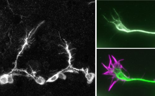
Cell adhesion molecules (CAMs) of the plasma membrane physically tie cells together into tissues, both via interaction of cells with other cells or with ECM (extracellular matrix); they also constitute hubs of information exchange with neighbouring cells and the environment. CAMs mediate a broad range of biological functions and their aberration contributes to a range of diseases, such as cancer progression or aberrant growth/regeneration of nerves.
On this project, you will study integrins, which are the major adhesion receptor class for extracellular matrices.Integrin signalling is performed by its adhesome, the dynamic population of proteins that link integrins both to the cytoskeleton and to signalling pathways via the cytoplasmic tails of the receptors. The composition and functional state of integrin-associated complexes essentially determines the adhesion state and signalling output.
To understand this, Martin Humphries has long-standing expertise in using proteomics based on mass spectrometry as a means to identify components of the integrin adhesome and study their function in mammalian cell culture [1]. On this project, you will extend such approaches to brain tissues in the living organism and study the function of identified proteins in the context of nerve growth.
For this, you will capitalise on the expertise of Andreas Prokop, who uses neurons of the fruit fly Drosophila to study mechanisms of nerve growth [2]. In this functional context, his team discovered and analysed important roles of integrins and some of their conventional adhesome components (new unpublished results). The small size and genetic tractability of Drosophila makes it feasible to perform proteomics on integrin adhesion complexes extracted from nerve cells of living organisms.
Once candidate components have been identified, you will then be able to capitalise on highly efficient genetic means provided by Drosophila to study their functional contributions during nerve growth [2] – potentially identifying promising drug targets for nerve regeneration therapies. A likely option will be the extension of such functional studies into mammalian models.
Your experimental skill training opportunities will include molecular biology, protein biochemistry, mass spectrometry, genetics, cell biology, a range of imaging techniques, bioinformatics, as well as insights into important concepts of the cell and neurobiology fields. In addition, AP is an expert in science communication providing further training opportunities important for your career development.
 (No Ratings Yet)
 Cell adhesion molecules (CAMs) of the plasma membrane physically tie cells together into tissues, both via interaction of cells with other cells or with ECM (extracellular matrix); they also constitute hubs of information exchange with neighbouring cells and the environment. CAMs mediate a broad range of biological functions and their aberration contributes to a range of diseases, such as cancer progression or aberrant growth/regeneration of nerves.