A technician/lab manager position is available in the laboratory led by Ricardo Mallarino, Department of Molecular Biology, Princeton University (www.mallarinolab.org). The lab focuses on uncovering the genetic and developmental mechanisms by which form and structure are generated during vertebrate embryogenesis. We combine the study of emerging and traditional model organisms to explore questions relating to patterning and evolution of novelty in the mammalian skin. The lab uses a variety of approaches, including experimental embryology, genetics, genomics, imaging, and mathematical modelling to uncover gene function and understand mechanisms of evolutionary change.
Responsibilities
The successful candidate will manage essential operating procedures for the lab and work with the PI and other lab members to design and perform experiments. Specifically, the candidate will develop in vivo functional and genome editing approaches in non-traditional model species and perform cell-culture based enhancer screens. Other duties will include molecular cloning, histology, in situ hybridization, nucleic acid extraction and library preparation, tissue culture and media preparation, as well as lab maintenance, organization, safety and ordering.
Essential Qualifications
Bachelor’s or Master’s degree in the biological sciences plus previous laboratory experience is required. Previous experience in cell culture techniques and rodent model systems is necessary (colony management, genotyping and dissections). Basic molecular biology methods and computer literacy are essential. Must be highly motivated and have demonstrated ability to plan, coordinate and carry out independent research. Excellent organization skills, ability to communicate effectively and willingness to learn new techniques are necessary traits. Applicants should be willing to commit to the position for at least two/three years.
Preferred Qualifications
A strong background in molecular biology and/or genetics is preferred. Knowledge of rodent reproductive biology and surgical skills are a plus. Rank and salary are dependent upon qualifications and experience.
To apply for this position please submit a CV and a cover letter describing research interests to rmallarino@princeton.edu.
My name is Sandra Treffkorn, and I recently finished my PhD in the department of zoology lead by Georg Mayer at the University of Kassel, Germany. In our research group, we focus on studying the evolution of animal diversity by investigating two very interesting but largely understudied taxa, the Onychophora and the Tardigrada. Tardigrades, commonly known as “water bears”, are well known to scientists and non-scientists alike due to their ability to survive extreme environmental conditions including exposure to space [1-3]. In contrast to tardigrades, onychophorans comprise a less known animal group even among biologists. Hence, I will use this opportunity to introduce you to these exciting animals and give you a short overview of our research.
When we get asked what onychophorans are, we sometimes tend to describe them as worms with legs and a smooth velvety skin because this is basically what onychophorans look like (Fig. 1). This is also reflected in their common name “velvet worms”, which refers to the velvety appearance of their skin. But actually, they are not worms at all. Onychophorans are terrestrial invertebrates that belong to the Ecdysozoa – the clade of molting animals [4, 5]. Together with the tardigrades, they are the closest living relatives of the arthropods (spiders, myriapods, crustaceans and hexapods), which is the most abundant and diverse animal group on our planet. Compared to arthropods, however, the anatomy of onychophorans has changed little since the Early Cambrian period (~520 million years ago) [6], and they resemble fossil lobopodians of that time, which represent stem-group panarthropods (onychophorans + tardigrades + arthropods).
Figure 1: World map showing the distribution of Peripatidae (orange) and Peripatopsidae (blue), the two major taxa of Onychophora, and photographs of the onychophoran species Principapillatus hitoyensis (Peripatidae) from Costa Rica and Euperipatoides rowelli (Peripatopsidae) from Australia. World map modified from ref. [10]. Photograph of P. hitoyensis modified from ref. [18]; image of E. rowelli was taken by Ivo de Sena Oliveira.
The onychophoran body comprises a head, a uniform worm-shaped trunk bearing a variable number of unjointed limbs, and a limbless posterior anal cone (Fig. 1). The head is composed of three segments, the limbs of which have been modified into three pairs of specialized appendages: the antennae, the jaws, and the slime papillae [7, 8]. The slime papillae are used in a fascinating way to capture the prey: two jets of a sticky secretion are ejected by the slime papillae, which immediately immobilizes the prey (woodlice, crickets and other small invertebrates) [6, 9]. Thus, even though onychophorans are relatively slow, they are very effective hunters. However, the slime is not only used for prey capture but also for defense against predators [6, 9].
Onychophorans typically inhabit humid microhabitats such as leaf litter, soil and decaying logs in tropical and temperate forests. There are currently about 200 described species of Onychophora which are classified into two major groups, the Peripatidae and the Peripatopsidae (Fig. 1) [6, 10]. While the Peripatidae can be found in equatorial areas such as the Neotropics, West Africa and South East Asia, the distribution areas of the Peripatopsidae are mainly restricted to the southern hemisphere, including Chile, South Africa and Australasia (Fig. 1) [6, 10].
But why do we study onychophorans? What’s so interesting about them? The close phylogenetic relationship of onychophorans to two of the most extensively studied model organisms – the nematode Caenorhabditis elegans and the fruit fly Drosophila melanogaster – makes them an attractive model for studying the evolution of animal diversity. Studies of onychophorans as an outgroup to arthropods can be used to polarize morphological and developmental characters of the arthropod ancestor. By extending this comparative approach to cycloneuralians (nematodes, priapulids, kinorhynchs and allies), we are able to make assumptions about the panarthropod ancestor, which in turn might help to clarify the ancestral characters in the ecdysozoan lineage when taking comparative data from spiralians and deuterostomes into account. Hence, onychophorans are a key taxon for understanding the evolution of animal diversity.
Our fields of research
In our lab, we study onychophorans in a wide variety of research fields, including taxonomy, phylogeography, population genetics, species conservation, biochemistry, physiology, behavior, evolutionary developmental biology, neuroanatomy and reproductive biology. Our major goal is to identify and understand the evolutionary changes that have taken place since the divergence of onychophorans from arthropods during the Cambrian radiation over 540 million years ago. Thus, even though working on an emerging model organism can be tricky, and we usually have to develop new methods and optimize existing protocols, the work is quite rewarding since we get the opportunity to look back in time and reconstruct evolutionary events that had taken place hundreds of millions of years ago.
A typical day in an onychophoran lab
Animal collection
For most of our work, we use the onychophoran species Euperipatoides rowelli from Australia, which is one of the most extensively studied onychophorans so far and has recently been established as a model for developmental studies (Figs 1; 2) [6]. Adults of E. rowelli are typically dark-blue colored with more or less prominent, reddish terracotta pattern (Figs 1; 2). Females reach up to 6 cm in length while males are usually smaller.
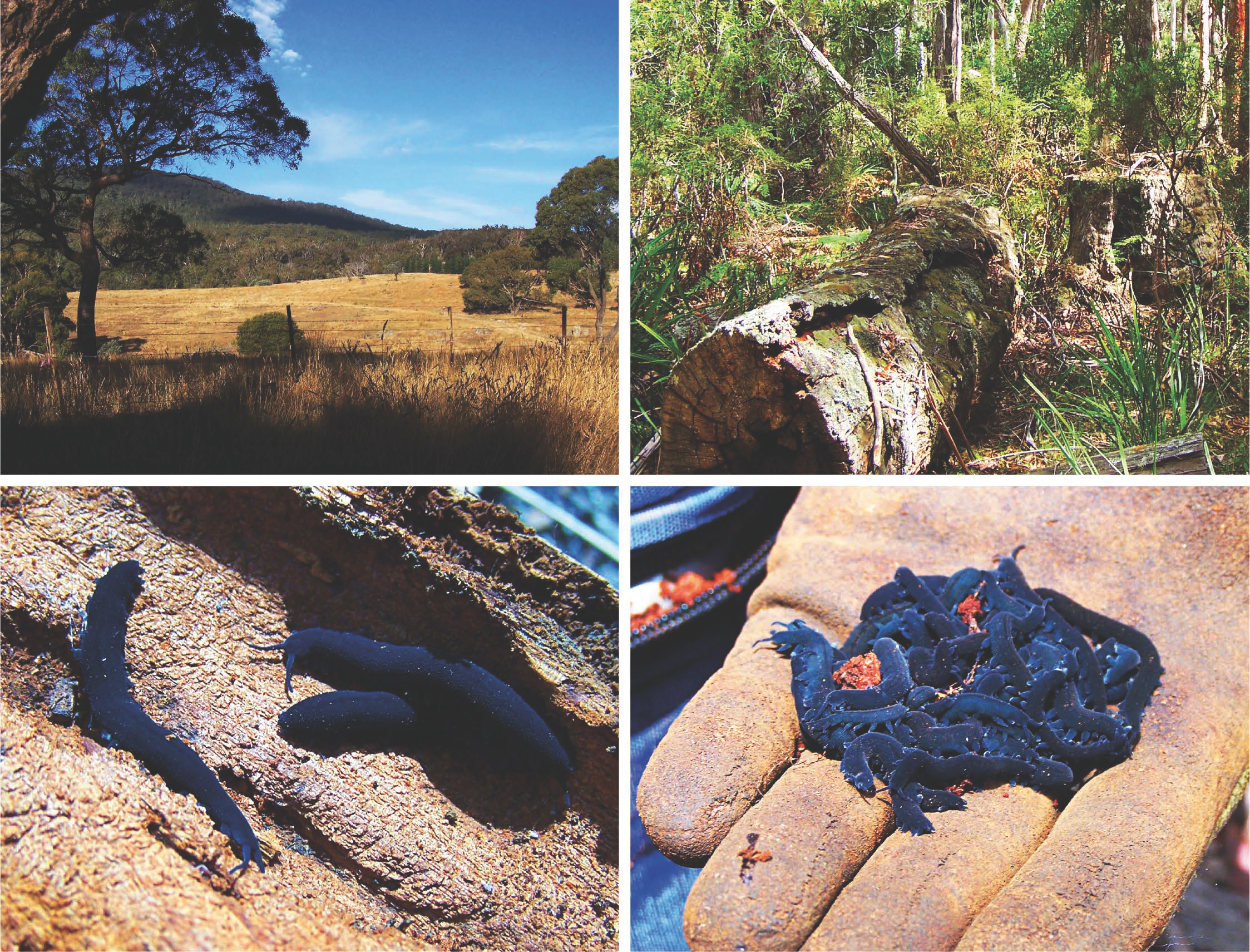
Figure 2: Before the lab work begins, there is the fun part. Members of our lab regularly set out for adventures Down Under to hunt onychophorans in the wild. The type locality of E. rowelli is in the Tallaganda State forest ([19]; top left), which is where we usually collect our animals. They are found in leaf litter (top right) as well as inside decaying logs (bottom left). After collecting the animals, we keep them in plastic jars with some earth and paper towels, in which they are transported to our lab in Germany (bottom right).
Before we can start our lab work, however, we actually have to go out and collect animals. Since E. rowelli lives in Australia but our lab is located in Germany, we are not able just to go out and collect animals from the wild whenever we need them. Thus, we have to go on collection trips from time to time to collect animals in Australia, which we then keep and breed in culture in our lab. We usually collect the animals in the Tallaganda State Forest situated about an hour’s drive southeast of Australia’s capital, Canberra (Fig. 2).
Like arthropods, onychophorans possess tracheae as respiratory organs. Unlike arthropods, however, they are not able to close their tracheal openings, which is why they prefer humid environments to avoid drying out (Fig. 2). Typically, onychophorans can be found in the humid leaf litter or under stones. The easiest way to collect them, however, is actually during dry periods when the leaf litter is too dry for the animals and they are all hiding in the decaying logs, which retain a decent level of humidity (Fig. 2). Instead of crawling around on the ground, turning every stone and searching the leaf litter for animals, we can just pry open the decaying logs and collect the animals inside. So, armed with crowbars, screwdrivers, shovels and other tools useful for prying open decaying wood, we head out and collect the animals, which we then put into plastic jars filled with some earth and damp paper towels to keep them safe for the journey around the world to their new home.
Maintenance in the lab
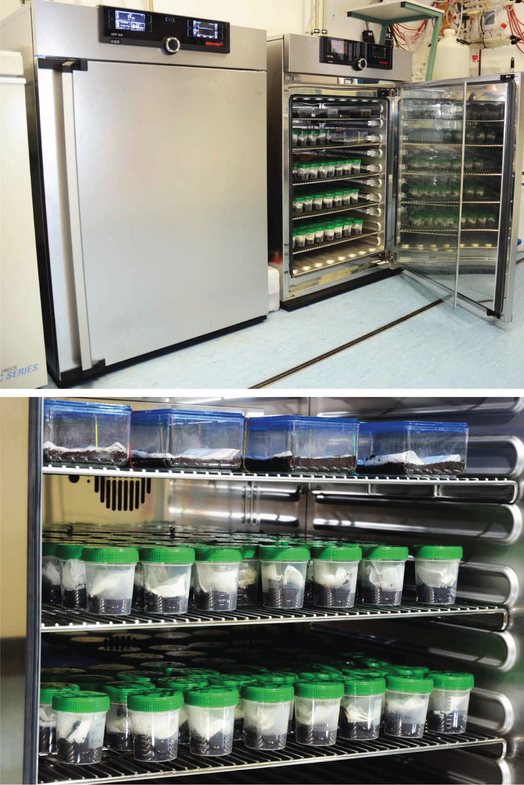
When back in Germany, the animals are cultured in plastic jars containing a layer of peat and damp paper towels, which we keep in climate cabinets under a constant temperature of 18 °C and 60% humidity (Fig. 3).
Figure 3: Back in the lab, we keep the animals in climate cabinets under specific light, temperature and humidity conditions (left). The animals are kept in plastic jars and plastic boxes with a layer of peat and damp paper towels (right).
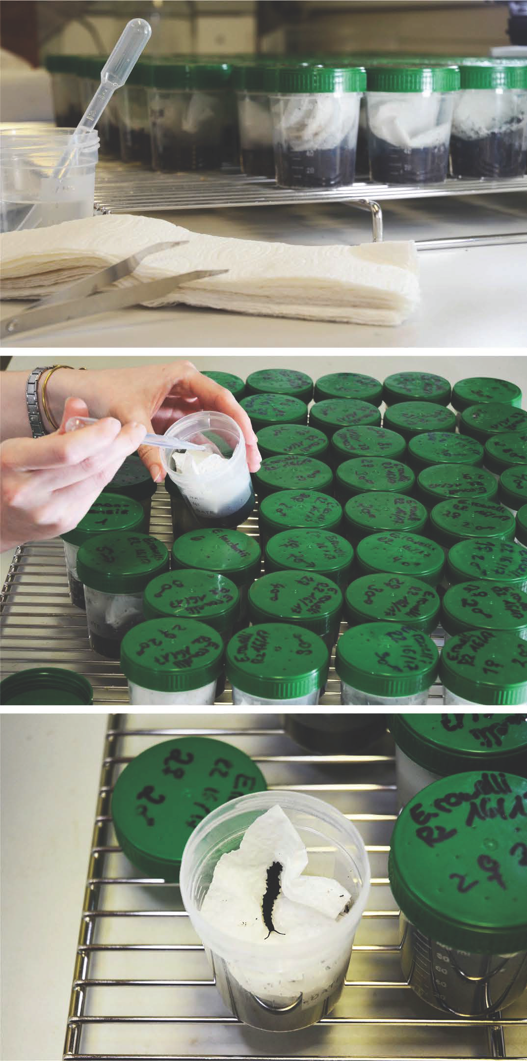
The first thing to do after a collection trip is to separate the animals. We keep around six specimens – usually three males and three females – together in each jar. And then the most laborious part of our work begins. Of course, we have to take care of the animals to keep them happy and thriving. The paper towels have to be kept damp at all times in order to provide a nice humid environment (Fig. 4). However, the humidity promotes fungal growth, which can harm the animals. So, in addition to maintaining a constant humidity, we replace the paper towels once a week (Fig. 4). Every three weeks, the animals are fed with crickets, which we leave in the jars for two days. Afterwards, we transfer the onychophorans to new jars with fresh peat and paper towels, again to avoid fungal growth due to the decaying cricket remains. Taken together, animal care has to be done at least twice a week and takes several hours each time. This is especially laborious when a new batch of babies is born, which we separate from the parents into new containers. But with a lot of teamwork, it is doable.
Figure 4: In order to keep the animals happy and thriving, we have to take care of them, which includes changing the paper towels every week (top) and dampening them regularly (middle). Using this procedure, the animals can be kept in their jars for several years (bottom).
Collecting embryos
One major part of our research includes studying embryonic development of onychophorans. For this, we apply a variety of different methods, including in situ hybridization, immunohistochemistry, fluorescence and confocal microscopy, and scanning electron microscopy [11-14]. During my PhD, I specifically analyzed expression patterns of different developmental genes by using in situ hybridization. So, the next step for me was to obtain as many embryos as possible, which is much trickier than for the commonly used model organisms, such as D. melanogaster and C. elegans. In contrast to these model organisms, where embryos are available constantly throughout the year [15, 16], E. rowelli has an annual reproductive cycle with females usually giving birth between November and February under laboratory conditions [17]. Furthermore, the entire embryonic development takes place within the uterus of the mother [6]. In order to obtain embryos of different developmental stages for the experiments (Fig. 5), they have to be dissected from the females manually – usually between September and January – to cover all developmental stages. Thus, we have to plan ahead and get as many embryos as possible during this time, which we then fix and store for long term usage. But since each female typically bears 20 to 40 embryos of different developmental stages, it is possible to cover most stages by dissecting only a few females.
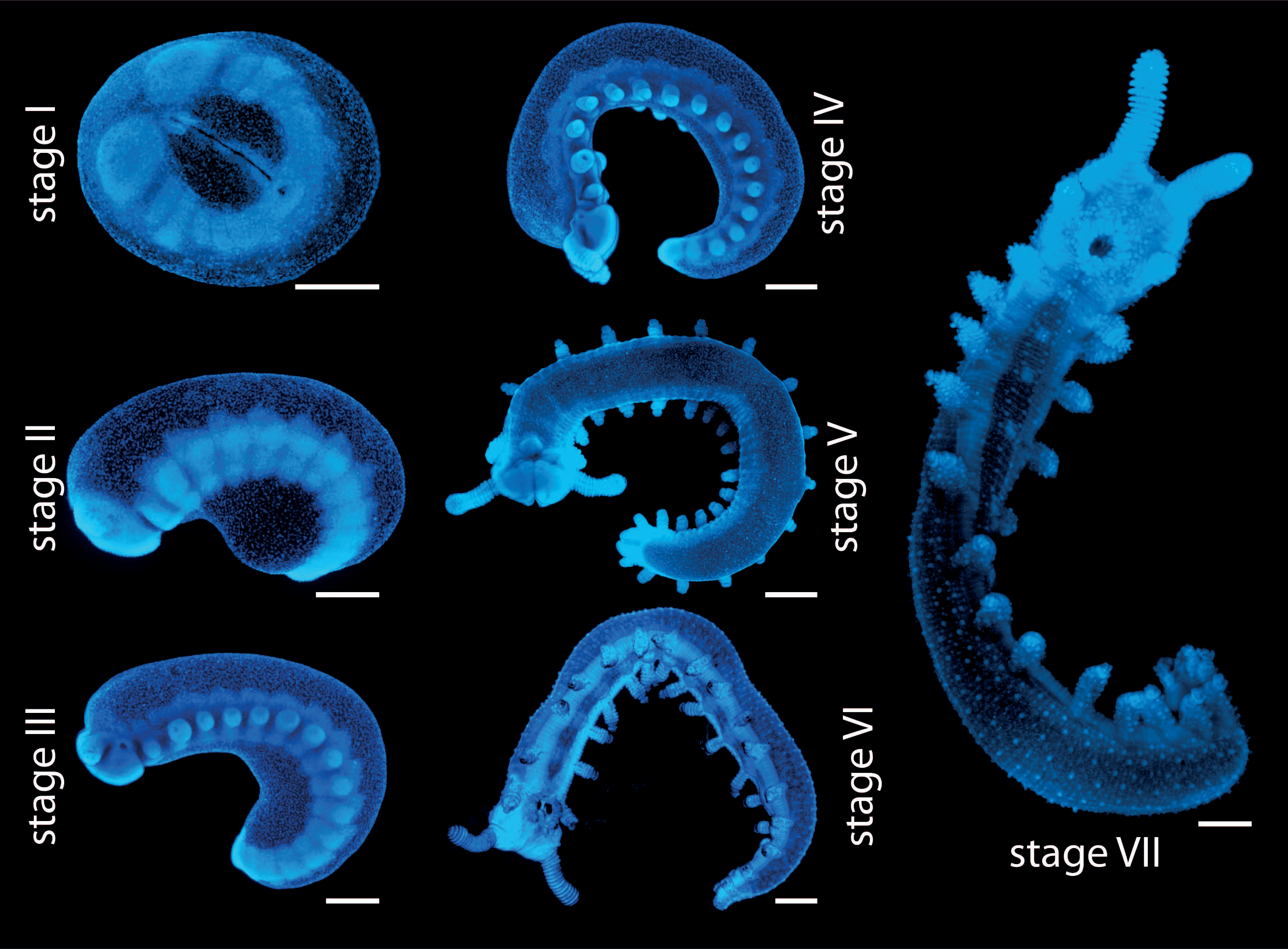
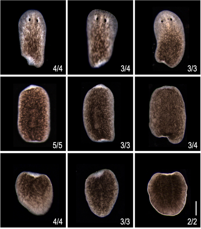
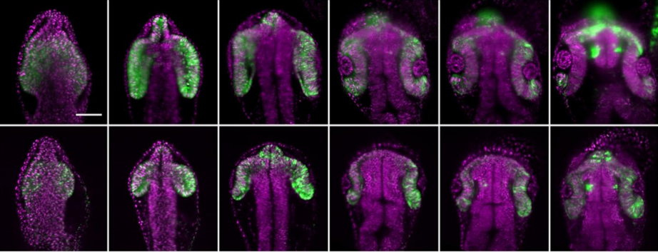
Figure 5: Summary of the seven developmental stages of E. rowelli according to Walker and Tait [17]. Anterior is left in all images. Stage I in ventral view. Stages II to IV in lateral view, dorsal is up; stage V in dorsal view; stages VI and VII in ventrolateral view. Scale bars: 500 μm.
To dissect the embryos, the female is first anaesthetized with chloroform vapor, the entire genital tract is then removed from the female, and the embryos are dissected from the uterus. The yolky embryos of E. rowelli are surrounded by two envelopes: an inner vitelline membrane and an outer chorion, which both persist until birth [6]. These envelopes are removed manually using fine forceps without damaging the embryo. This is especially difficult for the early developmental stages, which are extremely fragile, but with a bit of practice this becomes easier over time. After removing the envelopes, the embryos are fixed using different fixatives (depending on the experiments they are used for) and then stored until they are needed. Treated this way, the embryos can be stored for years.
Establishing a permanent culture of E. rowelli in the lab
Another problem with collecting embryos is that, thus far, we were unable to establish a stable culture in the lab that would reproduce over several generations. Newly collected females usually give birth for two or three years in culture and then stop reproducing. The F1 generation that is born in the lab from the females collected from the wild might also give birth in the lab, but beyond this, reproduction by the following generations has never been reported under laboratory conditions. So, in contrast to other model organisms, we need to collect new animals from the wild every couple of years for our research on embryonic development. One of our future goals is to establish a permanent culture of E. rowelli, which would reproduce in the lab by experimenting with different culturing conditions. However, due to the long gestation period of onychophorans, this is a long-term task. For now, our best option is to go on collection trips to Australia every once in a while.
I hope I could show you what a typical day in an onychophoran lab looks like. If you are interested in onychophorans, or are looking for further information about these fascinating animals, please feel free to check out our webpages: www.onychophora.com and http://www.uni-kassel.de/go/zoologie.
Mayer G, Franke FA, Treffkorn S, Gross V, Oliveira IS: In Evolutionary Developmental Biology of Invertebrates 3: Ecdysozoa I: Non- Tetraconata. Edited by Wanninger A. Wien: Springer; 2015: 53–98
Storch V, Ruhberg H: In Microscopic Anatomy of Invertebrates: Onychophora, Chilopoda, and Lesser Protostomata. Volume 12. Edited by Harrison FW, Rice ME. New York: Wiley-Liss; 1993: 11–56
Ruhberg H, Mayer G: Onychophora, Stummelfüßer. In Spezielle Zoologie Teil 1 Einzeller und Wirbellose Tiere. Edited by Westheide W, Rieger G. Berlin: Springer; 2013: 457–464
A short-term post-doc position (1st September until 20th July 2020, with potential extension) is available in Professor Kate Storey’s group to develop ongoing work with our human iPS cell derived neural differentiation assay to investigate cell biological mechanisms that regulate neurogenesis. This will include generating and characterising reporter hiPS cell lines and their use in live cell imaging experiments to monitor cell behaviour. Candidates require a PhD in a relevant biological subject and expertise in molecular and cell biology as well as imaging – although training in our specific imaging approaches can be provided. Please feel free to contact Professor Kate Storey k.g.storey@dundee.ac.uk for further information. Apply online here: https://www.lifesci.dundee.ac.uk/vacancies/2019/jul/31/postdoctoral-research-assistant The Dundee and School of Life Sciences are amazing places to live and work – world class life sciences research in a friendly and affordable city with a rich heritage in design and technology and the sea and mountains on your doorstep.
Welcome to our monthly trawl for developmental biology (and related) preprints. The preprints were hosted on bioRxiv, PeerJ, andarXiv. Let us know if we missed anything, and use these links to get to the section you want:
Blastula stage specification of avian neural crest
Maneeshi S. Prasad, Eileen Uribe-Querol, Jonathan Marquez, Stephanie Vadasz, Nathan Yardley, Patrick B. Shelar, Rebekah M. Charney, Martin I. Garcia-Castro
Mesenchymal Igf2 is a major paracrine regulator of pancreatic growth and function
Constanze M. Hammerle, Ionel Sandovici, Gemma V. Brierley, Nicola M. Smith, Warren E. Zimmer, Ilona Zvetkova, Haydn M. Prosser, Yoichi Sekita, Brian Y.H. Lam, Marcella Ma, Wendy N. Cooper, Antonio Vidal-Puig, Susan E. Ozanne, Gema Medina-Gómez, Miguel Constância
Molecular and genetic regulation of pig pancreatic islet cell development
Seokho Kim, Robert L. Whitener, Heshan Peiris, Xueying Gu, Charles A. Chang, Jonathan Y. Lam, Joan Camunas-Soler, Insung Park, Romina J. Bevacqua, Krissie Tellez, Stephen R. Quake, Jonathan R. T. Lakey, Rita Bottino, Pablo J. Ross, Seung K. Kim
HIPK4 is essential for murine spermiogenesis
J. Aaron Crapster, Paul G. Rack, Zane J. Hellmann, Joshua E. Elias, John J. Perrino, Barry Behr, Yanfeng Li, Jennifer Lin, Hong Zeng, James K. Chen
Gclc deletion in surface-ectoderm tissues induces microphthalmia
Brian Thompson, Ying Chen, Julien Philippe, David Anderson, Jaya Prakash Golla, Emily Davidson, Nicholas Apostolopoulos, Kevin Schey, Nicholas Katsanis, David J. Orlicky, David Thompson, Vasilis Vasiliou
Cell types of the human retina and its organoids at single-cell resolution: developmental convergence, transcriptomic identity, and disease map
Cameron S. Cowan, Magdalena Renner, Brigitte Gross-Scherf, David Goldblum, Martin Munz, Jacek Krol, Tamas Szikra, Panagiotis Papasaikas, Rachel Cuttat, Annick Waldt, Roland Diggelmann, Claudia P. Patino-Alvarez, Nadine Gerber-Hollbach, Sven Schuierer, Yanyan Hou, Aldin Srdanovic, Marton Balogh, Riccardo Panero, Pascal W. Hasler, Akos Kusnyerik, Arnold Szabo, Michael B. Stadler, Selim Orgül, Andreas Hierlemann, Hendrik P. N. Scholl, Guglielmo Roma, Florian Nigsch, Botond Roska
Longitudinal single cell transcriptomics reveals Krt8+ alveolar epithelial progenitors in lung regeneration
Maximilian Strunz, Lukas M. Simon, Meshal Ansari, Laura F. Mattner, Ilias Angelidis, Christoph H. Mayr, Jaymin Kathiriya, Min Yee, Paulina Ogar, Arunima Sengupta, Igor Kukhtevich, Robert Schneider, Zhongming Zhao, Jens H.L. Neumann, Jürgen Behr, Carola Voss, Tobias Stöger, Mareike Lehmann, Melanie Königshoff, Gerald Burgstaller, Michael O’Reilly, Harold A. Chapman, Fabian J. Theis, Herbert B. Schiller
Transcription factor NF-κB in a basal metazoan, the sponge, has conserved and unique sequences, activities, and regulation
Leah M. Williams, Melissa M. Inge, Katelyn M. Mansfield, Anna Rasmussen, Jamie Afghani, Mikhail Agrba, Colleen Albert, Cecilia Andersson, Milad Babaei, Mohammad Babaei, Abigail Bagdasaryants, Arianna Bonilla, Amanda Browne, Sheldon Carpenter, Tiffany Chen, Blake Christie, Andrew Cyr, Katie Dam, Nicholas Dulock, Galbadrakh Erdene, Lindsie Esau, Stephanie Esonwune, Anvita Hanchate, Xinli Huang, Timothy Jennings, Aarti Kasabwala, Leanne Kehoe, Ryan Kobayashi, Migi Lee, Andre LeVan, Yuekun Liu, Emily Murphy, Avanti Nambiar, Meagan Olive, Devansh Patel, Flaminio Pavesi, Christopher A. Petty, Yelena Samofalova, Selma Sanchez, Camilla Stejskal, Yinian Tang, Alia Yapo, John P. Cleary, Sarah A. Yunes, Trevor Siggers, Thomas D. Gilmore
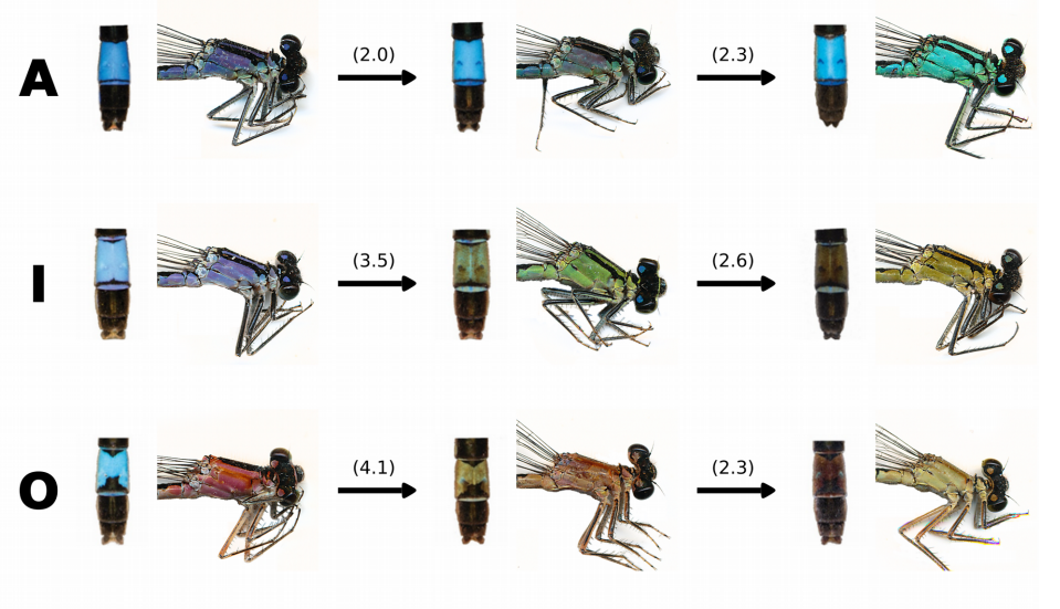
Common Bluetail damselflies from Willink, et al.’s preprint
Ins2 gene bursting activity defines a mature β-cell state
Honey Modi, Søs Skovsø, Cara Ellis, Nicole A.J. Krentz, Yiwei Bernie Zhao, Haoning Cen, N Noursadeghi, Evgeniy Panzhinskiy, Xiaoke Hu, Derek A. Dionne, Shouhong Xuan, Mark O. Huising, Timothy J. Kieffer, Francis C. Lynn, James D. Johnson
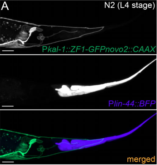
A Cre-amplifier to generate and detect genetic mosaics in vivo
Francesco Trovato, Riccardo Parra, Enrico Pracucci, Silvia Landi, Olga Cozzolino, Gabriele Nardi, Federica Cruciani, Laura Mosti, Andrzej Cwetsch, Laura Cancedda, Laura Gritti, Carlo Sala, Chiara Verpelli, Andrea Maset, Claudia Lodovichi, Gian Michele Ratto
Differential expansion microscopy
Sebastian P. Pernal, Asiri Liyanaarachchi, Domenico L. Gatti, Brent Formosa, Rishika Pulvender, Eric R. Kuhn, Rafael Ramos, Akshata R. Naik, Kathleen George, Suzan Arslanturk, Douglas J. Taatjes, Bhanu P. Jena
School of Life Sciences, University of Warwick, UK
A full-time, twelve-month Senior Research Technician position is available for an immediate start in the laboratory of Dr Kristen Panfilio, for molecular genetics and lab management tasks to support investigations of insect embryology for both biomedical and pest management research. Our lab uses a range of imaging, sequencing, transgenic, comparative genomic, and other approaches to examine the function of essential developmental genes. The primary focus is on cell and tissue structure in protective epithelial tissues, and how the tissues dynamically remodel during development, visualised with live imaging microscopy.
The technician’s principle role will be to conduct molecular biology and related research tasks in support of the PI and other members of the research team, and to do stock maintenance for our research organisms: the flour beetle, Tribolium castaneum, and the milkweed bug, Oncopeltus fasciatus. Tasks will support ongoing BBSRC- and MRC-aligned research on (a) the functional relevance of polyploidy in protective, barrier epithelial tissues, (b) tissue structure and integrity in relation to birth defects, and (c) testing the role of novel genes in lineages of pest insect species.
The candidate needs to have prior practical experience in standard molecular biology and developmental genetics techniques, and experience with insect cultures is desired. However, full training will be given in new techniques. The successful candidate is expected to be an active participant in a growing research group, so a record of successful time and project management is essential.
To view further particulars of the position and to apply, visit the University of Warwick Human Resources posting for this position. The closing date is 19th August.
Please submit your application including a CV and covering letter stating why you are interested in the topic and what you would bring to the project through the on-line application system. For informal enquires or further information about the project, please contact Kristen Panfilio (K.Panfilio@warwick.ac.uk).
In the interests of promoting transparency around the editorial process, Development will now be publishing a ‘Peer review history’ file alongside published papers, where the author has opted-in to such a file being published. All research papers submitted on or after 1 August 2019 are eligible. The file can be found on the ‘Info & metrics’ tab on the article page, and will include decision letters, referee reports and author point-by-point responses, along with a timeline of the submission and revision process, and the name of the handling editor. Reviewer reports will be published under a CC-BY license.
Authors will be able to opt out of having their peer review history files published, but we hope that most authors will be happy to include these as part of the published record. Referees, by accepting to review a paper for Development, will be indicating their willingness to have their comments published, although we will maintain anonymity unless a referee chooses to reveal their identity. Confidential comments to the editor will remain confidential (although we encourage referees to use these only under exceptional circumstances and would prefer all information to be included in the report to the authors), as will comments made through the cross-referee commenting process.
We hope that these files will provide additional insights into the published paper, by way of commentary from experts in the field, as well as opening up the process of peer review to interested readers.
Please note that Development reserves the right not to publish a ‘peer review history’ file relating to a paper in special cases, for example, if the tone or language used is inappropriate or defamatory or due to an ethical consideration.
For any queries or feedback about this policy, please contact the editorial office.
Development partners with Publons for peer reviewer recognition
Development is also pleased to announce a new partnership with Publons, part of the Web of Science Group (a Clarivate Analytics company). Publons gives reviewers formal recognition of their peer review contributions using the Reviewer Recognition Service.
Our partnership with Publons allows reviewers to easily track and verify every review by choosing to add the review to their Publons profile when completing the review submission form. Publons makes it simple for reviewers to showcase their peer review contributions in a format that can be included in job and funding applications (without breaking reviewer anonymity). Reviews completed previously can also be added by forwarding review receipts (i.e. ‘Review received’ emails) to reviews@publons.com.
We are genuinely grateful for the time and expertise volunteered by our reviewers each year. We regularly acknowledge their contributions and support by publishing a list of reviewers who have provided reports in the previous year.
The Kidney Regenerative Medicine Laboratory at the Rogosin Institute is searching for a talented and highly motivated individual interested in understanding how we can apply our understanding of kidney developmental biology to generating new tissue from stem- and primary cells. The goal of the lab is to generate kidney tissue that faithfully reproduces adult kidney functions when engrafted into experimental animals. Projects are flexible, and will take advantage of the applicant’s expertise and interests. Skills in stem cell culture, organ culture, physiological measurement, microscopy, and rodent surgery would be great assets. Strong cell culture skills are a requirement. The Rogosin Institute Kidney Regenerative Medicine Laboratory headed by Dr. Leif Oxburgh is located on the Upper East Side of Manhattan in the large biomedical campus consisting of New York-Presbyterian Hospital, Memorial Sloan-Kettering Cancer Center, New York Blood Center, Weill-Cornell Medical College and Rockefeller University. Facilities are outstanding and the local academic environment will provide top-notch training and networking opportunities. The lab works very collaboratively within the field of kidney development and regenerative medicine, both locally and nationally/internationally. The Rogosin Institute offers competitive employment packages adjusted for the cost of living in New York.
Please fill in our short listener survey, and you’ll be entered into a prize draw to win a signed copy of Kat Arney’s book, Herding Hemingway’s Cats.
The falling cost and rising speed of DNA technology has meant that genetic tests like those offered by 23andMe are now cheap enough to be advertised as fun family Christmas presents. But these tests only look at snapshots across the genome, rather than the whole thing.
The next frontier is direct to consumer whole genome sequencing, and the lower cost now makes it feasible for people with an interest in digging deeper into their genome and a bit of cash to spend. But is it a good idea?
If you enjoy the show, please do rate and review and spread the word. And you can always send feedback and suggestions for future episodes and guests to podcast@geneticsunzipped.com
The collaboration will study evolution of Wnt signalling and alternative transcript expression, splicing and function of TCF/LEF genes. This will be a close collaboration between the research group of David Ferrier at the Scottish Oceans Institute in St. Andrews and Hoppler group at the Institute of Medical Sciences in Aberdeen and will also involve Seb Shimeld and Nanopore in Oxford and Nori Satoh in Okinawa, Japan.
POSTDOCTORAL POSITION is available to study different aspects of the lymphatic vasculature in health and disease. Some of the projects include the characterization of the role of lymphatics in mammalian organogenesis in health and disease. Highly motivated individuals and have a strong background in vascular and molecular tools and are familiar with the use of mouse models are encouraged to apply. Interested individuals should send their curriculum vitae, a brief description of their research interests, and the names of three references to:
Guillermo Oliver, Ph.D
Thomas D Spies Professor of Lymphatic Metabolism
Director Center for Vascular and Developmental Biology
Northwestern University Feinberg School of Medicine
 (No Ratings Yet)
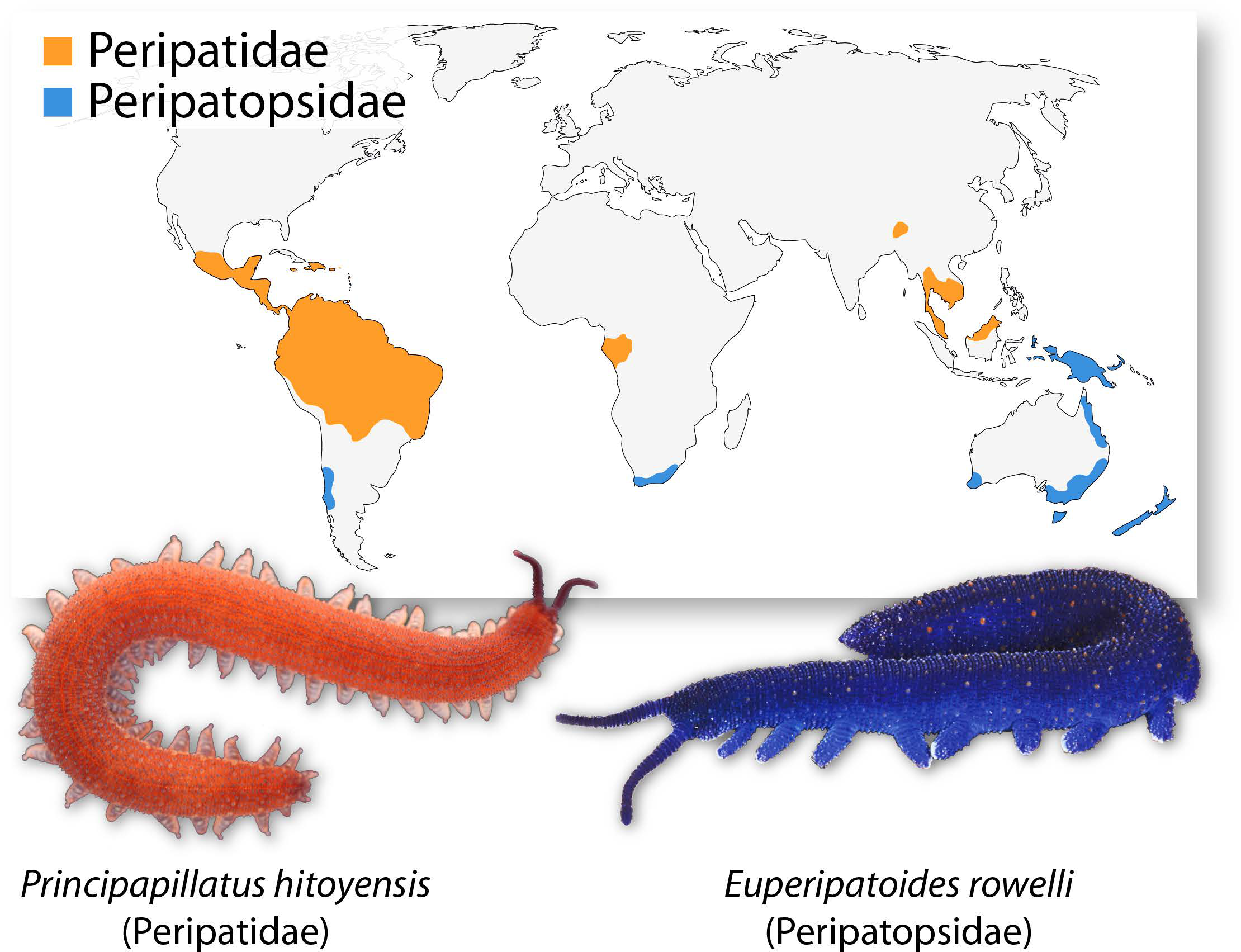
 (9 votes)


 (1 votes)