Join us in Athens 3-4 April 2025 for an unforgettable symposium, as global leaders in stem cell research come together to explore groundbreaking advancements in neural stem cells. From development to aging, disease, and repair, this event will dive deep into the complexities of stem cell plasticity, epigenetics, metabolism, and more. With a focus on neuron-glia interactions and brain disease modeling, this symposium offers a unique opportunity to connect, learn, and push the boundaries of science. Don’t miss out on this exciting event that promises to shape the future of neural research!
Visit our webpage to learn more and register today!
In this SciArt profile, we meet Harsh Kapoor, who has a background in genetics and molecular biology, but decided to switch gears from doing a PhD to starting his own visual science communication company.
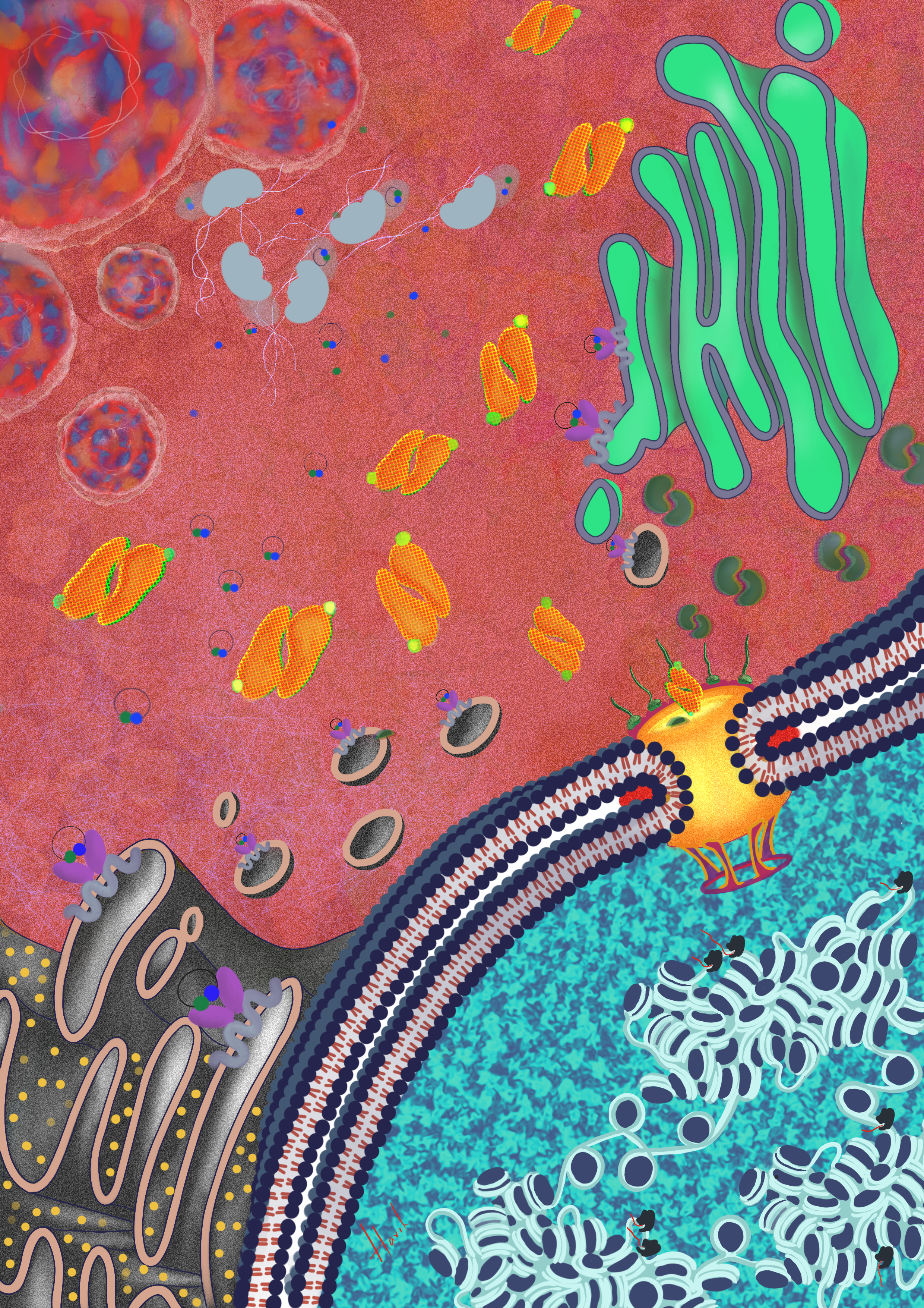
Cell – the molecular fingerprint of life A scientist is an investigator. Unlike crime scene investigators who examine the crime scene for fingerprints, a scientist seeks evidence inside a cell. Thus, I imagined a cell inside a fingerprint.
Can you tell us about your background and what you work on now?
I was born in a small town in India and first in my family to pursue science as a field of study and career. I did my bachelors in Microbiology from Madras Christian College, Chennai and my masters in Biotechnology from University of Hyderabad. I trained as a geneticist and molecular biologist during my years in PhD, but last year, I decided to leave the program to pursue my passion for science design and communication.
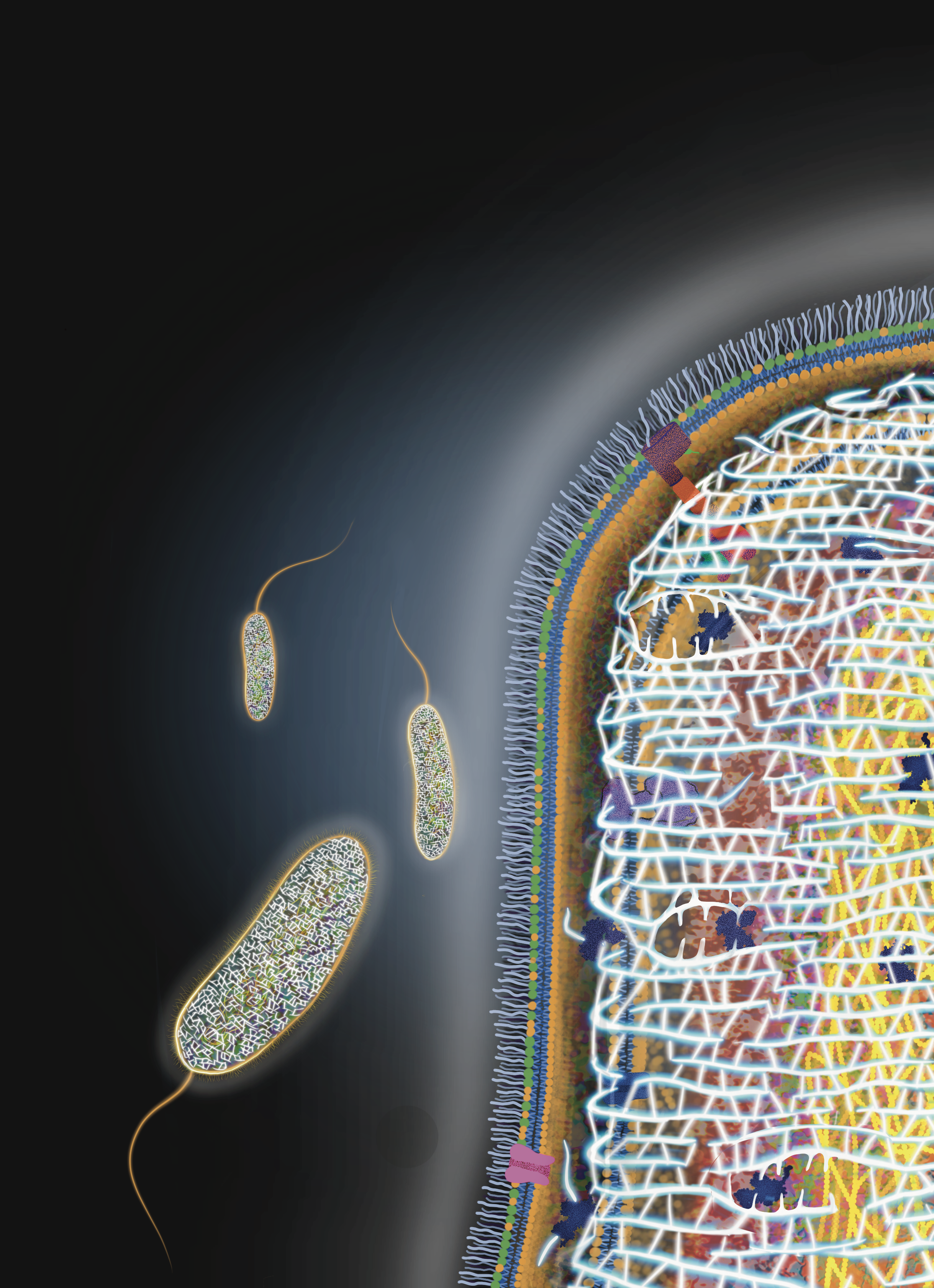
Peptidoglycan – the dynamic armour of the bacteria Cover art for Trends in Microbiology Journal for the month of May 2024 for Dr. Manjula Reddy lab, CCMB
Were you always going to be a scientist?
Haha, or so I thought, but life had other plans. During my PhD, I discovered that while I loved reading, discussing, and visualising science, the hands-on research itself wasn’t where my true passion lay. Instead of feeling fulfilled, I often found myself more stressed than excited. Accepting this and choosing a different path wasn’t easy, but in hindsight, I’m grateful I did.
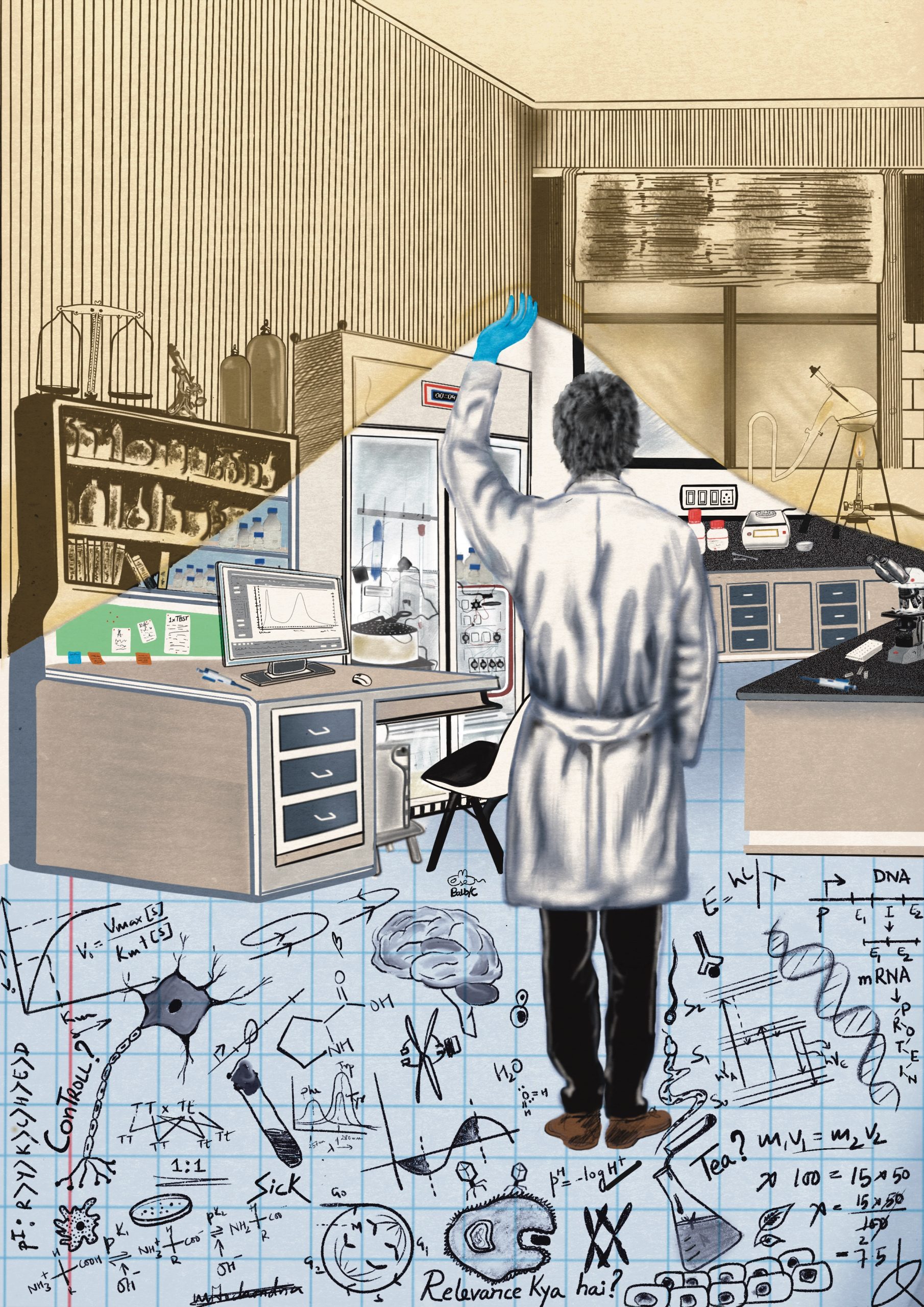
From past to progress Advancement in modern biological science
And what about art – have you always enjoyed it?
I have always loved art. While growing up I was the kind of kid who daydreamed in vivid colors, turned found objects into art, and saw potential in everything from leftover materials to doodles in the margins. In college, I have painted huge murals, designed T-shirts and logos, and participated in creative projects. Those days were filled with endless exploration, late night brainstorming sessions, and the thrill of bringing ideas to life.
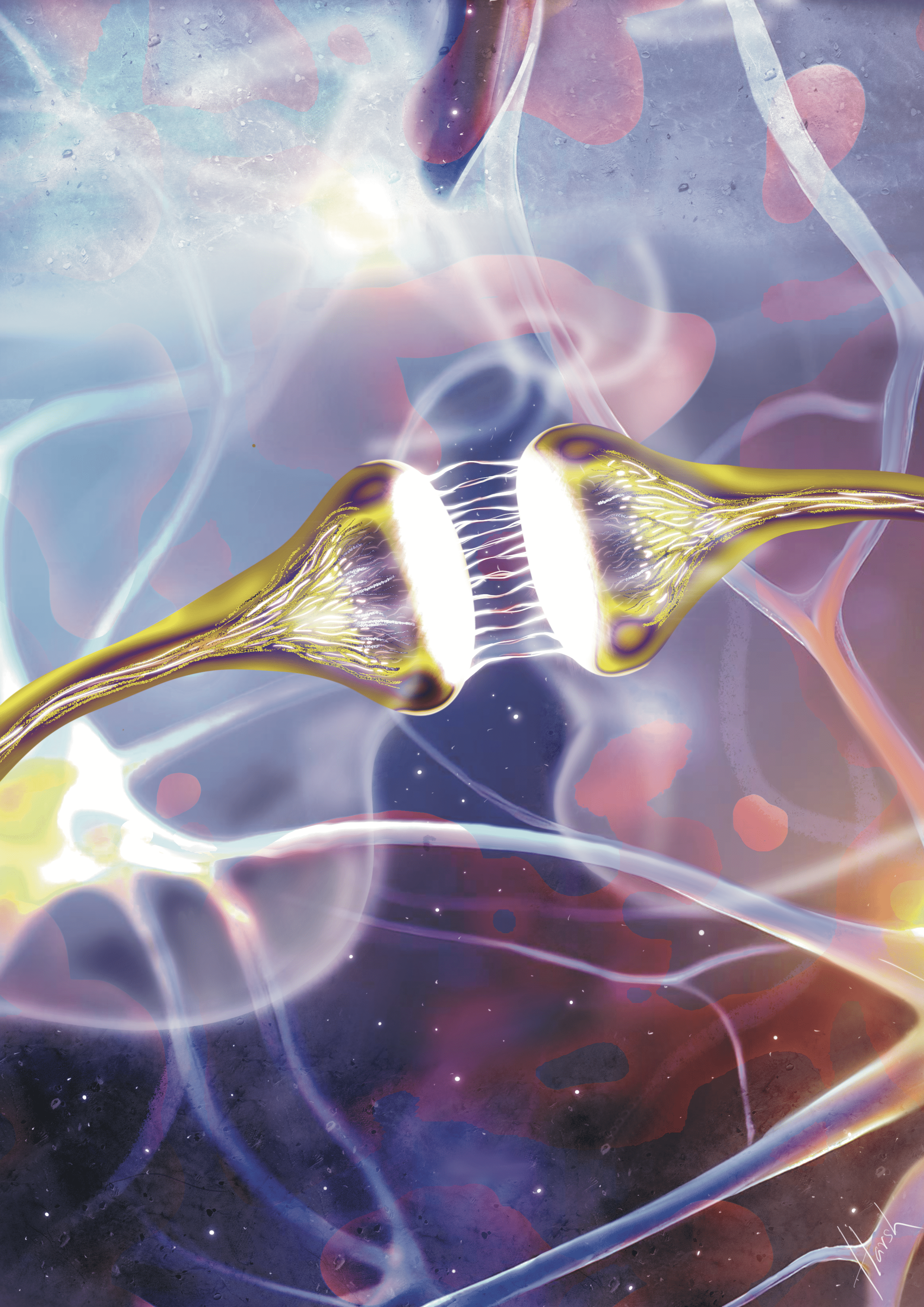
Dream a cell How do you dream a cell? Coloured or black and white?
What or who are your most important artistic influences?
I can’t think of a specific name, but my mother has been a major artistic influence throughout my life. She is incredibly creative and always encouraged me to explore and experiment with different artistic activities. Time during my bachelors degree was very inspiring for the creative in me. And Instagram, in particular, has been a great platform for inspiration lately.
Self A gentle reminder to love oneself first. and how much we need to communicate with our bodies and minds. listen to those whispers.
How do you make your art?
I have had some fair share of experience with acrylics in the past, but in recent years, all my artworks are digital. I like to start with reading about the concept I will be working on. I like to get inspired from scientific data and microscopy images. I initially started with Procreate on my iPad, and what an incredible experience that was! Eventually, I transitioned to more vector-based software like Adobe Illustrator. That said, I still rely on Procreate for rough sketches, storyboarding, and quick, fun artworks — it’s my go-to for spontaneous creativity.
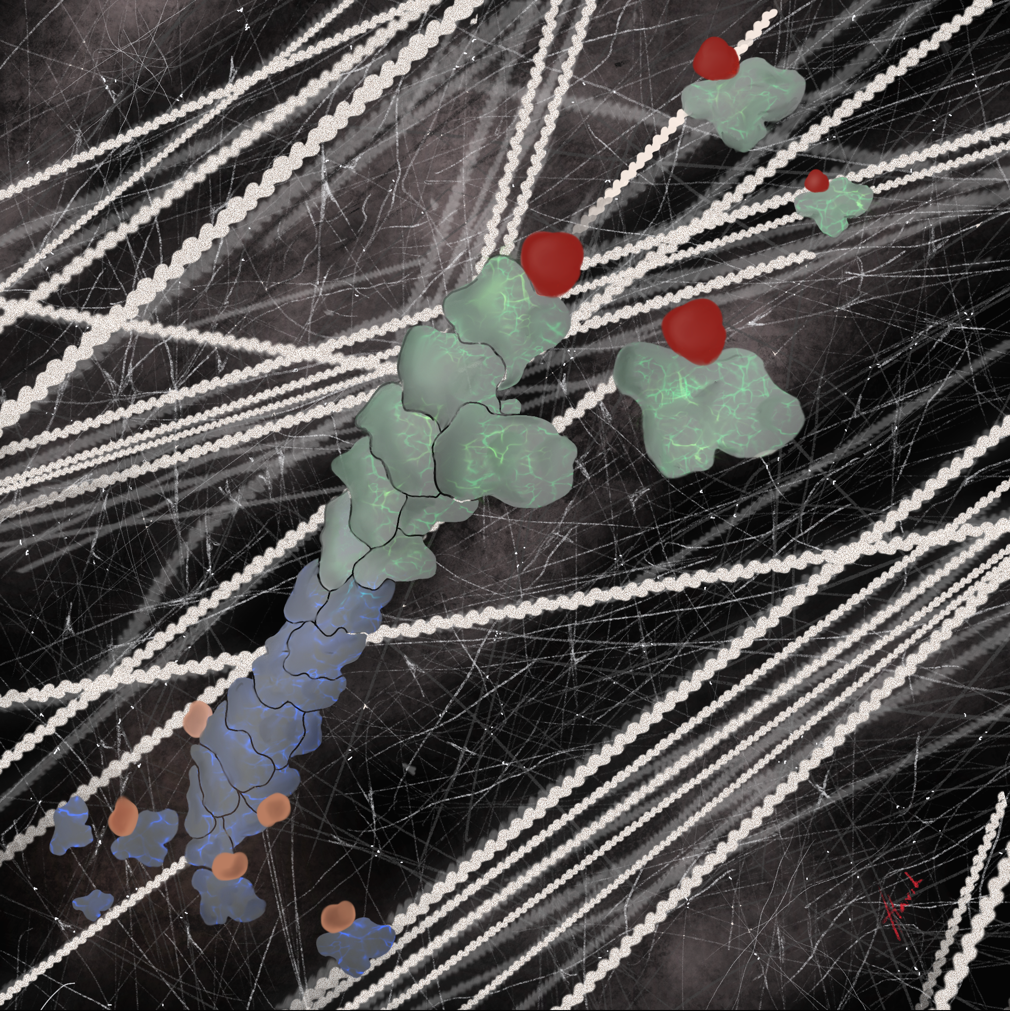
Actin polymerization Actin polymerization shown in 3 scales.
Does your background in science influence your art?
Absolutely! Science is my playground to explore the weird and wonderful. If it wasn’t for science I don’t know if I would have gotten back to doing art. And I don’t see why I should restrict myself to just digital illustration. Science can be communicated in so many different visual art formats.
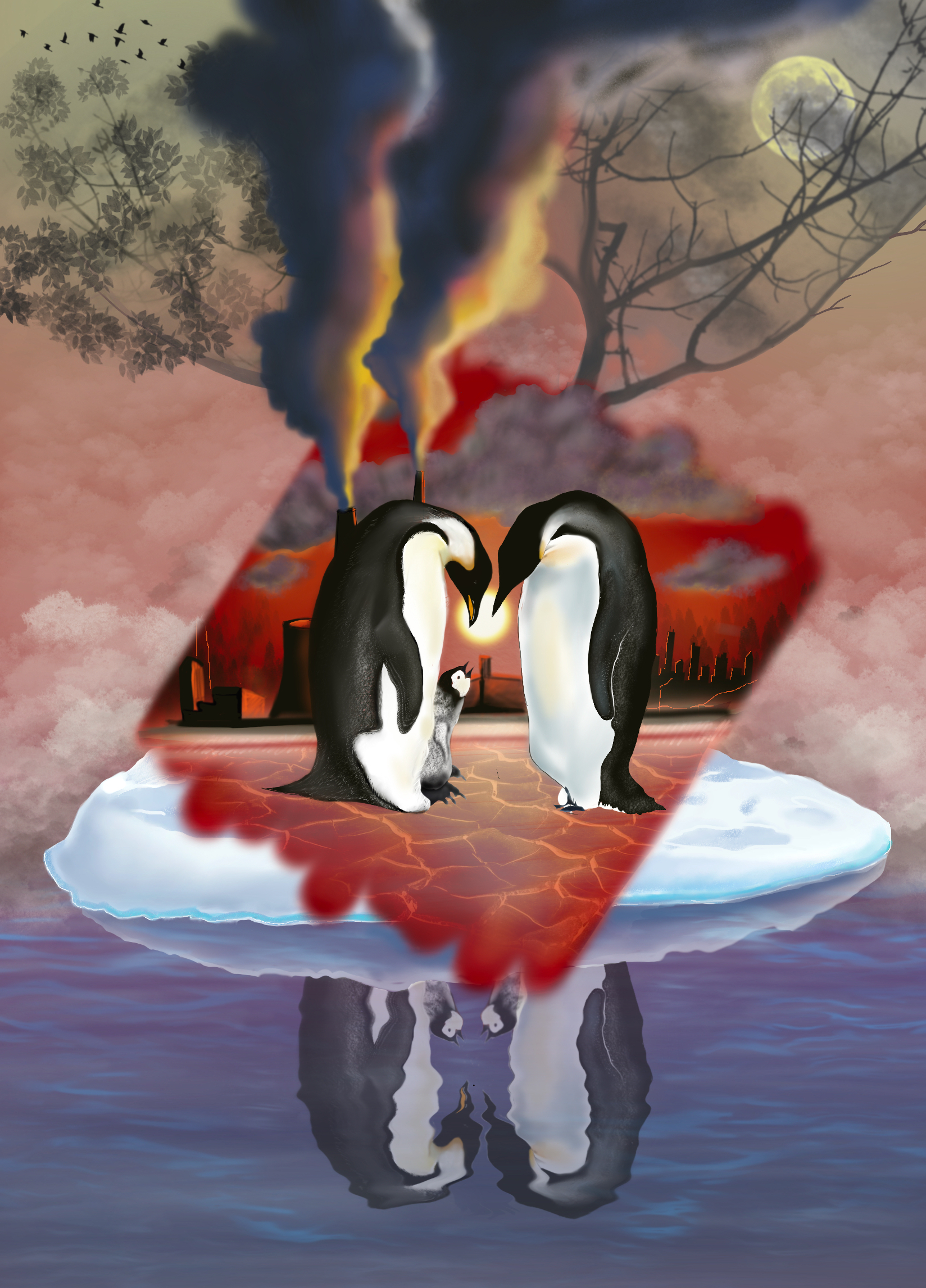
Vanishing Worlds Earth is experiencing extreme weather conditions reminding us of the escalating climate crisis. These shifts not only disrupt the lives of countless people but also disrupt ecosystems, having a vanishing effect and leading to biodiversity loss.
What are you thinking of working on next?
Since now this is what I do full time. My visual science communication agency, NERD, helps scientists, research institutes and biotech and healthcare companies communicate their science through various visual art formats and content creation. I have some interesting projects for this year and I am actively seeking for more exciting work. I’m currently working on a series, exploring the fascinating world of biomimicry through a unique digital content style, where we are highlighting 10 nature-inspired breakthroughs in science, tech, and sustainable design. This can be found on our Instagram and LinkedIn page.
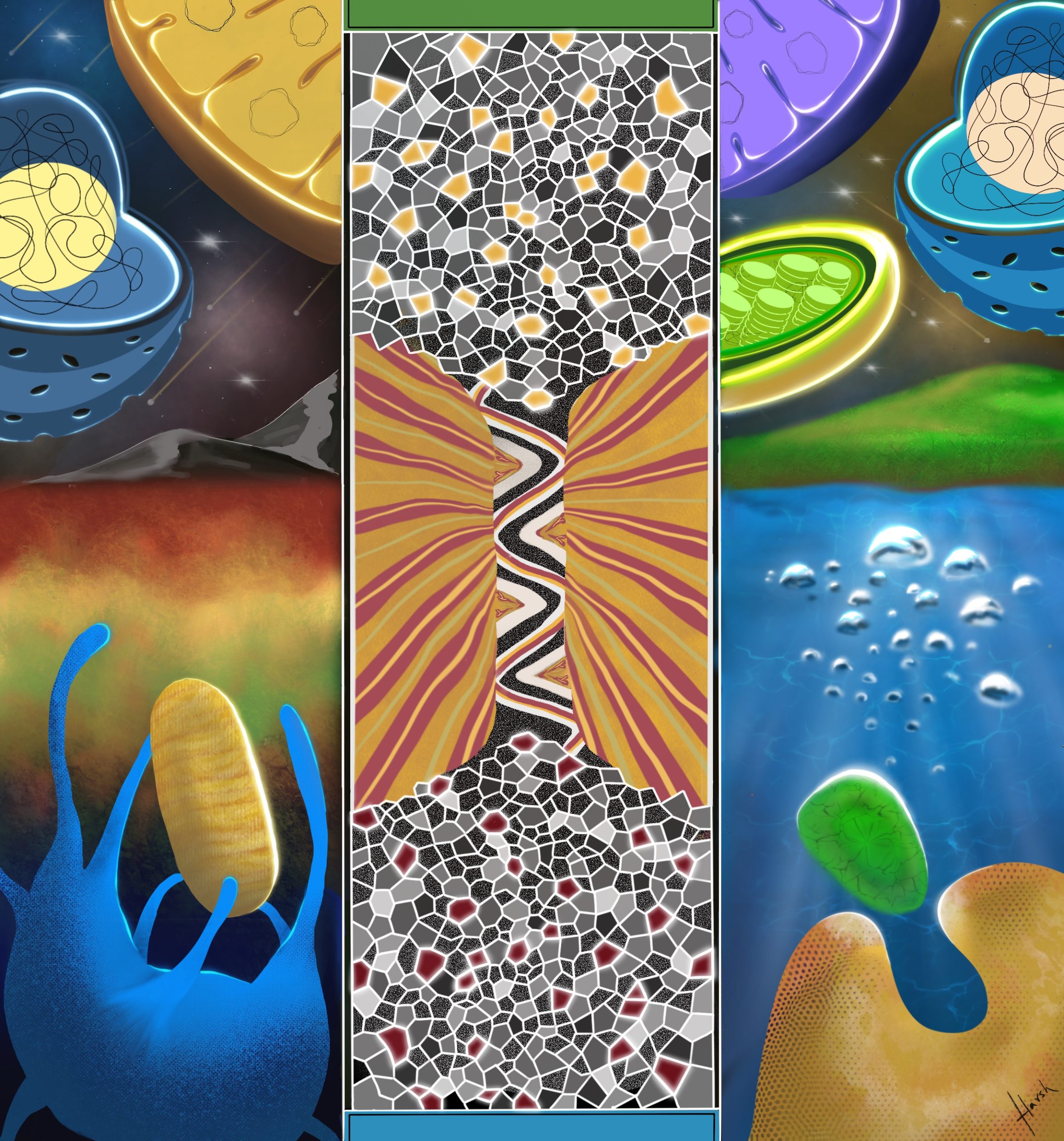
It takes two The journey of evolutionary fusion. It only takes two. Two organisms to fuse, two major organelles to form, and twice in the face of evolution for multicellular advanced life to emerge. It must have been so celestial.
Find out more about Harsh:
Website – for his visual science communication agency
Humans and other tetrapods evolved from aquatic fish. In making this leap, tetrapods evolved lungs to breathe air and lost respiratory gills. It is tempting to intuit that lungs evolved from gills. However, lungs and gills form in separate parts of the body, so they are unlikely to be evolutionarily related. Indeed, some living fish have both gills and lungs [1]. So, what became of fish gills? In work spanning the last 6 years and published in Nature, we show that gills may in fact have contributed to the origin of a functionally unrelated structure in humans – our outer ears.
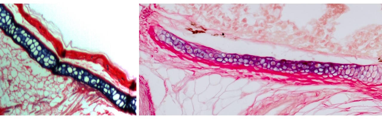
The outer ear, comprising the ear canal and flap-like pinna, is a unique feature of mammals with no known evolutionary precedent in reptiles or amphibians. At first, our research centered on the elastic cartilage that supports it, previously described only in mammals [2-5]. Elastic cartilage has cellular and mechanical properties distinct from the more widespread hyaline cartilage in the adult nose, joints, and embryonic skeleton. But we knew almost nothing about molecular drivers of these differences. In previous work in the Crump lab I had helped describe a zebrafish gill cartilage with gene expression distinct from hyaline cartilage. Ensuing discussions with Max Plikus at UC Irvine and a wonderful collaboration with Andrew Gillis at the MBL (in which he single-handedly sectioned and stained gills from multiple fish species!), helped established that cartilage in the gills of bony fishes, including zebrafish, is elastic in nature.
I next asked whether the elastic cartilage in fish gills and the mammalian outer ear might represent a homologous cell type. With help from the Evseenko lab at USC and cross-country shipments from collaborators in the Chen lab at Mount Sinai (once during a hurricane!), I acquired single-cell gene expression and open chromatin profiles from elastic cartilage of the human fetal outer ear and epiglottis, as well as hyaline cartilage from the human nose as a control and compared these with our zebrafish data. These analyses confirmed significant gene expression similarities between fish and human elastic cartilage.
Elastin staining on sections from a mouse outer ear (left) and Atlantic salmon gills (right)
At this point, two major conceptual ideas significantly expanded the focus of this work.
First, the activity of non-coding genomic elements called enhancers tends to be more tissue-specific than expression of associated genes, thus serving as better proxies for regulatory conservation between cell types. The problem is that unlike genes, only a tiny proportion of human enhancers have sequence-conserved counterparts in the zebrafish genome. However, previous work had shown that regulatory information encoded in enhancers can be recognized by similar sets of factors across species [6, 7]. This gave me the idea to sidestep the issue of DNA sequence conservation: if elastic cartilage is specified by a conserved regulatory program, then human elastic cartilage enhancers encoding this program might still be recognized specifically in fish gills.
I identified human genomic regions representing putative elastic cartilage-specific enhancers and, despite absence of similar fish sequences, tested their activity in zebrafish. It was the most incredible moment of my PhD when I looked through the microscope to see six of ten human outer ear elastic cartilage enhancers driving fluorescent activity specifically in the gills, reflecting shared biology across 400 million years of evolution. Equally amazing, a zebrafish gill elastic cartilage enhancer drove highly specific activity in the elastic cartilage of the mouse outer ear.
Second, I was influenced by work in other model systems showing that seemingly novel structures can represent re-emergence of an ancestral structure that apparently disappeared during evolution but was in fact retained in a cryptic form in intervening species [8]. Could a broader gill developmental circuitry have been retained in tetrapods and repurposed in mammals to drive outer ear evolution?
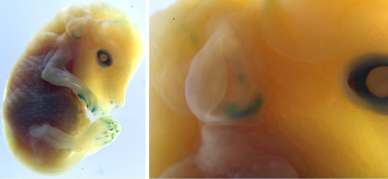
I realized we could use a similar enhancer-based approach, but with a focus on developmental timepoints when gills first grow out. This led to the discovery that one of the few enhancers well-conserved between zebrafish and humans is active in gill developmental populations, but critically not in the later-forming elastic cartilage. It thus represents a piece of the ancient instructions to make fish gills retained in our own DNA. In transgenic mice, the zebrafish version of this enhancer was faintly but consistently active in the developing outer ear. These findings demonstrated that it is not simply the elastic cartilage but also the early developmental outgrowth program that is evolutionarily conserved between fish gill filaments and the mammalian outer ear.
A fish gill enhancer is active in the mouse outer ear.
So, what was this program doing in amphibians and reptiles? In the case of our middle ear bones, fossil data have revealed their progressive evolution from fish jawbones through amphibian and reptile intermediates [9]. By contrast, cartilage does not preserve well, so the current fossil record provides little information on how gills may have transformed into outer ears.
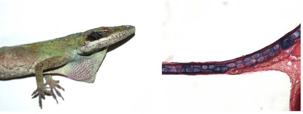
For answers, we again turned to enhancers. We formed a new, eleventh-hour collaboration with Helen Willsey at UCSF to test our enhancers in an amphibian – the frog. Helen and her postdoc Micaela gave me a crash course on staging, imaging, and staining Xenopus tadpoles in the final weeks of my PhD, patiently repeating injections to replace samples that I lost. The results were worth it: gill/outer ear outgrowth enhancers were active in developing tadpole gills, suggesting continuity in the developmental program from fish to tetrapods. Further, mature tadpole gills contained a little-characterized cartilage that activated both human and zebrafish elastic cartilage enhancers! Although transgenic testing was not feasible in reptiles, I collaborated with Tom Lozito at USC to examine histology of the ear in green anole lizards and found that the evolutionarily mysterious extracolumella is made of a permanent, elastic cartilage. These pieces of evidence helped put together a model through which fish gill developmental and cell type programs could have been repeatedly redeployed through the evolution of tetrapods.
Adult green anoles (left) have elastic cartilage in their outer ears (right)
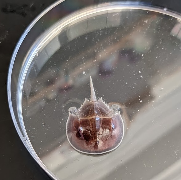
Finally, older reports had suggested that the horseshoe crab, an ancient invertebrate, makes a cellular type of cartilage-like tissue in its gills [10-13]. An exciting opportunity arose when I attended the wonderful Embryology Course held at the Marine Biological Laboratory (MBL), which happens to be in Atlantic horseshoe crab territory. From chats I had there with invertebrate evo-devo aficionado Heather Bruce (now at the University of British Columbia), I got in touch with the Marine Resource Center, acquired a horseshoe crab specimen, and performed single-nuclei multiomic analysis on its unique “book gills”. Amazingly, a horseshoe crab gill enhancer drove transgenic activity in the zebrafish gills! Although we can do only limited experimental work in horseshoe crabs, our data suggest that gills and their elastic cartilage support may have an ancient origin in early bilateria.
A juvenile Atlantic horseshoe crab
Traditionally, fish gills were thought to have played little role in the evolution of tetrapods. Now, our work reveals an unprecedent legacy for this complex ancestral structure. Our research supports the idea of “deep” or “cryptic” homology: the morphological disappearance of a structure does not imply the disappearance of the developmental field from which it emerged or the regulatory program that instructed its formation. These developmental fields and regulatory programs can instead be reused to make something new, contributing to the widespread innovation we observe in nature.
1. Cupello, C., et al., Allometric growth in the extant coelacanth lung during ontogenetic development. Nature communications, 2015. 6(1): p. 8222.
2. Sanzone, C.F. and E.J. Reith, The development of the elastic cartilage of the mouse pinna. American Journal of Anatomy, 1976. 146(1): p. 31-71.
3. Bradamante, Z., B. Levak-Svajger, and A. Svajger, Differentiation of the secondary elastic cartilage in the external ear of the rat. The International journal of developmental biology, 1991. 35(3): p. 311-320.
4. Cox, R. and M. Peacock, The fine structure of developing elastic cartilage. Journal of anatomy, 1977. 123(Pt 2): p. 283.
5. Kostović-Knežević, L., Ž. Bradamante, and A. Švajger, Ultrastructure of elastic cartilage in the rat external ear. Cell and Tissue Research, 1981. 218: p. 149-160.
6. Wong, E.S., et al., Deep conservation of the enhancer regulatory code in animals. Science, 2020. 370(6517): p. eaax8137.
7. Minnoye, L., et al., Cross-species analysis of enhancer logic using deep learning. Genome research, 2020. 30(12): p. 1815-1834.
8. Bruce, H.S. and N.H. Patel, The Daphnia carapace and other novel structures evolved via the cryptic persistence of serial homologs. Current Biology, 2022. 32(17): p. 3792-3799. e3.
9. Gould, S.J., An earful of jaw. Natural History, 1990. 99(3): p. 12-18.
10. Person, P. and D.E. Philpott, The biology of cartilage. I. Invertebrate cartilages: Limulus gill cartilage. Journal of Morphology, 1969. 128(1): p. 67-93.
11. Person, P. and D.E. Philpott, Invertebrate cartilages. Annals of the New York Academy of Sciences, 1963. 109(1): p. 113-126.
12. Cole, A.G. and B.K. Hall, The nature and significance of invertebrate cartilages revisited: distribution and histology of cartilage and cartilage-like tissues within the Metazoa. Zoology, 2004. 107(4): p. 261-273.
13. Tarazona, O.A., et al., The genetic program for cartilage development has deep homology within Bilateria. Nature, 2016. 533(7601): p. 86-89.
Applications to the new Master in Integrative Biological Sciences (iBioS) are opened
Dear student,
You are currently finishing, or already have, a Bachelor’s degree in Biological Sciences? You are highly motivated, and you are looking for an innovative, immersive and truly international master program? Then, iBioS is what you are searching for!
This immersive research-driven training program will train high-level international students in all areas of research developed in our ITI IMCBio+ with a strong emphasis on hands-on experience as well as innovation via internships and tutored projects. Training will be fully in English.
In a nutshell, iBioS (integrative Biological Sciences) is an advanced training to research through research that will offer you:
– A training fully in English
– A unique mentoring program in which each student will benefit from privileged interactions with 2 renown scientists that will provide personalized guidance and support all along the two years of training
– A personalized training. Each student will select a set of courses offered by the faculty of life sciences and that he/she thinks matches best his/her expectations and needs. Here again, the student will benefit from the guidance of his/her mentors right from the beginning of the program.
– 10 months of internship in labs belonging to 5 worldwide renown research institutes in which you will access the latest technologies and advanced facilities.
– A unique immersive tutored project spanning the three first semesters of the master. This group work will bring you to design a scientific project through literature survey. Your creativity will also be strongly stimulated since you will have to design and budget a set of experiments that the IMCBio graduate school will finance to enable you to put them in practice under the guidance of experts. Finally, you will be trained to exploit the produced data by writing a scientific publication and presenting your achievements in a conference.
– A training in intellectual property provided through dedicated courses, but also a workshop supervised by our technological transfer office and meetings with entrepreneurs and investors.
– A set of courses in biocomputing and ethics and other activities customized for this program.
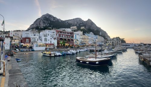
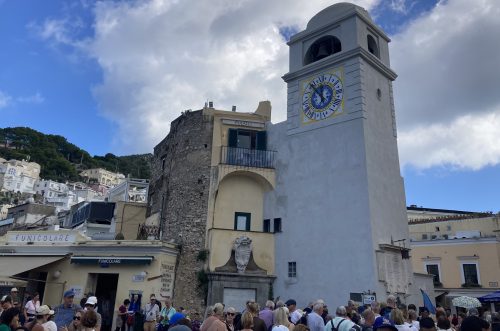
The island of Capri, lying south of Naples and West of Sorrento, is an Italian vacation destination known for its exquisite views and extravagant luxury shopping. So, it may come as a surprise to think that this could also be an ideal location where science-based education on human brain development might occur. Well, for one week in October 2024, this was indeed the case as the European Molecular Biology Organization (EMBO) hosted a workshop titled “Unlocking human brain complexity using 3D culture and single cell omics.”
Porto Turistico di Capri
EMBO workshops are typically smaller conferences, with an attendance size of about 100 or so registrants, to provide a more intimate atmosphere that promotes close interaction among scientists. The exotic setting of the host site also offers a more relaxed environment to foster networking and encourage collaborative discussions between research groups.
This EMBO workshop in Capri did not disappoint, and it served as a “Who’s Who” in the cerebral organoid research field, touting a roster of distinguished speakers highlighted by Sergiu Pasca, Madeline Lancaster, Jurgen Knoblich, Barbara Treutlein, and Paola Arlotta — a Mount Rushmore worthy collection of scientists in the in vitro stem cell brain development field.
Antonio Simeone, acting director of the Institute of Genetics and Biophysics (IGB-CNR), provides the welcome introduction.
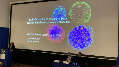
The workshop started off with a bang, led by a plenary lecture from Paola Arlotta, Ph.D., Professor and Chair of Harvard University’s Department of Stem Cell & Regenerative Biology (HSCRB), who detailed her lab’s work on the use of multi-donor stem cell villages to generate chimeric organoids to investigate population-wide differences in brain development and susceptibility to disease-causing environmental agents https://www.nature.com/articles/s41586-024-07578-8.
Dr. Paola Arlotta opens the conference with a plenary lecture on chimeric organoids
Later, we heard from Barbara Treutlein, Ph.D., Principal Investigator at ETH Zurich, on her lab’s use of multi-omics platforms and computational analysis pipeline to study human brain development. Recently, her team and others in the field have collaborated to compile transcriptomic datasets from neural organoid protocols while cross-referencing against existing brain atlases to generate an integrated organoid cell atlas https://www.nature.com/articles/s41586-024-08172-8.
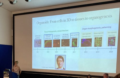
On the topic of neural organoids, we heard from Jurgen Knoblich, Ph.D., Professor of Synthetic Biology at the Medical University of Vienna and Scientific Director at IMBA, who presented a set of new protocols to generate diverse neural subtypes https://www.biorxiv.org/content/10.1101/2024.11.15.623576v1. Meanwhile, Madeline Lancaster, Ph.D., Professor at MRC Laboratory of Molecular Biology (UK), discussed the challenges with organoid reproducibility across biologically diverse human induced pluripotent stem cell lines (hiPSCs).
Dr. Madeline Lancaster provides a historical perspective of cerebral cortical organoid development
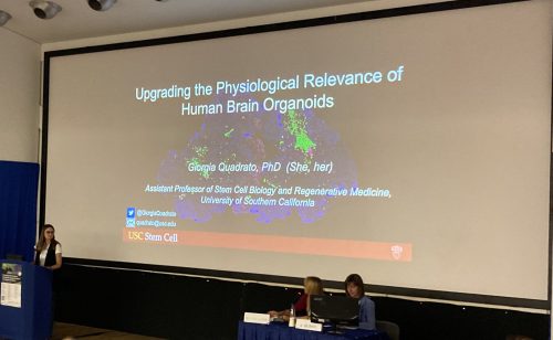
Dr. Giorgia Quadrato, Assistant Professor at USC Keck School of Medicine, details her lab’s work on autism and generating novel cerebellar organoid models
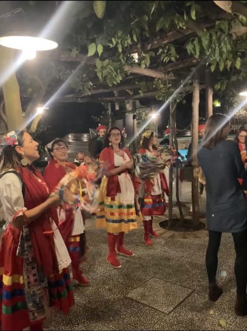
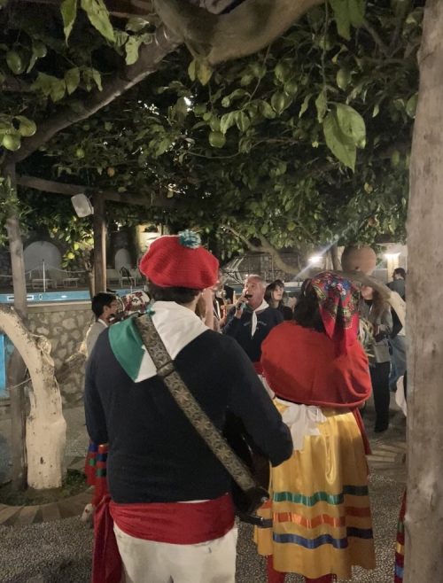
While you might think that all this fascinating science was the highlight of the conference, there were also several amazing extracurricular events such as free-periods to explore the island, evening dinners with the expert speakers, and a conference-ending gala featuring folk dancers, singers, and musicians playing traditional Neapolitan music.
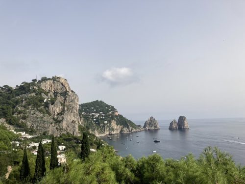
Spiaggia di Marina PiccolaNeapolitan Folk DancersNeapolitan Folk Singers and Musicians
So, the next time you find yourself vacationing on an island, either in Capri or elsewhere, imagine that a group of scientists could be congregating nearby to discuss the latest scientific discoveries on the development of our human brains.
As well as looking for the next Executive Editor for Development, we are also recruiting a Reviews Editor on the journal. If you love developmental biology but don’t want to be ‘doing’ it any more, this is a great opportunity to stay connected to the field and the community and to help shape the content of the journal. You’ll commission and edit review-type articles and perspectives, conduct interviews, write research highlights, travel to conferences and contribute to the strategic development of the journal. As a small publishing company, you’ll get great exposure to all aspects of the business – both the publishing and the charitable side – and you’ll also get to work for an organisation that really believes in supporting the academic community. All the details can be found in the job advert on our website.
You don’t need to have prior editorial experience (though obviously we’d be delighted to receive applications from people who do); what you do need are research experience and a broad interest in the field, excellent communication skills, and a willingness to learn! If this sounds like something you might be interested in, please feel free to get in touch with informal questions, or just apply directly (instructions in the job ad). The deadline for applications is 31 March 2025.
Spotted a preprint in this list that you love? If you’re keen to gain some science writing experience and be part of a friendly, diverse and international community, consider joining preLights and writing a preprint highlight article.
Michael G. Michalopulos, Yan Liu, Dinesh Ravindra Raju, John T. Lafin, Yanru Ma, Dhruv Gaur, Sadiksha Khadka, Chao Xing, Andrew P. McMahon, Thomas J. Carroll, Keri A. Drake
Arttu Junnila, Kalle T. Rytkönen, Guillermo Martinez-Nieto, Mats Perk, Hao Li, Jenni Airaksinen, Ida Hyötyläinen, Oliver Mehtovuori, Asta Laiho, Claes Ohlsson, Laura L. Elo, Satu Kuure, Matti Poutanen, Petra Sipilä
Péter Görög, Tibor Novák, Tamás F. Polgár, Péter Bíró, Adél Gutheil, Csaba Kozma, Tamás Gajdos, Krisztina Tóth, Alexandra Tóth, Miklós Erdélyi, József Mihály, Szilárd Szikora
Ismael Hernández-Núñez, Alaina Urman, Xiaodong Zhang, William Jacobs, Christy Hoffman, Sohini Rebba, Ellen G Harding, Qiang Li, Fengbiao Mao, Andi K Cani, Shiming Chen, Meelad M Dawlaty, Rajesh C Rao, Philip A Ruzycki, John R Edwards, Brian S Clark
Ridvan Cetin, Giulia Picco, Jente van Staalduinen, Eric Bindels, Remco Hoogenboezem, Gregory van Beek, Mathijs A Sanders, Yaren Fidan, Ahmet Korkmaz, Joost Gribnau, Jeffrey van Haren, Danny Huylebroeck, Eskeatnaf Mulugeta, Frank Grosveld
Esther Rodríguez-Correa, Florian Grünschläger, Tamar Nizharadze, Natasha Anstee, Jude Al-Sabah, Vojtech Kumpost, Anastasia Sedlmeier, Congxin Li, Melanie Ball, Foteini Fotopoulou, Jeyan Jayarajan, Ian Ghezzi, Julia Knoch, Megan Druce, Theo Aurich, Marleen Büchler-Schäff, Susanne Lux, Pablo Hernández-Malmierca, Julius Gräsel, Dominik Vonficht, Anna Mathioudaki, Judith Zaugg, Ralf Mikut, Andreas Trumpp, Thomas Höfer, Daniel Hübschmann, Simon Haas, Michael D. Milsom
Nakesha Agyapong, Leslie Dominguez-Ortega, Brian Macdonough, Patrick Mulluso, Sagar Patel, Briti Prajapati, Brian Saville, Andrew Shapiro, Ethan Trim, Kara Battaglia, Jocelyn Herrera, Gianna Garifo-MacPartland, Dianne Newcombe, Latoya Okundaye, Heather Paglia, Julia Paxson
Fatemeh Mazloumi Gavgani, Johanna E.M. Kraus, Joshua November, Layla Al-Shaer, Anna Cosima Seybold, Benjamin Lerstad, Harald Hausen, Michael J. Layden, Fabian Rentzsch
Jennifer T. Stocksdale, Matthew J. Leventhal, Stephanie Lam, Yu-Xin Xu, Yang Oliver Wang, Keona Q. Wang, Reuben Tomas, Zohreh Faghihmonzavi, Yogi Raghav, Charlene Smith, Jie Wu, Ricardo Miramontes, Kanchan Sarda, Heather Johnson, Min-Gyoung Shin, Terry Huang, Mikelle Foster, Mariya Barch, Naufa Armani, Chris Paiz, Lindsay Easter, Erse Duderstadt, Vineet Vaibhav, Niveda Sundararaman, Dan P. Felsenfeld, Thomas F. Vogt, Jennifer Van Eyk, Steve Finkbeiner, Julia A. Kaye, Ernest Fraenkel, Leslie M. Thompson
Tsuyoshi Fukushima, Trine Ahn Kristiansen, Lai Ping Wong, Samuel Keyes, Yosuke Tanaka, Michael Mazzola, Ting Zhao, Lingli He, Masaki Yagi, Konrad Hochedlinger, Satoshi Yamazaki, Ruslan I. Sadreyev, David T Scadden
Matthew K. Brachmann, Ana P. B. Costa, Shaun Robertson, Kate Donoghue, Natalie Pilakouta, Mark Whitehead, Xuan Liu, Bjarni Kristjansson, Skuli Skulason, Colin Selman, Kevin Parsons
T Cool, A Rodriguez y Baena, MGE Rommel, C Mattingly, E Bachinsky, S Saini, S Chattopadhyaya, BA Manso, S Rajendiran, AK Worthington, DM Poscablo, A Deguzman, T Berger-Cahn, DF Boyd, EC Forsberg
Alejo A. Morales, Vladimir Camarena, LéShon Peart, Sarah Smithson, Lindsay Shaw, Lucy Webber, Jose M. Negron, Juan E. Sola, Ann-Christina Brady, Katherina Walz, Gaofeng Wang, Mustafa Tekin
Sofia Makieva, Mara D. Saenz-de-Juano, Carmen Almiñana, Stefan Bauersachs, Sandra Bernal-Ulloa, Min Xie, Ana G. Velasco, Natalia Cervantes, Maike Sachs, Susanne E. Ulbrich, Brigitte Leeners
Maruthi K. Pabba, Miroslav Kuba, Tomáš Kraus, Kerem Celikay, Janis Meyer, Sunik Kumar Pradhan, Andreas Maiser, Hartmann Harz, Heinrich Leonhardt, Karl Rohr, Michal Hocek, M. Cristina Cardoso
Malika Nadour, Robert I. Valette Reveno Leatis, Marie Biard, Noémie Frébault, Lise Rivollet, Philippe St-Louis, Cassandra R. Blanchette, Andrea Thackeray, Paola Perrat, Carlo Bevilacqua, Robert Prevedel, Laurent Cappadocia, Georgia Rapti, Maria Doitsidou, Claire Y. Bénard
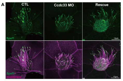
Juyeon Hong, Chanjae Lee, Ophelia Papoulas, Jiehong Pan, Maki Takagishi, Nadia Manzi, Daniel Dickinson, Amjad Horani, Steven Brody, Edward Marcotte, Tae Joo Park, John B Wallingford
Robert N. Rainey, Sam D. Houman, Louise Menendez, Ryan Chang, Litao Tao, Helena Bugacov, Andrew P. McMahon, Radha Kalluri, John S. Oghalai, Andrew K. Groves, Neil Segil
Zebrafish Rock! celebrates its 7th anniversary since its relaunch!
Dr. Sonya Neal receives US Presidential Early Career Award for Scientists and Engineers.
Prof. Jochen Guck awarded The Greve Prize from the German National Academy of Sciences Leopoldina.
Prof. Caren Norden awarded Pfizer Premio Award for Basic Research.
Prof. Iain Couzin awarded the 2024 Fyssen International Prize.
Dr.Itamar Harel promoted to Associate Professor at the The Hebrew University of Jerusalem.
Dr. Patrick Murphy to move to the Department of Molecular Biology and Genetics at Cornell University.
Dr. Philip D. Campbell started as an Assistant Professor at University of Pennsylvania.
Dr. Jason (Chang) Marvin awarded the 2024 MGH Center for Faculty Development (CFD) Outstanding Research Fellow Award.
Dr. Vincenzo Torraca receives the King’s College London Innovative Teaching Award.
Phoebe Reynolds wins the British Neuroscience Association (BNA) award for Public Engagement 2024.
PhDs awarded to: Dr. Caroline Zandecki of Eve Seuntjens Lab KU Leuven. Dr. Aaron Hickey of Emília Santos Lab University of Cambridge. Drs. Tuo Shi. & Chun-Che (Ted) Tseng of Gage Crump Lab at University of Southern California. Drs. Maya Wilde &Leandro Aluisio Scholz of Ethan Scott Lab at University of Melbourne.
Special thanks to Maddie Ryan, Charli Corcoran & Michaela Noskova Fairley for putting this digest together! If you would like to thank the Zebrafish Rock! team for their time & effort, you can buy us a strong cuppa at the link below. Every little bit keeps us caffeinated and motivated! We appreciate your support 🙂
The ISSCR Annual Meeting, taking place in Hong Kong on 11-14 June, is a gathering of the brightest minds in stem cell research and regenerative medicine across geographies and disciplines. Nearly 4,000 scientists from around the globe will convene at ISSCR 2025 to take part in a 4-day program comprising the year’s most significant new advances in the fields of stem cell research and regenerative medicine.
Visit https://www.isscr2025.org/ to learn more about registration, abstract submission, and to view the full scientific program. Do not miss the final opportunity to present your work at ISSCR 2025 – late-breaking abstracts are due 19 March, and there will be no deadline extensions.
The scientific program is organized around 6 tracks:
Clinical Applications
Disease Modeling and Drug Discovery
Global Stakeholder Initiatives
Organ Generation and Regeneration
Pluripotency and Development
Somatic Stem Cells and Cancer
The ISSCR 2025 Annual Meeting is the place to meet potential collaborators and network with stem cell and regenerative medicine researchers from around the world. One-on-One Partnering and ample networking opportunities help attendees make vital connections that drive new discoveries and accelerate progress. More than 250 scientific talks will be presented across the 4-day program, as well as 1,400+ scientific poster presentations.
Meeting Co-Chairs:
Kathryn Cheah, PhD, The University of Hong Kong, Hong Kong
Eugenia Piddini, PhD, University of Bristol, UK
Meeting Organizing Committee:
Alessandro Aiuti, MD, PhD, San Raffaele Telethon Institute for Gene Therapy, Italy
Vivian Gama, PhD, Vanderbilt University, USA
Richard J. Gilbertson, MD PhD, CRUK Cambridge Institute, UK
Valentina Greco, PhD, Yale School of Medicine, Genetics Department & Yale Stem Cell Center, USA
Lijian Hui, PhD, Shanghai Institute of Biochemistry and Cell Biology (SIBCB), China
Tina Mukherjee, PhD, Institute for Stem Cell Science and Regenerative Medicine (inStem), India
Lygia da Veiga Pereira, PhD, Universidade de São Paulo, Brazil
Kirstin Sadler Edepli, PhD, NYU Abu Dhabi, United Arab Emirates
Takanori Takebe, MD, PhD, Cincinnati Children’s Hospital Medical Center, USA and Osaka University and Tokyo Medical and Dental University, Japan
Angela R. Wu, PhD, Hong Kong University of Science and Technology, Hong Kong
At the end of each month, I pick the same month from a random year from the past 15 years of the Node, and take a look at what people were talking about back then.
Since this is the first time I’m travelling via the Time Machine, I’ve picked the first ever February that the Node experienced, back in 2011. Let’s step into the machine, and turn the dial to February 2011…
If you scroll down to the comments section, you might find a few familiar names discussing the definitions of stem cells vs progenitors. 14 years on, what are people’s views on this?
Here, Sarah Gibb told the story of how she took the leap and applied for a job at the Glasgow Science Centre after her PhD. Since that blog post, we have featured many interesting career stories on the Node. Browse through the collection.
How amazing is this? Dresses that represent different human embryonic developmental stages. Head over to the website about the exhibit to find out more!
Emma Kemp from EuroStemCell was very active on the Node from 2011-2012. In 2020, EuroStemCell was superseded by EuroGCT, but people can still find useful resources around stem cell research on their website.
 (No Ratings Yet)
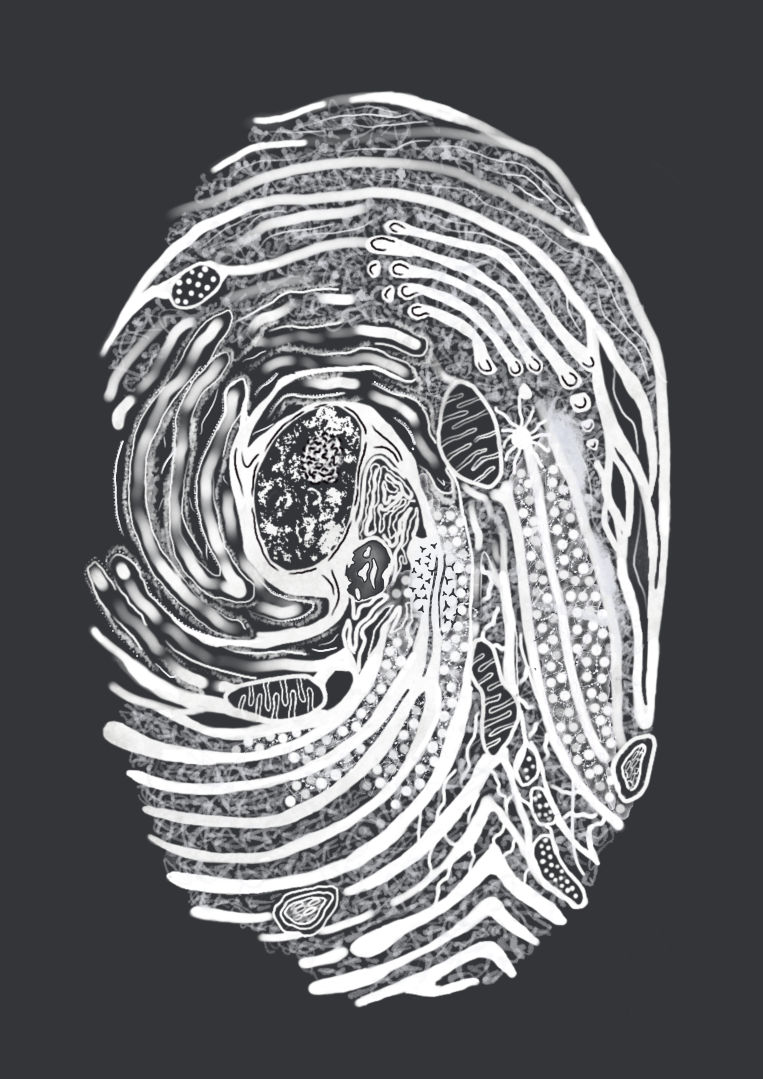

 (7 votes)