We are seeking a highly motivated and talented postdoctoral researcher initially for 1 year, starting 1 October 2017, to develop nanomaterial-based gene delivery strategies for the topical treatment of genetic skin conditions. We are looking for someone with experience in gene transfer and cell biology. Further experience with optical microscopy and drug delivery would be advantageous.
New fellowships from SDB for students from USA and Canada to attend the International Course on Developmental Biology on January 9-21, 2018 in Quintay Chile. Fellowship for Latin American students will be available as well.
Nipam Patel, Alejandro Sanchez-Alvarado, Ray Keller, Claudio Stern, Corinne Huart, Maria Leptin, Andrea Streit, among other will teach, hands-on, the paradigms, problems and technologies of modern Developmental Biology.
More Information here:
DEADLINE 31st August 2017
For the student opinion’s about previous version of the course: 2014 2012
Post doctoral and/or Research Associate position available to study the genetic and epigenetic control of stem cell attributes and pluripotency, focusing on the neural crest gene regulatory network (NC-GRN). Neural crest cells are stem cell-like progenitors that migrate extensively and whose genesis was central to the evolution of vertebrates. Misregulation of components of the NC-GRN underlies numerous human diseases and congenital disorders.
Candidates should have a background in bioinformatics and/or computational biology, strong analytical skills, and significant experience in the analysis of NGS data (ChIP-seq, RNA-seq, ATACseq etc) and proficiency in programming languages (R, Python, PERL etc). Projects may also involve quantitative image analysis. Candidates should be highly motivated, detail oriented, possess excellent communication skills and the ability to work collaboratively as part of an interdisciplinary team. The LaBonne lab is located on Northwestern University’s beautiful lakeside campus close to the amazing city of Chicago.
Please send a CV, brief description of research interests, and the names of three references to:
Carole LaBonne, PhD (clabonne@northwestern.edu)
Department of Molecular Biosciences
Northwestern University, Evanston, IL 602028
Last month I went to Boston for the Annual Meeting of the International Society for Stem Cell Research (ISSCR), which was celebrating its 15th birthday. Sally Temple handed over the Presidential reins to Hans Clevers with a reminder of what a remarkable 15 years it had been for the field – induced pluripotent cells (iPSCs), an explosion of differentiation protocols, the arrival of organoids, CRISPR and single cell sequencing, new windows onto human development, clinical trials and drug screening. She encouraged the audience to think “This is our century” – that is, the century of harnessing the power of stem cells in tackling disease. She also reiterated the ISSCR’s commitment to keep basic science at the core of what it does (you can hear more of her thoughts on the ISSCR in Caroline Hendry’s video interview for Development). In fact the whole meeting (or at least what I got out of it) was a healthy mix of basic and applied, from model organisms to drug trials, and developmental biology was repeatedly heralded as a vital foundation for any future clinical advance; my worries about wandering into a wall-to-wall differentiation protocol fest turned out to be unfounded.
So here’s my belated diary of highlights and impressions from the meeting. There were a thousand talks and posters so of course I missed a lot: if you went and want to write something about the meeting, just register and you’re free to post. Also check out the highlights from Agnes Soos, Cátia Bandeiras and RegenMedNet.
Tuesday, 13th June
Over the Atlantic the in-flight pasta meal tasted like it was cooked in popcorn butter. In Boston it was sweltering until out of nowhere a storm cooled the air down, and in the evening I went to a basement bar where Twitter had assembled a dozen strangers going to the meeting to meet in real life (a ‘Tweet Up’). For someone who knew hardly anyone at the meeting, it was a great way to meet new people and something I’ll try to do again (thanks to Samantha Yammine for instigating this!). After some local IPAs packed with enough hops to blow your head off, I stopped in at a Wallgreen’s on my way back to the hotel and had one of the more depressing late night dinners of my life: Salsitas followed by a flapjack.
Wednesday, 14th June
In the morning I went to a focus session on the ethics of organoids, which was chaired by Megan Munsie (more on Megan’s work below) and paired scientists with ethicists to discuss the impact and meaning of this new technology on wider society.
On the scientific side of things, Hans Clevers gave us the latest update on how organoids derived from adult (often patient) stem cells were helping to understand various pathologies and inform treatments. In an almost throwaway comment he told us that he could make lung organoids from patients’ sputum, and kidney organoids from their urine; you just need to spin the cells down and start the protocol. It struck me as one of those ‘living in the future’ moments. Melissa Little reminded us how critical an understanding normal development is for organoid derivation – you need a guide for which factors to add and in which order, something echoed for iPSC differentiation later in the meeting by George Daley. Indeed most iPSC-derived organoids are models for embryonic rather than mature tissues, which can complicate interpretations. Where the technology is particularly helping Little’s lab is to validate variants of unknown function in genetic kidney diseases – derive iPSCs from patients, ‘fix’ the sequence with gene editing, and see if organoids can form normally; this is such a cool clinical use of organoids that I hadn’t appreciated before.
On the ethics side, Annelien Bredenoord emphasised the inevitable collision of organoid technology and ethics – we have to decide what the moral and legal status of organoids are. Do we treat organoids like any other patient-derived tissue? Are they most akin to cell lines, or something else? What types of consent do we expect patients to give? How do patients see their own oragnoids? And what about if private companies seek to use organoids?
How we answer these questions might really depend on definitions: for Melissa Little, organoids are really just an extension of iPSC differentiation protocols, and if so we need not radically rethink the guidelines that exist for iPSCs (for instance, those written up by the ISSCR). There is also a lot of difference between different kinds of organoids, depending on how complex an organ they are modelling. Plus, according to Jeurgen Knoeblich, the ethical concerns around those most provocative of organoids, cortical ones – “when will they think?” – are based on a perceived rather than a real risk to societal ethics, given that we are light years away from making something capable of a thought or a feeling. Later in the meeting Knoeblich told us about his lab’s efforts to make more complex and representative brain organoids. You can dorsalise one organoid, and ventralise another, culture them together until they fuse, and then observe neural migration from ventral to dorsal, a feature that previous cortical organoids lacked.
Bredenoord’s work involves asking patients what they think about organoids derived from their cells, and revealed ambivalence in one particular cystic fibrosis patient community. Melissa Little had a different experience with kidney patients: they want the opportunity to donate tissue or cells to any initiative that might help them, and organoids are no different; I heard similar things from Jayaraj Rajagopal. Bredenoord also described a reluctance to donate tissue if it was to be commercialised by a private company, and this was echoed by others and presumably a feeling not unique to organoids. Hans Clevers described a fix for this, which was to establish not-for-profits to run the biobanks and profit from commercial customers. The question of consent in biobanking has recently been explored by Tim Caulfield and Blake Murdoch in PLoS Biology.
Insoo Hyun, who sits on the ISSCR’s ethics committee, made the distinction between two types of research ethics: philosophical and practical. Philosophical ethics cover the questions of “should we be doing this?” and “is it wise?”, while practical ethics emphasises the regulatory guidelines in place once we’ve said yes to the philosophical questions. I guess we’ve pretty much gone into the practical side of things – no one in the session questioned the use or potential of organoids, though some thought the hype might catch up with us soon – but he reminded us of the need to really educate the public and justify what we are doing to cover the philosophical part. To paraphrase Hyun, “don’t just forget the public and leave it to committee”.
It was quite a compelling session, though I didn’t exactly come out of it with a clear feeling for where organoids stand from an ethical perspective; I guess that was the point of the session, perhaps the field is too young. For a more coherent discussion of the various ethical issues in hand I’ll send you to a couple of reviews by the speakers, one in Development and one in Science.
The rest of the day was spent in the aircraft-hangar-sized hall where the plenaries were held. It was funny seeing the great and the good walk up to the podium to the snippets of EDM bangers or Coldplay, though I was disappointed that none of them danced their way up. A series of research talks was followed by Sanford Greenberg, who, as a roommate of Art Garfunkel in college (we can thank Greenberg for financing Art’s initial union with Paul Simon), went blind and has since dedicated much of his life to ending blindness, “this wretched curse”. You can check out the website detailing his 3 million dollar prize for blindness research at endblindnessby2020.com.
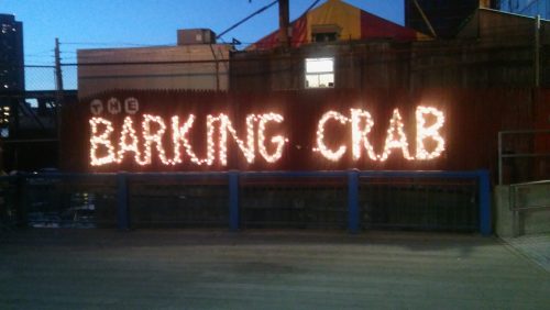
That evening I actually managed to have dinner, at the Barking Crab – a raucous place on the water, with snow crab and another great IPA (Boston’s a wonderful place for a beer drinker).
Thursday 15th June
In the afternoon I had the mindhurt of trying to choose from one of seven (!) concurrent sessions. The ‘Single Cell Heterogeneity’ session featured so much single cell sequencing of organs and organoids that one almost came out of it feeling blasé about the whole enterprise – just to think of telling a developmental biologist from the eighties that she could sequence the genome of every cell in a developing organ! It wasn’t all sequencing though: David Scadden explored the role of the stem cell niche in blood development, starting with some history (read more here). In the seventies, Raymond Schofield had proposed the niche hypothesis but apparently was at loggerheads with Ernest McCulloch about it – he ended up leaving science and becoming a sheep farmer in Wales, where Scadden met him for a pint in the village pub to discuss his ideas. Scadden’s approach was to test the role of the niche by taking stem cells from one animal and place them into another. Under these new conditions, they behaved pretty much the same. HSCs are very heterogeneous, but this is intrinsic rather than defined by the niche, and Scadden used a game-ey metaphor: rather than highly adaptive ‘transformers’, HSCs are like a collection of chess pieces with distinct functional attributes.
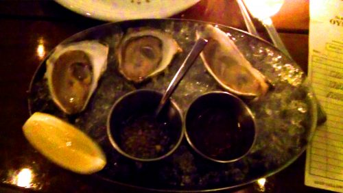
In the evening, dinner at an oyster restaurant with the Twitterati, and my first taste of these opinion-splitting molluscs (verdict: yup, with some hot sauce and lemon juice).
Friday 16th June
The highlight of the day was interviewing George Daley and Jayaraj Rajagopal for Development. Both are residents of Boston who come from clinical backgrounds but are in love with research, and are truly optimistic and excited about the wave of clinical translation of research into treatments. Both are also wonderful people who were generous in conversation with me; watch out for the final interviews in the coming months.
In a stimulating ‘Tissue Regeneration and Homeostasis’ session, Ben Simons began by apologising for being a physicist (though later he proudly showed us some of the first wet data from his lab). Simons uses mathematical approaches to understand developmental problems, and his talk focussed on the statistics of proliferation versus termination in mammary epithelial stem cells during pubertal morphogenesis. I’ve previously talked to Bill Harris about how Simons’ approach helped him understand the retina, and like the idea that sometimes we can be too burdened by knowledge, so a clear eye from a different field is needed to look at a problem. Given the number of collaborations Simons is involved in, his eye is in high demand.
In the evening I took the metro up to Cambridge though it was too rainy really to see much of it, and ate at a Tex Mex place with an old friend from old Cambridge (accompanied this time by Margaritas).
Saturday 17th June
I’d never been so moved at a scientific meeting – in the ‘Road to the Clinic’ session, Michele De Luca, well known for his promotion of responsible stem cell treatments and efforts to shut down charlatans, told us about epidermolysis bullosa, a terrible disease caused by a defective link between the dermis and the epidermis that can lead to skin blistering at even the slightest touch. De Luca described a patient with a severe genetic form of the disease, a child refugee from Syria who on his arrival to Germany had lost much of his epidermis to an infection and was in effect skinless, and placed in an induced coma for the pain it caused. De Luca’s solution was to grow skin from gene-corrected epidermal stem cells, and then graft this skin onto the child’s body (these were really huge grafts). And a couple of years on it seems to have worked – essentially the entire body is covered by transgenic skin that is now functioning like normal skin (faring better, indeed, than skin grafts used to treat burns, as the disease does not affect the underlying dermis like burns do). He ended his talk with a picture of the boy – standing slightly awkwardly in an ill-fitting suit in a hospital corridor, the most heart-wrenching contrast to the pre-treatment images De Luca had shown before. A kid in a coma to a kid standing and smiling.
An uplifting, miraculous story such as this shows us the potential of stem cells and gene therapy for treatment. As Megan Munsie then described, shadowing these remarkable advances is the growth of unregulated stem cell treatments. I remember a few years ago hearing that Texas Governor and GOP presidential candidate Rick Perry had had stem cells injected for back pain, and since we’ve seen an explosion in the number of clinics in the US and beyond offering stem cell treatments for just about anything (these are often covered by Paul Knoepfler’s excellent blog, the Niche). Not only might such treatments be expensive, and useless, they could of course also be dangerous. Munsie interviewed a number of patients who had sought such treatment – even if they understood the risk, for some it was really about the hope that it gave them, however small. Many of the patients (all based in Australia) felt let down by their own health care practitioners, which suggests we could do more to engage with patients who see no other way out than unregulated treatments.
A brief trip to see the tall ships that had gathered in the harbour, and that was that – back on the night flight to Heathrow, with the image of de Luca’s patient, smiling awkwardly, still in my head.
A 3-year funded PhD position is available at the Institut de Biologie Paris Seine to investigate the role of mechanical forces in the construction of a neuronal circuit in vivo.
During neuronal circuit formation, neurons move towards their final location while growing axons towards their target. While the biochemical guidance cues involved in neuronal migration and axon elongation are extensively studied, the contribution of mechanical forces in these processes remains largely unexplored in vivo.
In the lab we address this question using the zebrafish olfactory circuit as a model system. Its location underneath the skin of the embryo makes it amenable to live imaging and mechanical perturbation. We already obtained imaging and functional data suggesting an important function for mechanical cues in the formation of the circuit: olfactory axons extend through the effect of extrinsic mechanical forces that drive the passive displacement of neuronal cell bodies away from their axon tips (Breau et al., Nat Comm, in press).
The purpose of the PhD project is to further identify the origin and contribution of mechanical forces in the construction of the circuit, and the molecular mechanisms involved in force propagation and sensing. To achieve this goal, the student will use a pluridisciplinary strategy combining multiscale live imaging, genetic/optogenetic tools and physical approaches to measure and perturb forces in vivo.
We are looking for a highly motivated student willing to join an interdisciplinary environment involving strong interactions between biologists and physicists.
Requirements:
– Master degree in cell/developmental biology or in biophysics
– Strong interest towards interdisciplinary work
Additional beneficial skills:
– Experience with zebrafish
– Skills in confocal, biphoton or light sheet microscopy
– Experience in image analysis (Image J, Matlab)
Starting date: between October 2017 and January 2018. To apply, please send your CV and references to Marie Breau, Institut de Biologie Paris Seine:
A Postdoctoral Research Associate position is available in the Hoffman Lab at Yale University (www.hoffmanlab.net). We use zebrafish as a translational tool to investigate the function of genes that are strongly associated with autism spectrum disorders (Hoffman et al. 2016 Neuron). Specifically, we use CRISPR-generated zebrafish mutants to study how disruption of autism risk genes affects the developing brain and the neural circuitry underlying simple behaviors. Our goal is to utilize this system to identify basic mechanisms underlying autism and potential new pharmacotherapies.
Candidates must have a Ph.D., M.D., or M.D./Ph.D. in Neuroscience, Genetics, or Cell and Developmental Biology. The ideal candidate will have demonstrated expertise in standard and advanced molecular biology techniques, developmental neurobiology, and microscopy. Special consideration will be given to applicants with experience using animal models of human genetic disorders.
Candidates should be highly motivated, enthusiastic, learn quickly, have a strong work ethic, and a high degree of independence.
Please send your CV, a cover letter stating your research interests and professional goals, and the contact information for three (3) references to:
Ellen J. Hoffman, M.D., Ph.D.
Assistant Professor
Yale Child Study Center and Department of Neuroscience
General Purpose: Function as a Lab Manager overseeing a Molecular Biology Lab using a zebrafish model system. Research in the Hoffman Lab at Yale focuses on using zebrafish as a model system for the functional analysis of autism risk genes (www.hoffmanlab.net). Work on an independent research project, master generation of zebrafish mutants using CRISPR/Cas9 technique, maintain wild-type and mutant fish lines, characterize new mutant fish lines by genotypic and phenotypic analysis, supervise undergraduates and post-graduate associates in the laboratory and fish facility.
Required Education and Experience: Master’s Degree in a scientific discipline and one year experience or an equivalent combination of education and experience.
Qualifications:
Demonstrated knowledge and ability with zebrafish husbandry and maintenance.
Demonstrated proficiency in molecular biology, including cloning, PCR, in vitro transcription, in situ hybridization, western blotting, and immunohistochemistry.
Demonstrated ability in zebrafish analysis and mRNA injection.
Demonstrated excellent interpersonal skills.
Demonstrated strong work ethic.
Preferred Education, Experience and Skills: Master’s Degree in the Biological Sciences, Neuroscience, Genetics or a related discipline, and one year experience or an equivalent combination of education and experience. 5 or more years of experience. Ability to lead and provide oversight in a lab setting.
Application: For more information and immediate consideration, please apply online at http://bit.ly/2txRpIY. Please be sure to reference this website when applying for this position.
Here at Development we are very sad to be saying goodbye to two of our editors: Ottoline Leyser and Geraldine Seydoux. Both Ottoline and Geraldine have been valued members of the editorial team since 2011, and we are hugely grateful for the time and effort they have put in to handling research papers, helping to shape the journal’s future plans and, in Ottoline’s case, coordinating our 2016 Special Issue on Plant Development. They will both be greatly missed, but will maintain connections with and continue to provide input on the journal as members of our Editorial Advisory Board.
Taking their places, we are delighted to announce two new editors: Yka Helariutta and Susan Strome. Yka is taking on Ottoline’s role as Development’s plant editor. After a PhD in plant genetics at the University of Helsinki, Finland, Yka undertook postdoctoral research at New York University and New York Botanical Garden with Philip Benfey before moving back to Helsinki in 1998 to establish his own lab. From 2003 to 2014 he was also affiliated with the Umeå Plant Science Centre in Sweden, and was elected as an EMBO Member in 2008. In 2014, Yka became Professor of Plant Developmental Biology at the University of Cambridge, UK, and moved to the Sainsbury Laboratory, although he still runs an active group in Finland. Yka’s work focusses on vascular development in plants, from patterning to cell differentiation. He primarily uses Arabidopsis as a research model, but is also interested in how the basic molecular mechanisms operate in species with an extensive vascular domain, such as trees.
Susan Strome began her career as a biochemist working on bacteriophages at the University of Washington, before moving to the University of Colorado for a postdoc with William Wood. There, she started working on the C. elegans germline – an interest that has continued throughout her career. Susan gained an independent position at Indiana University in 1984, staying there until 2007, when she moved to the Department of Molecular, Cell and Developmental Biology at the University of California, Santa Cruz, where she currently holds the position of Distinguished Professor. A Member of the American Academy of Arts and Sciences, Susan’s current research focusses on how cells are instructed to develop as germ or soma, and particularly on how chromatin regulators act to promote and maintain germline fate.
One other significant piece of news is that I [Olivier] will be stepping down as Development’s Editor in Chief in September 2018, after 8 years in the role. During this time, I have tried to adapt the journal to the new challenges faced by developmental biology. Notably, I have tried to engage the journal more actively with the stem cell field and, more recently, with human developmental biology. I have also sought to promote emerging fields such as evo-devo and quantitative biology. We have added new sections to the journal: the ʻTechniques and Resources’ and ʻStem Cells and Regeneration’ sections, which have proved to be very popular. We have also tried to listen to the community and implement a number of changes to the submission and review processes. My time as Editor in Chief has been a fantastic experience and I am pleased I had such a great opportunity to serve our community. However, I think turnover is important to maintain journal dynamism and I have chosen to end my tenure at the 2018 Development/The Company of Biologists meeting ʻFrom Stem Cells to Human Development’, which I am co-organising (for details, please see http://www. biologists.com/meetings/from-stem-cells-to-human-developmentseptember-2018/).
Both the journal, which has undergone significant changes during the past 8 years, and the field as a whole are, right now, in an exciting place. We are of course aware of the challenges that researchers face in today’s funding and publishing environment, but we continue to believe that developmental biology has a bright future. Not only can we now use stem cells, genomic and other techniques to analyse human development as never before, but traditional and new model systems are also being exploited in evermore innovative ways to understand the molecular, cellular and physical bases of development across evolution in unprecedented spatiotemporal detail.
We (The Company of Biologists and Development) are enthusiastic about the prospects for developmental biology, and part of our mission is to support you, the research community. We are seeking a new Editor in Chief with a strong vision for Development at the heart of the field. This search will be led by members of The Company of Biologists’ Board of Directors: Sarah Bray, James Briscoe and Kate Storey. As a key part of the process, we want to consult a broad cross-section of the developmental biology community about the strengths of Development, where we can improve, into which (new or existing) areas the journal should expand and what more we can do to support our field. We therefore encourage you to get in touch with any feedback that you may have, particularly where you think it might be helpful in directing our search for a new Editor in Chief. Please contact Sarah, James and Kate – along with Development’s Executive Editor Katherine Brown and the Company’s Publisher Claire Moulton – via dev.feedback@biologists.com, or get in touch with any of us individually. With over a year to go before Olivier steps down, we are happy to have the time to gather and digest community input to help us make the right choice for the next Editor in Chief and ensure that Development continues to hold an important place in the community long into the future.
Evan Brooks is a rising senior at North Carolina State University. For the past two years, he has worked in the lab of Nanette Nascone-Yoder studying the developmental mechanisms of cardiac left-right asymmetry using Xenopus as his model system. Evan was awarded a Choose Development! Fellowship from the Society of Developmental Biology to conduct two summers of mentored research in developmental biology. He was selected to spend one week with us at the Embryology Course this year. I had the pleasure of working next to Evan for the zebrafish and Xenopus module. I thought I would take this opportunity to check in with Evan and ask him a few questions!
Were you told any stories of Woods Hole or did you know anything about the course before you arrived?
Before getting my invitation to join the course, I had only heard of Woods Hole and the Embryology course in passing in the lab. I had never heard of anyone’s stories or experiences with Woods Hole or the Embryology course.
When I got my invitation in May from SDB to join the course for a week, Nanette talked to me about her experience in the course when she was a graduate student. She not only told me how much she enjoyed the course, but also how it allowed her to think more about the evolutionary similarity between model organisms. She also told me how much fun she had in Woods Hole when she wasn’t in the lab, whether it was looking at bioluminescence in Eel Pond or staying up late with her classmates at Captain Kidd. With the knowledge about her experience and its impact on her, I was super excited to join the course for a week.
Did your time in the Embryology course match what you had anticipated?
I will say that I was expecting to be immersed in science while I was there. I looked at the schedule before arriving and was excited to see so many prominent developmental biologists, like Richard Harland and Ray Keller, participating in the course in some way. I was totally expecting to be thinking about possible new experiments all the time.
However, I was not expecting to dive headfirst into the course on my first day in the lab and stay up super late conducting experiments. I was up past 1am every night and woke up every morning around 7am to get back into the lab and check my experiments. On the day I left Woods Hole to return home, I got up at 5am to finish an in situ hybridization before catching a 7:20am bus ride to Boston Logan airport. I was committed to performing so many experiments and seeing them all pan out during my one week and I do not regret it.
What was the most memorable part of your week?
I’d probably say the camaraderie within the program and how integrated I felt in the course is what I will remember most from my week. Everyone was so welcoming and I was frequently included in microscope demos down in the basement of Loeb Laboratory, on coffee runs before lectures, and during meals in Swope Hall. Also, people would encourage me to stay up into the wee hours of the morning with them to try a new experiment. Everyone that I talked to was interested in my research at NC State and what I was doing in the lab during the course. As the week progressed, I was told by more and more people to talk to the course directors, Rich Schneider and Dave Sherwood, to see if there was any way to extend my stay. I really wish that I could’ve extended my stay as I was just getting comfortable with working in the lab and getting to know everyone when it was time for me to leave. I’m still connected to everyone in the course through WhatsApp and I know that I’ve made at least twenty-four new friends.
What did your learn during the week?
Scientifically, I learned so many new things in the course. I learned so much about the current research going on in Xenopus and zebrafish labs around the world, like work on liver regeneration and development in Elke Ober’s lab and the roles of planar cell polarity in development in John Wallingford’s lab. The one lecture that I was most intrigued by was Andrew Gillis’ lecture about gill arch development in skates and its comparative development to limb buds. The ideas, techniques, and approaches presented in the lectures that week showed me how broad the field of developmental biology really is.
From the course, I’ve also learned to take more risks not only in the lab, but in life in general. Taking a week off from my research at NC State to come to Woods Hole was somewhat of a risk as I had to get more data and prepare for my presentation at the SDB Annual Meeting a few weeks after my return. The risk ended up being well worth it in the end as I learned a lot more about myself and developmental biology in that one week.
How did this course change how you approach your current research at NCSU? Did it impact your future plans?
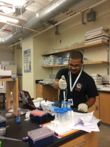
Evan working in lab in Loeb
In terms of working with my hands in the lab, I can say that I have a greater confidence with microinjecting Xenopus embryos. Before the course, all of my embryos would die after microinjections. I can’t really say what caused my embryos to die, but I moved away from microinjection experiments for a bit in the lab. During my week in the course, John Young, one of the Xenopus TAs, showed me how he calibrated his needles and microinjected his embryos. After employing his method, I had quite a few successful morpholino microinjection experiments that I used as the basis for my show-n-tell at the end of the week. I even had the chance to microinject zebrafish embryos and those survived injections too! With my new confidence in microinjecting, I’m looking to incorporate that technique back into my lab repertoire.
Additionally, one thing that I realized that I did not take advantage of during my time in the Embryology course was the cutting-edge microscopy. Before the course, I had only used light microscopes in my lab. During the show-n-tell at the end of my stay, there were so many amazing time-lapse movies and images of cells, tissues, and embryos that I was in awe! I can say that this alone has changed the next steps of my project as I’ve talked to Nanette about designing experiments that will allow me to use other types of microscopes available. I know that there is an expansive microscopy core at NC State, so I may ask for a guided tour in there soon. In terms of my next steps in the Nascone-Yoder lab, I’m planning on performing some antibody stains and confocal microscopy for a few proteins to understand their left-right distributions during developing heart tube stages. I hope to get some pretty images that may be used in a future publication from the Nascone-Yoder lab!
This opportunity is given yearly by SDB, is there any advice you would give to next year’s Choose Development! Fellow who will come to the Embryology Course?
Do not be afraid to jump right in by planning experiments and making friends! The course directors, the instructors and TAs, and fellow course participants are all there to help and get you acclimated to the course and Woods Hole. I remember staying up until 2am with Ray Keller one night because I wanted to learn how to create a two-headed Xenopus embryo. Even though I failed miserably (probably due to exhaustion), he was super patient with me and made sure that I gave it my best effort.
Do you have anything else you would like to include?
I thought I was super excited about embryology and developmental biology before the course. My level of excitement for the discipline has grown so much to where I feel that putting my excitement into words will not accurately describe it. I’m glad that I had the opportunity to experience one week of the course. I am totally looking forward getting into a Ph.D. program and reaching candidacy so that I can be eligible to return to Woods Hole to experience the full six-week course!
Lastly, I honestly cannot thank SDB, Dave Sherwood, and Rich Schneider enough for allowing me to experience one week of the Embryology course this year and for past course directors, Richard Behringer and Alejandro Sánchez Alvarado, for spearheading the idea of inviting Choose Development! Fellows to the course for a week. Past Fellows that I’ve met that have participated in the course absolutely love the experience and I am glad we can partake in this course. The course has helped me affirm my desire to pursue a career in developmental biology!
To check out all the cool things we did during the course follow us on Twitter #embryo2017 or instagram #embryology2017
The Drosophila tracheal system is a powerful model for understanding the genetic and cell biological control of tubulogenesis. In their new PLoS Genetics paper, Ivette Olivares-Castiñeira and her PI Marta Llimargas of the Molecular Biology Institute of Barcelona connect EGFR signalling to intracellular cell trafficking during tracheal morphogenesis. We caught up with Marta and Ivette to hear the story behind the paper.
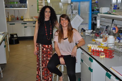
Marta and Ivette in the lab
Marta, can you give us your scientific biography and the main questions your lab is trying to answer?
MA I studied Biology at the University of Barcelona where I took a course on developmental biology and embryology, given by Dr. Jaume Baguña that made me enthusiastic about development. Then I was very lucky because Dr. Jordi Casanova, a well-known Drosophila developmental biologist, just got a position in Barcelona and he accepted me as a PhD student in his lab. By chance we started to work on tracheal development, and I became fascinated by the development of such a beautiful and interesting tissue. The analysis of tracheal development was just an emerging field by then and this allowed me to follow and contribute to the field during all these years.
In 1997 I moved for a postdoctoral stay in Dr. Peter Lawrence’s lab, at the LMB-MRC in Cambridge. There I had an incredible scientific and personal experience, and I was encouraged by Peter to develop my own projects. Because I was really thrilled by tracheal development and Peter allowed me the freedom, I continued working on the subject while there. In 2002 I moved back to Barcelona where I started my lab and later, in 2007, I got a permanent position at the Molecular Biology Institute of Barcelona. My lab has always been, and still is, interested in tracheal formation as a model to understand the morphogenesis of branched tubular organs. We are interested in the genetic mechanisms that drive tracheal formation and in the molecular mechanisms used by these genetic networks to instruct the cellular changes that underlie organ formation.
With the recent announcement of a new EMBL site and a number of universities and research institutes, Barcelona seems to be an exciting place for life sciences at the moment?
MA Yes, Barcelona has become a real hub for science in the past few years and at the moment it is an excellent place for researchers. Barcelona is attracting many senior and young researchers, from many different areas of research. This provides an exciting and encouraging atmosphere for interdisciplinary research. Moreover, many pharmaceutical and biotech companies are set in Barcelona, which also encourages translational research and transfer of knowledge. Obviously the lifestyle, the climate and being a medium sized city also positively contribute to the success of Barcelona as a scientific hotspot.
Barcelona has a number of very good institutions and the production of high quality science in the region is high. Some institutions are very good and very visible, but there are other good institutions and talented people that are less known and overall people struggle with lack of consistent funding to properly develop their research. The general funding situation for science is still below what would be required to fully take advantage of the people and resources.
And Ivette, how did you come to join the Llimargas lab?
IO-C I joined Marta’s lab in 2013, when I had just finished my master’s degree and I realised that I would like to do a PhD. I was looking for an opportunity and Marta gave me the chance to join her lab. After the first year, I became interested in analysing how EGFR was controlling the development of the tracheal system. Being in a developmental biology lab gave me the opportunity to investigate the mechanisms of organ and tissue formation. I find Drosophila tracheal development particularly interesting and ideal to analyse the genetic control of morphogenesis and the cellular mechanisms at play. Doing my PhD with Marta is a great experience for my future career and allows me to get insights into the morphogenesis of epithelial tissues.
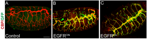
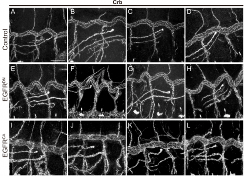
Lateral views of stage 16 embryos expressing EGFR constructs, from Fig. 1, Olivares-Castiñeira & Llimargas 2017.
What was known about the role of EGFR signalling in tracheal development before your paper?
MA & IO-C We already knew a few things. Our lab had published work on the role of EGFR during embryonic tracheal development, in which we showed that it is required for invagination of tracheal cells and to maintain epithelial integrity. In addition, other labs, like Dr. Casanova’s and Dr. Hayashi’s, had also investigated the role of EGFR in tracheal invagination and identified an EGFR-dependent regulation of Myosin-II.
However, a role for EGFR in tube growth was not previously reported. It was actually, back in 2006, during our analysis of EGFR signalling on epithelial integrity that we noticed the effect of EGFR on tube length. However, this observation coincided with the end of the PhD of the student working on this project and it was not followed at the time. When Ivette started her PhD we thought that investigating EGFR requirement on tracheal elongation was a good project to explore, also because there was much more information about tracheal tube growth at the moment and we felt it was the right time to approach the issue. In addition we were also interested in analysing in more detail the possible different molecular mechanisms of EGFR underlying the different tracheal requirements such as invagination, integrity and tube length mainly at the cell biology level.
After analysing EGFR activity in our and other labs, we now know that, as it happens in many other organs and tissues in Drosophila and in other organisms, EGFR is used reiteratively during tracheal formation, but the exact molecular mechanisms behind each of these requirements still need to be fully clarified.
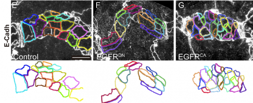
Apical cell shape in tracheal metameres, from Fig. 1, Olivares-Castiñeira & Llimargas 2017.
Can you give us key results of the paper in a paragraph?
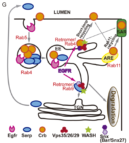
MA & IO-C We have found that EGFR is required to prevent an excessive growth of the tracheal tubes. EGFR is required for the proper accumulation and localisation of two previously known regulators of tube length, the apical determinant Crumbs and the apical extracellular matrix (aECM) regulator Serpentine. This positions EGFR as a hub coordinating cell intrinsic properties (Crumbs-mediated apical expansion) and cell-extrinsic mechanisms (Serpentine-mediated modification of the aECM) regulating tube length. Interestingly we found that these two proteins accumulate in common sorting endosomes, and that EGFR is required for the proper organisation of these common endosomes, likely regulating the correct delivery of both cargoes to their final destination. We observed that the two cargoes are partitioned into different discrete domains within this common sorting endosome, consistent with the hypothesis that they use different retrieval pathways to recycle. We also observed that, during tracheal development, Crumbs undergoes a complex pattern of recycling, which involves internalisation and different sorting pathways. Our results illustrate a role for EGFR in endocytic trafficking, a molecular mechanism that could potentially underlie different developmental and pathogenic EGFR activities.
What do you think the EGFR receptor is doing in the endosomes?
MA / IO-C That is what we would like to unravel! Many reports in the literature describe the internalisation and trafficking of EGFR receptor, the many factors involved in the process and the consequences for EGFR activity. So obviously EGFR is a cargo of endocytic trafficking, and this trafficking regulates its signal termination and the recycling of the receptor to the membrane. Much less is known about a possible role of the receptor itself in controlling its own or other protein’s trafficking. Our results point to a role of EGFR in organising the endosome, because when EGFR is downregulated endosomes are bigger, cargoes are mis-sorted and WASH accumulation, which is recruited by the Retromer complex, is also affected. Whether this is a direct or an indirect effect of EGFR downregulation is still unclear. However, the fact that we find EGFR itself in Crumbs/Serp sorting endosomes and the fact that endosomes are affected leads us to speculate that EGFR regulates targets in the endosome. In fact, EGFR cytoplasmic targets and endosomal targets have been identified, suggesting that intracellular organelles can act as EGFR signalling platforms. To get further insights into the molecular mechanism of EGFR in tube elongation we are working with the hypothesis that EGFR (or EGFR downstream effectors) signals from the endosome to organise this organelle.
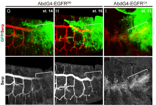
Lateral views of embryos at the indicated stages carrying AbdGal4-UASGFP stained with Serp antibody, from Fig. 2, Olivares-Castiñeira & Llimargas 2017.
Do you have any clues as to how the apical ECM instructs the underlying tracheal epithelium during tube elongation?
MA / IO-C This is an outstanding question in the field. It is clear that the aECM and the underlying tracheal cells cross-talk and that this cross-talk is required for the correct tube growth in length and diameter. But the exact molecular mechanism is still unclear. The aECM can potentially instruct tracheal cells by exerting a mechanical force that is received by mechanosensors in the tracheal cells. It could also trap or expose chemical signal/s that could be received by tracheal cells. Or it could both act as a mechanical and a signalling platform. Bo Dong and Shigeo Hayashi have put forward a model where the apical membrane expansion force in tracheal cells that drives tube elongation is balanced with the resistance of an elastic aECM that restricts overelongation. The aECM mechanical tension and the apical expansion force are coupled through proteins attaching or coordinating the apical membrane to the aECM, such as Zona Pellucida proteins (as proposed by Hayashi lab) or Src42A (as proposed by Sofia Araújo and Jordi Casanova). Hopefully the dedicated work of several labs in the subject will soon shed light into this issue and we will better understand not only the growth of tubular structures but also how the cross-talk of extracellular matrices and organ/tissue/cells instructs morphogenesis.
Model for EGFR’s role in intracellular trafficking, from Fig. 7, Olivares-Castiñeira & Llimargas 2017.
When doing the research, did you have any particular result or eureka moment that has stuck with you?
IO-C From the beginning of the work, our main goal was to identify the target/s of EGFR that control tracheal tube length. We found that Crumbs and Serpentine were affected in the tracheal tubes upon modulation of the EGFR activity. My eureka moment was when one afternoon, we realized that these two protein seemed to be more related than expected and we observed that they were loaded in common vesicles (we did not know that these were endosomes at the moment). It was a moment of euphoria as this was shedding light into a complicated puzzle of many pieces, although this also raised many other questions.
And what about the flipside: any moments of frustration or despair?
IO-C Science always has moments of frustration. It was particularly frustrating to repeat many times some of the stainings before we got good samples. In general, I find that technical details can often be really time-consuming and despairing. It was also frustrating to use several RNAi or dominant negative lines that did not produce phenotype, although I knew that the RNAi technique does not always work well for embryonic development. And finally, I found it very frustrating when we had different results but we were not able to understand what was happening. It is not always easy to put the pieces of the puzzle in the right place to get the final picture.
What are your career plans following this work?
IO-C First, I need to finish my PhD, which I plan to do this year. In the lab I am trying to get more details on the molecular mechanisms of EGFR and on the role of Crumbs in tube size. I am particularly interested in the connection of these proteins with intracellular trafficking pathways. After I finish my PhD, I will probably continue in the research world, as I find it fascinating.
Crb expression in different conditions, from Fig. S4, Olivares-Castiñeira & Llimargas 2017.
And what next for the Llimargas lab?
MA Well, we are continuing different aspects derived from this work. For instance, we are now very interested in understanding Crumbs localisation in different apical subcellular domains (in trachea and other tissues) and whether this correlates with different Crumbs activities. Obviously this aspect is not only relevant for tracheal development, but may help to better understand the complexities of Crumbs. We are also interested in understanding how Crumbs, Serpentine, Vermiform, Src, EGFR and Dumpy, all of them known regulators of tube length, interact and regulate each other. Furthermore, we are really curious to understand how the retromer complex selects the different cargoes, Crumbs and Serpentine in this case, and sorts them into different recycling pathways. For this particular question we have identified a putative regulator of retromer trafficking, a nexin, that could provide cargo and itinerary specificity for the specific sorting of Serpentine and Crumbs. We are now investigating this aspect, generating mutants for this nexin and characterising its role. But obviously, the main question we are trying to answer is how EGFR regulates endocytic trafficking.
Besides this project, in the lab we are also interested in other aspects of tracheal formation, related with chitin deposition and with adhesion and polarity maintenance and remodelling.
Finally, what do you like to do when you are not in the lab?
MA I spend as much time as I can with my family. I try to get some time to read, to go to the theatre, to dance and to go out with friends. I love cooking for friends and family and I love eating, particularly new and surprising foods. I also exercise, particularly gym and running, in the nature when possible.
IO-C I try to switch off my mind from the lab issues and I like to go for run and do sport, which is good to clear my mind. Also, I like to read or to be with my friends. If I had more time I would like to travel more.
 (1 votes)
 (4 votes)
 (No Ratings Yet)