In Development this week (Vol. 144, Issue 9)
Posted by Seema Grewal, on 2 May 2017
Here are the highlights from the current issue of Development:
SEGGA: new software for analysing cell shape and dynamics

Changes in epithelial cell shape and organisation are essential for tissue morphogenesis, regeneration and repair. In recent years, advances in microscopy have made it possible to capture these changes in living animals, but the quantitative analysis of changes in cell shape, behaviour and polarity in large cell populations remains a significant challenge. Here, on p. 1725, Jennifer Zallen and colleagues describe new image analysis software that allows automated image processing, image segmentation, cell tracking, data analysis and data visualization. Using this software, which they term SEGGA (for image SEGmentation, Graphical visualization and Analysis), the team analysed cell behaviours during convergent extension in the Drosophila embryo. Their analyses reveal that cell intercalation is a key mechanism that drives convergent extension and that planar polarity is rapidly established, prior to the onset of elongation, and is dynamically remodelled as cells intercalate. The researchers also demonstrate the general utility of this software by analysing images of epithelial cells from other tissues and organisms. This software, which is freely available and can run on Mac, Windows and Linux operating systems, promises to be a valuable tool for the community.
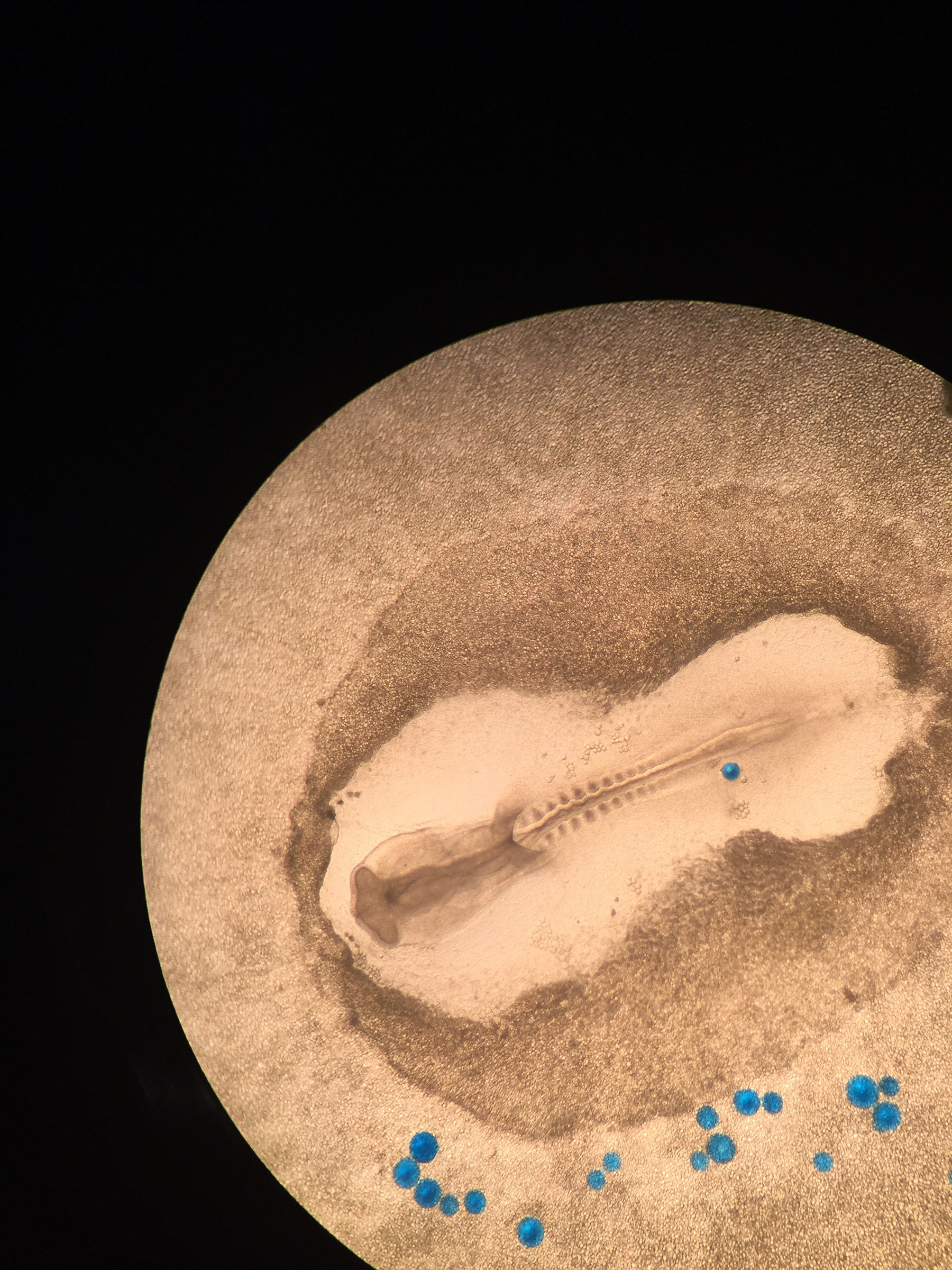
CDC42 shapes up the epicardium

The epicardium – a single layer of cells that covers the heart – contributes to multiple cardiac lineages and is essential for heart development and regeneration. During development, the epicardium arises from a transient structure known as the pro-epicardium (PE): pro-epicardial cells (PECs) dissociate from the PE, translocate across the pericardial cavity and attach to the heart surface. The molecular and cellular mechanisms that control this process have thus far been elusive but, now, Mingfu Wu and co-workers reveal four different mechanisms of PEC translocation to myocardium and a key role for the small GTPase CDC42 during epicardial development in mice (p. 1635). They first report that the conditional knockout of Cdc42 in PECs results in epicardial defects, with PECs failing to form villous protrusions and floating cysts and failing to translocate to the myocardium. The authors further demonstrate that Cdc42 null PECs exhibit loss of polarity and impaired cell dynamics. Finally, they reveal that CDC42 also regulates the trafficking of FGFR1 in PECs and hence is likely to be required for FGF2-mediated signalling activity in these cells. Overall, these findings provide new insights into the mechanism by which the epicardium forms and highlight a pivotal role for CDC42 in epicardial development.
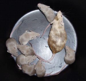
Controlling somatic cell NUMB-ers during gonadogenesis

During gonadogenesis in mice, cells within the coelomic endothelium (CE) proliferate to give rise to additional CE cells as well as somatic cells of the gonads. But how is this cell fate decision – CE versus gonadal – determined? In this issue, Blanche Capel and colleagues show that NUMB, a known antagonist of Notch signalling, plays a central role (p. 1607). They show that, during early gonadogenesis in mice, NUMB is asymmetrically localised to the basolateral domain of CE cells, which also exhibit high levels of Notch signalling. Importantly, the authors report that the temporal deletion of Numb results in gonadal defects; mutant gonads contain patches of undifferentiated cells, reduced numbers of differentiated somatic cells and, curiously, reduced numbers of germ cells. The polarity of CE cells in mutant gonads is also disrupted. Finally, the researchers demonstrate that blocking Notch signalling (using the g-secretase inhibitor DAPT) can rescue the somatic cell defects. Based on their findings, the authors propose that asymmetric divisions in the CE give rise to one daughter that remains in the CE, and one daughter that inherits NUMB and the competence to differentiate.
PLUS:
An interview with John Gurdon
 John Gurdon is a Distinguished Group Leader in the Wellcome Trust/Cancer Research UK Gurdon Institute and Professor Emeritus in the Department of Zoology at the University of Cambridge. In 2012, he was awarded the Nobel Prize in Physiology or Medicine jointly with Shinya Yamanaka for work on the reprogramming of mature cells to pluripotency. We met John in his Cambridge office to discuss his career and hear his thoughts on the past, present and future of reprogramming. Read the Spotlight article on p. 1581
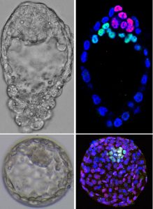
Human organomics: a fresh approach to understanding human development using single-cell transcriptomics
 Innovative methods designed to recapitulate human organogenesis from pluripotent stem cells provide a means to explore human developmental biology. New technologies to sequence and analyze single-cell transcriptomes can deconstruct these ‘organoids’ into constituent parts, and reconstruct lineage trajectories during cell differentiation. In their Spotlight article, summarize the different approaches to performing single-cell transcriptomics on organoids, and discuss the opportunities and challenges of applying these techniques to generate organ-level, mechanistic models of human development and disease.
Development of the hypothalamus: conservation, modification and innovation
 eview the factors involved in the induction, patterning and neuronal differentiation of the hypothalamus, highlighting recent evidence that illustrates how changes in Wnt/β-catenin signaling during development may lead to species-specific form and function of this important brain structure. Read the Review article on p. 1588.
 (No Ratings Yet)


 (8 votes)
 Jenny’s main research interests are the mechanisms that establish and maintain pluri-potency in the early embryo and during the formation of embryonic stem cells in mammals. She also uses animal models to understand defects which lead to type 1 diabetes. Jenny started her career at Oxford University where she worked as a research assistant to Prof. Richard Gardner (1981-90). In 1990 she moved to the University of Edinburgh to carry out her PhD project in the group of Prof. Austin Smith. She obtained her PhD in 1995 for her thesis entitled ‘A Study of the Expression and Function of Differentiation Inhibiting Activity and its Receptor in the Early Mouse Embryo‘. She stayed as a post-doctoral research fellow in the group of Austin Smith in Edinburgh, until she became a group leader at the Wellcome Trust-MRC Stem Cell Institute of the University of Cambridge, where she has stayed since then.