Tiny titans: fantastic worms and their powerful regenerative abilities
Posted by Rannyele Passos Ribeiro, on 19 January 2025
In their paper recently published in Evolution & Development, Vanessa Spieß, Rannyele P. Ribeiro and colleagues explore the regenerative abilities of the marine segmented worm Syllis malaquini. Their research reveals that a small piece of this tiny worm can regenerate its entire body forming a whole new individual. Now, co-first and co-corresponding author Rannyele P. Ribeiro offers insights behind this fascinating discovery.
How did the project get started?
During my PhD, supervised by Dr. M. Teresa Aguado, professor at University of Gottingen, I discovered a new species of segmented worm, Syllis malaquini, living in an aquarium. This worm has a segmented body, which means that between the head and tail there is a trunk formed by repeated body units called segments. The trunk of the worm also has a regionalized digestive tube, with foregut and gut regions. My thesis characterized morphological and cellular dynamics of S. malaquini regeneration, showing that the worm could restore the missing body part after amputation of half of its body1. That means splitting a worm into two pieces generates two new worms that are clones of one another. This breakthrough revealed a critical research challenge: identifying the smallest body fragment that retains the potential for whole-body regeneration. To answer this question, Master’s student Vanessa Spieß conducted many experiments isolating body fragments with different segment numbers, and from different gut regions along the antero-posterior axis of the worm. At that time, I transitioned to do my postdoctoral research with Dr. Duygu Özpolat, assistant professor at Washington University in St. Louis. However, I continued to collaborate with Vanessa Spieß and Dr. Aguado to analyze and interpret the acquired data, culminating in the recently published paper in Evolution & Development2.
Why did you choose Syllis malaquini as your research organism?
Syllis malaquini was discovered serendipitously while performing experiments to investigate regeneration in segmented worms collected from an aquarium located at the University of Leipzig, Germany3. While working with what was thought to be Typosyllis antoni, I observed an unexpected ability to regenerate the anterior body, of which T. antoni is incapable4,5. This observation led to a careful examination of the morphology and DNA sequence of my experimental worms, revealing a new species that we named S. malaquini. This species amazed me in many ways. During an experiment, I successfully cultured multiple fragments from a single individual in a Petri dish, with each fragment regenerating into a complete clone, multiplying the worm culture. This astonishing ability suggests a form of near immortality by continuous regeneration and self-cloning. I think that their regenerative mechanisms probably rely on powerful mechanisms that maintain cellular health and proliferative capacity. Therefore, this worm is an excellent model for studying not only whole-body regeneration but also fundamental mechanisms of cellular integrity maintenance.
Can you summarize the key findings of the paper in one paragraph?
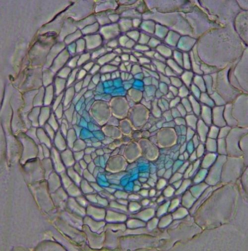
Our research revealed the minimum body size for whole-body regeneration in Syllis malaquini, assessing regenerative success and failure. We not only confirmed the worm’s remarkable regenerative capacity but also demonstrated its ability to achieve whole-body regeneration from extremely small fragments of the trunk, specifically from the intestinal region. We discovered that, while a piece of trunk with just two segments (that is around 300 μm long) can initiate head and tail regeneration, it cannot restore the whole-body. However, a fragment with three segments can successfully regenerate an entire new individual (Fig. 1). The research also uncovered that regenerative capacity varies depending on the gut region, with fragments of the foregut being less regenerative than of the ones from the intestinal region. This discovery opens new avenues for understanding the influence of the gut regions in successful regeneration in segmented organisms.
Were there any other unexpected results and challenges, especially associated with working with a non-model research organism?
Working with Syllis malaquini presented unique challenges, particularly in controlling sexual reproduction, which hindered our ability to perform transgenesis and genome editing. While we successfully maintain asexual reproduction in aquariums, the lack of control over egg and embryo production limits genetic tractability in this species, as generating transgenic segmented worms typically relies on egg injection techniques. To overcome these obstacles, I’m currently developing gene knockdown systems using RNAi in segmented worms. This approach offers a promising avenue for investigating the molecular basis of S. malaquini’s regenerative ability, despite limitations in traditional genetic editing techniques. By adapting and refining these methods, we aim to unlock new insights into the mechanisms underlying whole-body regeneration in this fascinating species.
What’s next for this story?
A direction to be followed next in S. malaquini research is identifying the crucial factors that enable successful regeneration in amputated fragments with three segments but are absent in fragments with only two segments. We will keep this question in mind exploring anterior-posterior molecular patterning and specific cell-cell signaling as candidate components playing a vital role in successful regeneration. My current postdoctoral research on Platynereis dumerilii explores the systemic effects of regeneration on developmental transitions, gametogenesis, and lifespan, potentially revealing parallels with S. malaquini. Leveraging comparative research across multiple species, we aim to unravel unique regeneration mechanisms and adaptations in segmented worms.
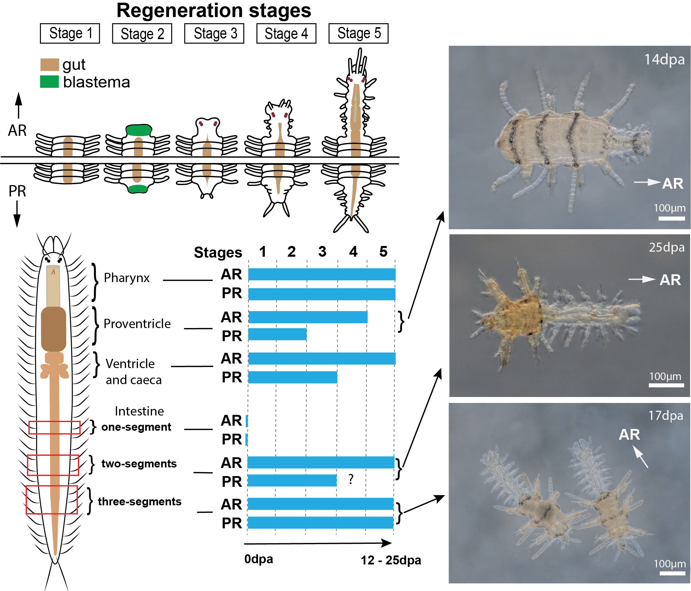
Figure 1. Regenerative capabilities of Syllis malaquini. A body fragment containing two segments can regenerate both head and tail structures but does not exhibit further growth. Body fragments with three or more segments can regenerate into complete individuals. This demonstrates the remarkable regenerative plasticity of S. malaquini, with segment number being a critical factor in determining regenerative outcomes. Figure obtained from Spieß et al. 20242.
References
1. Ribeiro, R. P., Egger, B., Ponz-Segrelles, G. & Aguado, M. T. Cellular proliferation dynamics during regeneration in Syllis malaquini (Syllidae, Annelida). Front. Zool. 18, 27 (2021).
2. Spieß, V., Ribeiro, R. P., Helm, C. & Aguado, M. T. From two segments and beyond: Investigating the onset of regeneration in Syllis malaquini. Evol. Dev. 26, e12492 (2024).
3. Ribeiro, R. P., Ponz-Segrelles, G., Helm, C., Egger, B. & Aguado, M. T. A new species of Syllis including transcriptomic data and an updated phylogeny of Syllinae (Annelida: Syllidae). Mar. Biodivers. 50, 31 (2020).
4. Weidhase, M., Beckers, P., Bleidorn, C. & Aguado, M. T. On the role of the proventricle region in reproduction and regeneration in Typosyllis antoni (Annelida: Syllidae). BMC Evol. Biol. 16, 196 (2016).
5. Weidhase, M., Beckers, P., Bleidorn, C. & Aguado, M. T. Nervous system regeneration in Typosyllis antoni (Annelida: Syllidae). Zool. Anz. 269, 57–67 (2017).
 (2 votes)


 (No Ratings Yet)