The Inaugural Sainsbury Lab Symposium on Induced Plant Development 2016
Posted by Erin Sparks, on 9 May 2016
By: Erin Sparks and Matthias Benoit
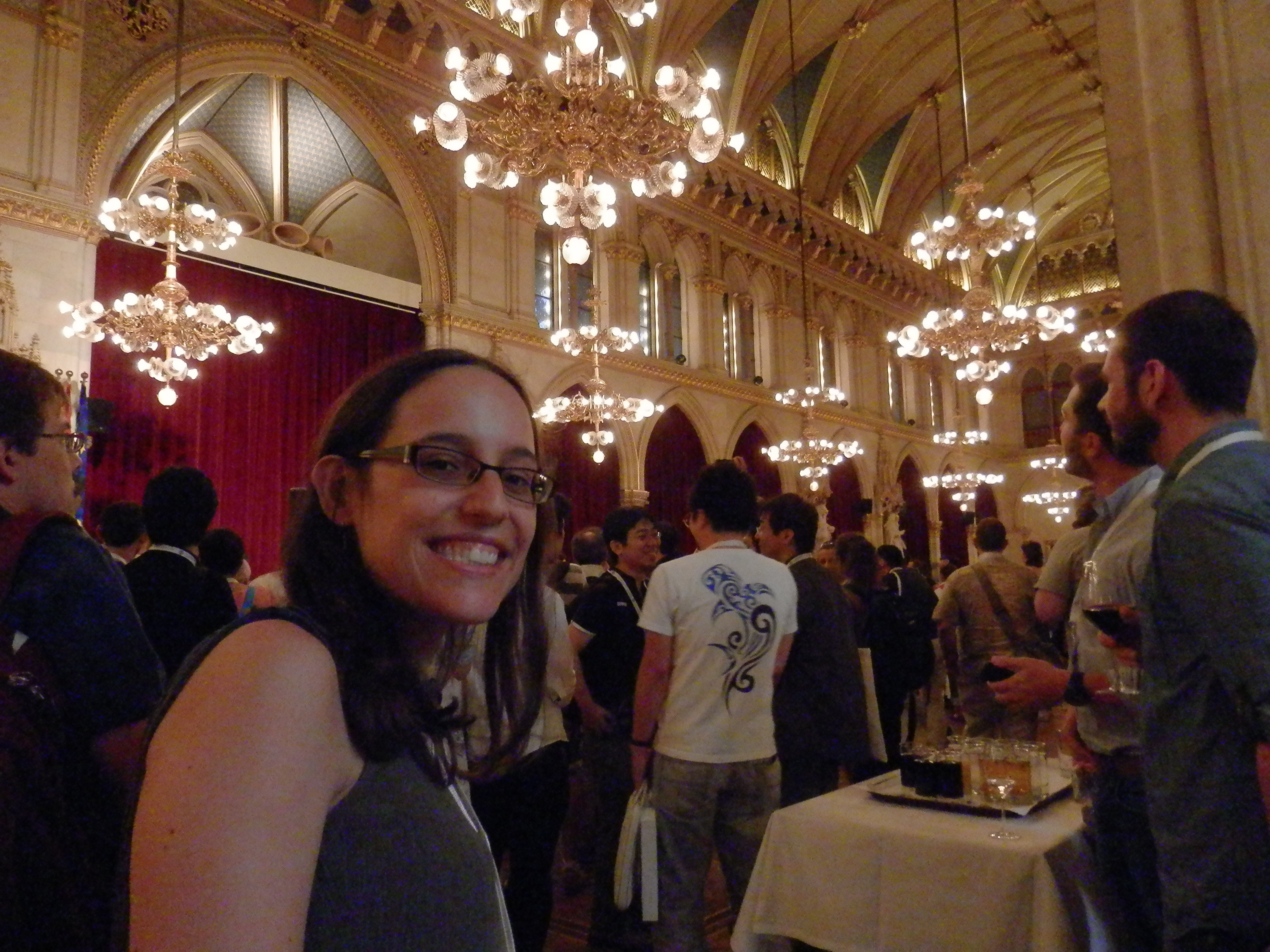
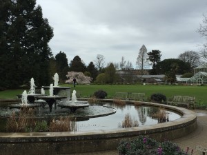
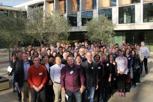
Set in the idyllic location of the Sainsbury Laboratory Cambridge University (SLCU) building adjacent to the Cambridge University Botanic Garden, the inaugural Sainsbury Lab Symposium (#SLS16 on Twitter) attracted over 100 researchers from across the world. This three-day symposium focused on the topic of Induced Plant Development and featured 18 speakers, 10 poster flash talks and 36 posters, covering a variety of research topics. Several themes emerged from this meeting that we highlight in this report.
The symposium kicked off with a Keynote Address by Sofie Goormachtig (VIB Department of Plant Systems Biology – Ghent University), discussing her elegant work on the role of Strigolactones in controlling root architecture. The importance of Strigolactone signalling was further supported by, Thomas Greb (COS – Heidelberg University) presenting his results on the role of Strigolactones in primary energy metabolism and growth regulation. The implication of hormones in induced plant development was repeatedly discussed during the symposium, including several posters emphasizing the role of Auxin, Cytokinins and Abscisic Acid signalling in the regulation of cell elongation, regeneration and differentiation.
In addition to hormones, the importance of abiotic environmental signals in controlling plant development was highlighted. The environmental cues discussed included light, water, calcium, sugar and mechanical induction. Seisuke Kimura (Kyoto Sangyo University) presented his exciting results on phenotypic plasticity and heterophylly in the herbaceous semiaquatic plant Rorippa aquatica. Discussing his latest results, Seisuke showed that leaf form in R. aquatica is also affected by light and temperature. Further, Elena Baena-González (IGC – Portugal) underlined the importance of monitoring plant carbon status by SNF1-related Protein Kinase1 (SnRK1) in the restoration of post-stress homeostasis.
Biotic regulation of plant development was another central feature of the symposium. This included the discussion of parasitic plants, insects and fungi. Of particular interest was work from Melissa Mitchum (University of Missouri) on the signalling mechanisms of cyst nematode parasitism. In the classic co-option of host mechanisms, the cyst nematode delivers CLE-like effector proteins to mimic the plant CLE peptides. The subsequent signalling through the CLE pathway is essential for the establishment of nematode feeding sites. In considering parasitic plants, Ken Shirasu (RIKEN) reported the draft genome sequence of Striga asiatica and revealed a recent whole genome duplication as a potential driving force for the adaptation to host plants. In parallel, he presented a new model for forward and reverse genetics in parasitic plants, Phtheirospermum japonicum. Recent results from his lab provide evidence for the cooption of a lateral root developmental program for haustorium (penetration structure required for host invasion) formation.
Each of these areas of induced plant development revealed the inherent problems with studying environmental influences on plant development and the need for improved technology. Several researchers presented their emerging technologies for improving plant phenotyping and environmental control. For example, Olivier Loudet (INRA) discussed his development of a high throughput phenotyping robot called the Phenoscope. This impetus for this design was the importance of controlling water content per pot and the growth room position, which are vital for his drought response studies. Along these same lines, Christian Fankhauser (University of Lausanne) also developed a phenotyping platform to analyse leaf growth and positioning with high spatial and temporal resolution. Least we forget about the belowground portion of plants, Malcolm Bennett (University of Nottingham) updated us on the progress of the Hounsfield Facility for 3D x-ray imaging of root systems. With a particular interest in how plants find and respond to water, Malcolm discussed his latest work on the molecular control of hydropatterning.
The link between fundamental sciences and agronomics has never been so tight as it was during this symposium. Many speakers have emphasized how their research can be applied to agronomics and industry. Kerry Franklin (University of Bristol) described a nice industrial partnership with a company interested in UV-B treatment of their plants undergoing shade avoidance and thermomorphogenesis problems. Christian Fankhauser (University of Lausanne) also revealed industrial interest in his research on light-induced morphogenesis and accessibility to sunlight.
It is noteworthy to mention the extreme diversity of plant species used in the topics discussed during the symposium, reflecting the vitality of the plant induced development field. From Arabidopsis thaliana to Rorippa aquatica, to the parasitic plant Striga asiatica (aka witchweed) to a variety of crops, not less than a dozen different species were represented, pushing the boundaries of plant development to new and exciting topics.
All together the inaugural Sainsbury Laboratory Symposium was a rousing success. In our humble opinion, this symposium highlighted the reason many of us attend meetings – there was stimulating discussions, new ideas, cutting edge research and a great diversity of topics. Congratulations to Dana MacGregor, postdoctoral scientist in Steve Penfield’s lab (John Innes Centre), awarded best poster of the Symposium for her work on natural variation and environmental regulators of seed dormance. The success of this symposium is due in large part to the organizer, Sebastian Schornack, who did a wonderful job and has our gratitude for the substantial effort required in organizing such an event. This symposium will definitely be included on the “must attend” list for future years.
 (3 votes)
 (No Ratings Yet)

 During the last three years I have been involved in a lot of projects on the Node, which I hope you have enjoyed. The most obvious change was the