The community site for and by
developmental and stem cell biologists

EmbryoMaker: a general modeling framework to simulate developing systems and perform experiments in silico.

Posted by , on 4 October 2015

One of the main challenges of Developmental Biology is to understand the complex developmental mechanisms giving rise to different organs or whole organisms. In most cases, these involve the interplay between cell-cell signalling and cell and tissue movements driven by one or several cell behaviours (such as cell proliferation, cell migration, etc.). Cell signalling will affect how surrounding tissues grow or change in shape, which in turn will change the spatial context in which signalling is taking place. Such complexity in the developmental dynamics can account for the formation of quite complex organs, such as mammalian teeth1 or vertebrate limbs2, but understanding how perturbations on those developmental processes will affect the resulting phenotype is not trivial.

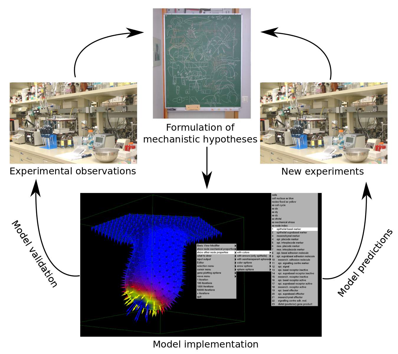
Multi-scale computational models can help in better understanding the dynamics of developmental processes both qualitatively and quantitatively. They should be built upon explicit mechanistic hypotheses about how development takes place. Computational models can then provide explicit quantitative predictions on how the morphology of the organ or embryo or the expression pattern of certain gene products change during development (Figure 1).

Mathematical models need to be based on experimental observations. These are interpreted and a mechanistic hypothesis is formulated (i.e. how the system is supposed to work). The model is implemented by translating the hypothesis into a mathematical formulation and solved through computational methods. Model validation consists on testing how accurately the model can reproduce experimental observations. A validated model can then be used to predict the behaviour of the system under conditions that haven't been yet reproduced experimentally.
Figure 1. Model design and validation. Mathematical models need to be based on experimental observations. These are interpreted and a mechanistic hypothesis is formulated (i.e. how the system is supposed to work). The model is implemented by translating the hypothesis into a mathematical formulation and solved through computational methods. Model validation consists on testing how accurately the model can reproduce experimental observations. A validated model can then be used to predict the behaviour of the system under conditions that haven’t been yet reproduced experimentally.


In the Salazar-Ciudad lab we work on the design of computational models of development in order to study how the complexity of developmental dynamics may give rise to complex structures and how the presence of different types of developmental mechanisms in different lineages may affect their evolution. For some time we have been using a tooth development model1 in order to approach those questions. However, if one wants to tackle those questions from a more general point of view, organ-specific models of development are not enough. For that purpose we need a modelling framework that implements a general developmental toolkit, that is: 1) the ensemble of cell behaviours known to happen in animal development (cell growth, division, death, cell migration, adhesion, epithelial-mesenchymal transition, cell signalling and extracellular matrix modification), 2) the basic mechanical properties of epithelial and mesenchymal cells and extracellular matrix and 3) the freedom to design gene regulatory networks (GRN) that dynamically control the mechanical properties of cells and their behaviours. Such a modelling tool should be able to simulate the development of, say a tooth or a limb, given that we correctly choose the gene networks and initial conditions in each case. Most interestingly, this would allow to study in silico how to transform one organ into the other by replacing, for example, the tooth forming GRN by the limb forming GRN and vice versa. In a similar way evolutionary transitions between different organs or structures could be inferred by rewiring the GRN step-wise.

Although there already exist modelling frameworks of development3,4,5 none explicitly implements the differential mechanical properties of both epithelial and mesenchymal cells or the whole range of cell behaviours we enumerated above. Thus, we decided to develop the most general developmental modelling framework up to date with all the elements described above.

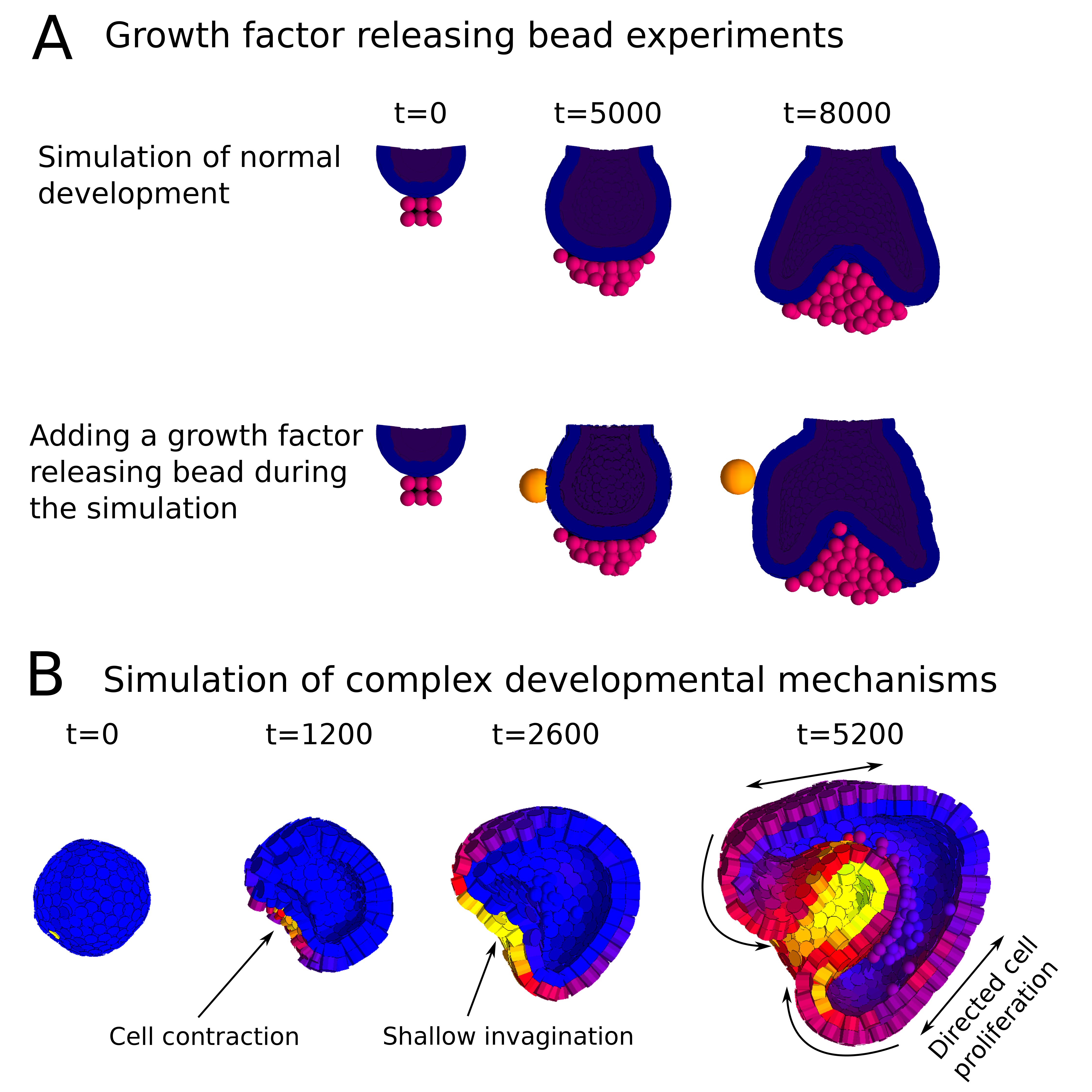
In a paper recently published in Bioinformatics we present this new modelling framework implemented in the open source software EmbryoMaker, freely available for download at our lab’s website. The software provides a graphical interface to visualize the progress of a simulation in real time. In addition, it comes with a user-friendly editing tools in order to design the spatial initial conditions of any developmental system made of either epithelia, mesenchyme and/or extracellular matrix (the size and shape of the tissues at time 0 and their mechanical properties and gene expression profiles) and the GRN (that is defining the number of genes that will participate, their regulatory interactions and their regulation of different cell behaviours). The editing tools can be further used during the simulation of development in order to manipulate the system in real time. For instance, groups of cells can be removed or replaced in the fashion of a graft experiment, or growth factor releasing beads can be placed in any point in the developing system (Figure 2A). Thus, EmbryoMaker may work as an in silico wet lab that allows to predict the outcome of possible experiments on the system of study before carrying them out in vivo or in vitro (Figure 1).

We also show how the modelling framework is able generate complex morphologies from rather simple initial conditions by combining different cell behaviours in a dynamic way (Figure 2B). In this case, the joint action of localized cell contraction, cell polarization and polarized cell division drive the invagination of a spheric epithelium by epiboly.

Overall we expect this new modelling framework to contribute positively to the advance of the Developmental Biology and Evolution fields by providing powerful predictive tools to aid experimental design but also as a means to systematically study the capacity of Development to generate complex and disparate structures and how those might evolve.


A, Simulation of a growth factor releasing bead experiment in which. The first row shows a developmental sequence in which an epithelial bud (blue and purple) grows over a mesenchymal condensate (pink). The second row shows the same process, but in this case a growth factor releasing bead is place at a certain time during the simulation. The growth factor diffuses towards the epithelial cells and increases their proliferation rate, thus making the left side grow larger than the right side. B, Simulation of a complex developmental system by combining several cell behaviours on a hollow spheric epithelium. The contraction of a localized group of cells causes a shallow invagination of the epithelium. Meanwhile, a molecular gradient is being formed towards the invaginated region, and cells across the embryo are instructed to proliferate in the direction of the gradient. The directional growth of the embryo pushes cells deeper into the embryo in a fashion reminiscent of the process of gastrulation by epiboly.
Figure2. Examples of simulations performed with the EmbryoMaker software. A, Simulation of a growth factor releasing bead experiment. The first row shows a developmental sequence in which an epithelial bud (blue and purple) grows over a mesenchymal condensate (pink). The second row shows the same process, but in this case a growth factor releasing bead is placed at a certain time during the simulation. The diffusible growth factor will only reach the cells closest to the bead and will increases their proliferation rate, thus making the left side grow larger than the right side. B, Simulation of a complex developmental system by combining several cell behaviours on a hollow spheric epithelium. The contraction of a localized group of cells causes a shallow invagination of the epithelium. Meanwhile, a molecular gradient is being formed towards the invaginated region (colors depict the concentration of the molecule: yellow is high and blue is low), and cells across the embryo are instructed to proliferate in the direction of the gradient. The directional growth of the embryo pushes cells deeper into the embryo in a fashion reminiscent of the process of gastrulation by epiboly.


Main reference: 

Marin-Riera M, Brun-Usan M, Zimm R, Välikangas T, Salazar-Ciudad I (2015) Computational modeling of development by epithelia, mesenchyme and their interactions: a unified model. Bioinformatics


Other references:

1- Salazar-Ciudad I and Jernvall J (2010) A computational model of teeth and the developmental origins of morphological variation. Nature 464, 583-586.

2- Hentschel HGE et al. (2004) Dynamical mechanisms for skeletal pattern formation in the vertebrate limb. Proc. Royal Soc. B. 271, 1713-1722.

3- Pitt-Francis J et al. (2009) Chaste: a test-driven approach to software development for biological modeling. Comput Phys Commun. 180: 2452-2471

4- Izaguirre JA et al. (2004) CompuCell, a multi-model framework for simulation of morphogenesis. Bioinformatics 20, 1129-1137

5- Delile J et al. (2013) Computational modeling and simulation of animal early embryogenesis with the MecaGen platform. In: Kriete A, Eils R editors. Computational Systems Biology, 2nd ed. Academic Press. Elsevier pp. 359-405


Thumbs up (6 votes)
Loading...

Tags: ,
Categories: Research, Resources

Navigate the archive

Use our Advanced Search tool to search and filter posts by date, category, tags and authors.

DMM Travel Grants for 2015

Posted by , on 2 October 2015

Are you interested in attending a meeting or course in a DMM-relevant field during 2015? Apply for one of our new travel grants, valid for travel before the end of the year.

Applicants will usually be PhD students and postdoctoral researchers at the beginning of their research careers, who will use the funding to support their travel to relevant scientific meetings. We also welcome applications from independent group leaders and PIs with no independent funding, seeking support to attend meetings, conferences, workshops, practical courses, PI laboratory management courses and courses to re-train.

For further information, and to download an application form, go to http://dmm.biologists.org/site/misc/DMM%20travel%20grants.xhtml

Thumbs up (1 votes)
Loading...

Categories: Funding, News

Upcoming Free Webinars sponsored by Abcam

Posted by , on 1 October 2015

Save the Dates!  

Abcam is pleased to announce that our November calendar of free live webinars, presented by expert guest speakers, is now LIVE!

Register for a webinar (or three!) today and come prepared with your questions for the live Q&A session at the end of every webinar.

Can’t attend the live webinar? Not a problem! Register for the event today and simply look for an email from Abcam Events the day after the live webinar with the on-demand recording.


Developing Durable miRNA Biomarker Technologies for Microbial Carcinogenesis in Resource Poor Settings

Register Here: http://bit.ly/mirnaprofilingwebinar 


Benefits and Limitations of Primary and Secondary Antibody Conjugates

  • Thursday, November 12th
  • 15:30-16:30 CET/ 14:30-15:30 GMT/ 09:30-10:30 EST/ 06:30-07:30 PST
  • Presenter: Dr Javier Conde Vancells of Abcam

Register Here: http://bit.ly/antibodyconjugateswebinar


Intestinal Organoids as a Model System and Tools for Genetic Manipulation

Register Here: http://bit.ly/intestinalorganoidswebinar


Questions? Feel free to contact Abcam Events at events@abcam.com

Thumbs up (No Ratings Yet)
Loading...

Categories: Education, Events

This month on the Node- September 2015

Posted by , on 1 October 2015


Research:

– Are cellular movements essential for the formation of Turing patterns? Two theoretical chemists posted about their recent paper in Nature Communications!

– David wrote his recent paper in Disease Models & Mechanisms, where he developed two novel and complementary strains that facilitate genetic studies in the mouse.


Discussion:

– Are PhD internships a valuable exposure to careers outside academia or a harmful distraction from research? Share your thoughts in the latest question of the month!

– What are the books that every developmental biologist should read? Check out the growing list and add your suggestions!


books Flickr


Meeting reports:

ZDM8 square– Alfonso considered the  10th edition of the Physics of Living Matter conference and the history of how physical sciences came to shed light onto biological problems

– Check out this joint report on the recent FASEB meeting on ‘Mechanisms in Plant Development‘.

– and Amrita wrote about the 8th Annual Zebrafish Disease Model conference.


Also on the Node:

DEV2861

– You may have presented posters and given talks about your research, but how about a rap? Check out this Rapstract on a paper using the direct differentiation of motor neurons from mouse ESCs as a tool to identify genes that direct phrenic neuron identity

– Two interviews featured on the Node this month: an interview with Didier Stainier and an interview with Philip Zegerman

A tour of grant hell– a scientific parody of Dante’s Divine Comedy!


Happy reading!


Thumbs up (1 votes)
Loading...

Categories: Highlights

Postdoctoral position in Developmental Biology – University of Manchester, UK

Posted by , on 1 October 2015

Closing Date: 15 March 2021

Postdoctoral Research Associate in Developmental Biology

Closing date : 25/10/2015

Reference : LSX-07097

Employment type : Fixed term for 3 years (full time)

Salary : £30,434 to £37,394 per annum

Location : Takahashi lab, Faculty of Life Sciences, University of Manchester, UK

Our laboratory is looking for a highly motivated and skilful developmental biologist to work on a full-time Leverhulme Trust funded project. The overall aim of this project is to study vertebrate head development with particular interest in its evolutionary origin. Using the zebrafish (Danio rerio) model system, you will investigate the genomic cis-regulatory mechanisms of the genes controlling craniofacial development. In particular, you will integrate this in vivo experimental approach with evolutionary research using amphioxus – a marine invertebrate closely related to vertebrate ancestors – in collaboration with Professor Peter Holland at Oxford.

You should hold (or shortly expect to gain) a PhD in a relevant subject, along with a research background in developmental biology or related fields. Excellent laboratory skills are essential. You will be required to establish zebrafish transgenic lines and conduct real-time image analysis in this project. Previous experience with any of these techniques will be an advantage.

The post is available for up to 36 months in the first instance, with an expected appointment date of 1 December 2015.

To apply online for this vacancy, please go to the website (https://www.jobs.manchester.ac.uk/displayjob.aspx?jobid=10397) and click on the ‘Apply for job’ button below. This will lead you to the University’s Job Application System, where you can complete and submit the online application form. A full role specification can be obtained from the same website. The closing date for applications is midnight on Sunday 25 October 2015.


Informal enquiries can be made to Dr Tokiharu Takahashi.

Tel : 0161 275 5538

Email : Tokiharu.Takahashi@manchester.ac.uk

Thumbs up (1 votes)
Loading...

Categories: Jobs

Postdoctoral position in Neuroscience University of Oxford, United Kingdom

Posted by , on 1 October 2015

Closing Date: 15 March 2021

Postdoctoral Research Scientist in Neuroscience : Oxford, United Kingdom

Salary: Grade 7: £30,434 – £37,394 p.a.

Stephen Goodwin’s lab, based at the Centre for Neural Circuits and Behaviour, are looking to appoint a postdoctoral research scientist to work on a project entitled “Genetic Dissection of Sexual Behaviour”.

Professor Goodwin and his group use Drosophila courtship behaviour to study how sex-specific neural circuitry and behaviours are established during development by the action of complex networks of genes. The aim of the study is to understand how activity in functioning dimorphic neural circuits gives rise to a different sex-specific behavioural repertoire in the male and female fly.

The successful applicant will have a PhD in neuroscience, and experience with the study of behaviour would be desirable. Candidates must have an emerging publication record and a proven ability to communicate their work. Good organisational skills and a willingness to work as part of a team are essential.

The post is fixed-term and Wellcome Trust funded for 3 years and is based at the Centre for Neural Circuits and Behaviour, University of Oxford, Tinsley Building, Mansfield Road, Oxford, OX1 3SR.

The closing date for applications is 12.00 noon on 2 November 2015.

Please send a CV, letter of motivation, and at least two letters of recommendation to Prof Stephen Goodwin (stephen.goodwin@cncb.ox.ac.uk).


Thumbs up (No Ratings Yet)
Loading...

Categories: Jobs

Postdoctoral positions in mammalian epigenetics

Posted by , on 30 September 2015

Closing Date: 15 March 2021

Applications are invited for postdoctoral positions in the laboratory “Epigenetic Regulation of Cell Identity” headed by Dr. Michael WEBER at the CNRS in Strasbourg (France). The positions are funded for up to three years by an ERC Consolidator grant and the INCa (National Cancer Institute).

The research in our laboratory aims at better understanding the function and regulation of DNA methylation in mammalian development by combining molecular biology, high-throughput sequencing, bioinformatics and mouse genetics (Auclair G et al Genome Biol 2014; Guibert S et al Genome Res 2012; Borgel J et al Nature Genet 2010). The successful applicants will uncover and study novel regulatory pathways of DNA methylation in normal and malignant cells.

Our Institute is located on the Illkirch research campus in Strasbourg. The campus offers an international environment with access to state-of-the-art facilities (NGS sequencing, animal housing, proteomics, imaging). The successful candidates will join a young and dynamic team. For more information see our website: http://irebs.u-strasbg.fr/spip.php?rubrique186&lang=en.


We are looking for candidates with a Ph.D. in Molecular or Developmental Biology. Prior experience with epigenetics, NGS datasets or mouse embryology will be positively considered. We expect the candidates to be motivated, creative, excellent team players, and preferably with at least one publication as first author. Applicants with less than two years after obtaining their PhD will be favored.

Starting date: end 2015 or early 2016

Salary: Competitive salary following the CNRS guidelines (> 2,500 € brut / month)

Please send a CV, letter of motivation, and at least one letter of recommendation to Dr Michael Weber (michael.weber@unistra.fr).


Thumbs up (No Ratings Yet)
Loading...

Categories: Jobs

Postdoctoral Position – Developmental Biology and Biophysics

Posted by , on 29 September 2015

Closing Date: 15 March 2021

Postdoc_AD_HFSP

We are seeking outstanding and highly motivated postdoctoral candidates to join an interdisciplinary collaboration between the Gros lab (“Imaging and Regulation of
Morphogenesis in Higher Vertebrates” lab) at the Pasteur Institute in Paris, France, and the Campas lab (“Morphogenesis and Self-Organization of Living Matter” lab) at the University of California, Santa Barbara. This specific position would be based at the Pasteur Institute in Paris, with occasional stays at the University of California, Santa Barbara.

The overall aims of this collaborative project are to (1) quantify the spatial and temporal distributions of physical forces in developing mouse limb buds using novel force transducers developed recently by the Campas lab and, (2) elucidate their role in maintaining skeletal progenitor differentiation/maintenance balance. For more information about projects and the labs please visit:
www.jgroslab.com ,
https://research.pasteur.fr/en/team/morphogenesis-regulation-in-higher-vertebrates/
and http://www.engineering.ucsb.edu/~campas/.

The position is a 3-year postdoctoral position funded by the Human Frontier Science Program (HFSP), available December 1st, although the starting date is flexible. Candidates should have expertise in developmental and/or cellular biology, be willing to extend their knowledge to quantitative biophysics approaches and collaborate with biophysicists and engineers. Experience in live imaging, mouse development, biophysics and/or quantitative biology will be positively considered.

The Pasteur Institute, located in the vibrant city of Paris, has a longstanding history of excellence in developmental biology and in science in general, with access to excellent core facilities.

Applicants should send a cover letter (describing briefly research interests), a C.V and contact information for up to 3 academic references to jgros@pasteur.fr and
campas@engineering.ucsb.edu.

Thumbs up (No Ratings Yet)
Loading...

Categories: Jobs

Question of the month- PhD internships

Posted by , on 28 September 2015

There is an increased awareness that only a small fraction of PhD students will ultimately secure a tenure-track position in academia. This has led to a discussion on whether graduate schools have a responsibility to help PhD students prepare for a career outside academia, either by providing training on a broader range of transferable skills, or simply by increasing awareness of the various career paths available.

One of the ways PhD programmes have started to support students in this way is by providing access to internship opportunities during their PhDs. A recent article in Nature Jobs discussed the efforts in this direction in the US, where internship options are available for PhD students and postdocs. In the UK, 3 month internships are now part of the compulsory training of PhD students in certain graduate programs (most notably the PIPS scheme, part of the BBSRC PhD studentships). So this month we are asking:


Are PhD internships a valuable exposure to careers outside academia or a harmful distraction from research?


Share your thoughts by leaving a comment below! You can comment anonymously if you prefer. We are also collating answers on social media via this Storify. And if you have any ideas for future questions please drop us an email!

Thumbs up (3 votes)
Loading...

Tags:
Categories: Discussion, Education, Lab Life

From our sister journals- September 2015

Posted by , on 28 September 2015

Here is some developmental biology related content from other journals published by The Company of Biologists.


CoB_DisModMech_AW_RGB


Auditory hair cell defects in Wolf-Hirschhorn syndrome

DEVDMMWHSWolf-Hirschhorn syndrome (WHS) is a rare genetic disorder associated with sensorineural deafness. In this study, Andrea Streit and colleagues show that, although cochlear hair cells are specified normally in a WHS mouse model, they are disorganised and display sterocilia defects. Read the paper here [OPEN ACCESS].


Studying Huntington disease using iPSCs

DMMHdiseaseFigiel and colleagues examined putative signaling pathways and processes involved in Hutington disease pathogenesis in pluripotent cells. They show that dysregulation of signaling pathways is a very early event in the pathogenesis of Huntington disease and that these pathways are already dysregulated in cells at the stage of pluripotency. Read the paper here [OPEN ACCESS].


Sequential mutagenesis in the mouse

mouse mutagenesis squareZhang and Kirsch generated two mouse strains expressing Cre-ERT2 under Flp-FRT regulation. These tools enable sequential mutagenesis in the same or different cells to study development, tissue homeostasis and diseases such as cancer. Read the paper here [OPEN ACCESS].


Journal typography


KCNK1 on osteoclastogenesis

osteoclastKCNK1 (K+ channel, subfamily K, member 1) is a member of the inwardly rectifying K+ channel family, which drives the membrane potential towards the K+ balance potential. Kim, Choi and colleagues show that KCNK1 negatively regulates osteoclast differentiation. Read the paper here.


A role for periostin on Schwann cell migration

Schwann cells 2Riethmacher and colleagues performed comparative gene expression analysis of dorsal root ganglia explant cultures from ErbB3-deficient and wild-type mice, in order to identify genes that are involved in Schwann cell development and migration. Their results demonstrate that the expression of periostin is stimulated by the ErbB ligand NRG1 and promotes the migration of Schwann cell precursors. Read the paper here.


Journal typography


Daphnia sniff out doom with first antennae

DaphniaWeiss and colleagues analysed the developmental characteristics of the inducible defences formed by Daphnia in response to the warning odours (kairmones) exuded by predators. Read the paper here [OPEN ACCESS].


Developmental remodeling in response to hypoxia/anoxia

hypoxia graphHarrison and colleagues observe that Drosophila larvae, which live and feed in severely hypoxic conditions under normal laboratory conditions, show strikingly different behavioral and physiological responses to anoxia from those of adults. Read the paper here.


Toadfish hearing improves with age

toadfishVocal differentiation is widely documented in birds and mammals but has been poorly investigated in other vertebrates, including fish. Vasconcelos and colleagues examined how closely hearing development in the toadfish matches the development of their vocal repertoire. Read the paper here.


Thumbs up (No Ratings Yet)
Loading...

Tags: , , , , , ,
Categories: Research