The community site for and by
developmental and stem cell biologists

BUILDING, REPAIRING AND EVOLVING BIOLOGICAL TISSUES, Roscoff

Posted by , on 11 May 2015

Deadline for application: 15 May 2015.

BUILDING, REPAIRING AND EVOLVING BIOLOGICAL TISSUES

Roscoff (Brittany), France, September 13-17, 2015

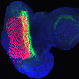
This conference will bring together Developmental Biology, Evolutionary Biology and Maths/Physics, disciplines that are often combined pair-wise, but rarely all together. This will elicit creative discussions, not only on classical questions in developmental biology and morphogenesis but also about regeneration, repair and the behavior of stem cells in their natural environment.

There will be 5 thematic sessions:

(1) Specifying and evolving patterns, size and shape; (2) Collective behavior; (3) Self-organisation; (4) Regeneration, repatterning and repair; and (5) Tissue homeostasis.

The conference will be held in picturesque Roscoff, on the north coast of Brittany. The atmosphere will be convivial, allowing junior researchers and students to interact with world class scientists. The number of attendees will be limited to 100.

Deadline for application: 15 May 2015.

Details at http://www.cnrs.fr/insb/cjm/2015/Vincent_e.html and http://jpvincentlab.com/news/

Thumbs up (No Ratings Yet)
Loading...

Categories: Events

Navigate the archive

Use our Advanced Search tool to search and filter posts by date, category, tags and authors.

Developmental Gene Regulatory Networks Course at Woods Hole

Posted by , on 11 May 2015

Print
GRN Course PosterAn advanced short course “Gene Regulatory Networks for Development” will be given at the Marine Biological Laboratory at Woods Hole, MA, October 11-24, 2015. This will be the 8th edition of this unique course, which is open to graduate students, postdocs, PIs or anyone else who might profit from a fast moving treatment of this new field. The Network Course is an intense and always interesting experience. The course covers structure and function of genomically encoded gene regulatory networks (GRNs) controlling many developmental processes, from biological, modeling, and quantitative viewpoints. Its subject matter ranges from informational and mechanistic aspects of transcription control systems to applications of GRNs in diverse developmental and evolutionary contexts. The course, which is supported by NICHD, has drawn rave reviews in all of its previous incarnations. This year’s faculty is a stellar lineup:
 
Scott Barolo, University of Michigan
James Briscoe, MRC National Institute for Medical Research, London
Martha Bulyk, Harvard Med. School
Justin Kumar, Indiana University
Arthur Lander, University of California Irvine
Bill Longabaugh, Institute for Systems Biology
Roger Patient, University of Oxford
Ellen Rothenberg, Caltech
Harinder Singh, Cincinnnati Childrens Hospital Medical Center
Isabelle Peter, Caltech, Course Assistant Director
David McClay, Duke University, Course Co-Director 
Eric Davidson, Caltech, Course Co-Director
GRN Course Poster
Applications are due July 29, 2015  (Some fellowships and travel assistance are available on request). 
 
Thumbs up (No Ratings Yet)
Loading...

Tags: , , ,
Categories: Events

Young Embryologist Network USA: Frog brains and mouse hearts

Posted by , on 8 May 2015

YENMay2015

The first Boston Young Embryologist Network talks event will be held in the Warren Alpert Building at Harvard Medical School Thursday May 14th, 6-8pm. Food and drink will be provided for discussions and mingling afterwards.

Please register with eventbrite to give an estimate of numbers for food.

Two postdocs, Vaibhav Pai from Tufts, and Stan Artap from Beth Israel, will give short talks about their work investigating bioelectric patterning in Xenopus laevis brain development, and the role of transcription factors in facilitating endocardial-myocardial interactions in the developing mouse heart.

We look forward to seeing you there, and stay tuned for the next meeting on June 18th!

Thumbs up (1 votes)
Loading...

Categories: Events

The SDB-BSDB Interview Chain: Niteace Whittington interviews Wendy Gu

Posted by , on 8 May 2015

Last year we interviewed Niteace Whittington, who won the Society for Developmental Biology (SDB) poster prize at the 2014 meeting in Seattle. Niteace’s prize was attendance at the joint meeting of the British Societies for Cell Biology and Developmental Biology (BSCB and BSDB). Continuing the interview chain, Niteace interviewed Wendy Gu, who won the BSDB poster prize there. As a prize, Wendy will be attending the 2015 SDB meeting this July, in Snowbird, Utah. Unfortunately they did not get a chance to meet in person, so Niteace interviewed Wendy over Skype a few days later.


NW: Congratulations on your achievement. You weren’t at the conference dinner when your prize was announced, so how did you feel when you found out?

WG: I found out at breakfast the following morning. The first person who told me was a PI who used to be based in my department. She sat down and said ‘Congratulations!’ I thought ‘For what?’ I thought she meant ‘Congratulations, you are about to submit your dissertation next week’. So I said ‘Yes, I am very relieved’. She had a strange look on her face, thinking ‘Why are you relieved that you won a poster prize?’ It was only afterwards that she explained to me what had happened the night previously, and it was then that it hit and the news made any sense to me. I’m sure it was the same for you when you won your poster. The standards are so high, and it could have gone to any number of other equally capable scientists, so I know how lucky I am!


NW: Yes, when I won I was in such a state of shock. I was thinking ‘Are you sure that you said the right name?’!

In which lab did you do your PhD and what does your lab work on?

WG: I am based in the lab of Matthias Landgraf in the Department of Zoology at the University of Cambridge. As a group we are interested in how neural circuits are specified, how they function, and the behavior of the animal once the nervous system is built. The model organism we work on is the fruit fly, Drosophila melanogaster, an insect that undergoes complete metamorphosis. But most of our work focuses on the embryonic, and larval stages of development.


NW: What was the title of your poster?

WG: The role of Wnt5 ligand and the Ryk family Wnt receptors in positioning neurites along the anteroposterior axis of the developing Drosophila ventral nerve cord. It is a very technical title!


NW: Could you give a brief summary of what you presented?

WG: The biological question I addressed during my PhD is how axons make a choice between growing anteriorly or posteriorly in the developing nerve cord. The ventral nerve cord of Drosophila is analogous to the vertebrate spinal cord, and within it neurons have to decide where to terminate within a 3-Dimensional space. We know from work done previously in both vertebrate and invertebrate systems that these decisions are axon guidance mechanisms, which involve guidance molecules and the receptors expressed in the neurons. In the medio-lateral axis,the positional cue system Slit/Robo determine the extent to which axons grow medially or laterally. Another positional cue system dictates how the dorso-ventral axis is specified: the Sema/Plexins.

When I started four and a half years ago we didn’t know what positional cue system, i.e. which signal and receptors, was acting in the anterior-posterior axis. What I managed to do in the last few years was to show that Wnt5 is the ligand, or the positional cue, that provides information to the sensory neurons when they grow into the central nervous system. One class of partner receptors of Wnt5 are Ryks, and these include Derailed (Drl), Derailed-2 (Drl-2) and Doughnut on 2 (Dnt). I have shown that sensory neurons express Dnt receptors but not Drl or Drl-2 receptors. Dnt receptors are required for the afferent terminals that project posteriorly. So, DNT needs to be expressed in those neurons in order for them to grow in the proper direction. However, the molecular mechanism that underpins selective growth of axon terminals either anteriorly or posteriorly is unclear. The second biggest finding is that although the other two receptors are not normally expressed, if you exogenously provide either Derailed or Derailed 2 you can also force them to grow and shift their terminals more anteriorly. So one of the receptors is necessary, while the other two are sufficient, in a developmental context.


NW: Had you presented your data previously?

WG: I presented a less complete story at the 2013 Neurobiology of Drosophila meeting at Cold Spring Harbor Laboratory. My current poster has a complete narrative, or as much as I can do within the time frame of a UK PhD. It contains some of the data that will go into a publication.


NW: Do you plan to submit a publication soon?

WG: I would like to but I have to write it first! I think I am going to treat myself to a week or two of break after the intensive writing of a PhD (I submitted on Tuesday!). Once I have celebrated properly I will write it in a journal format. The difficulty is deciding where to submit! Luckily, my topic can be published either in a neuroscience journal or a development biology journal, so in a way I am lucky that I have more choices in that respect.


NW: So you are ready to finish up?

WG: I am. I’m interviewing for postdoc positions. My first interview is happening in less than two weeks and then I have two other ongoing applications. In fact, I met one of the PIs during the BSDB meeting in Warwick, so it was a very productive meeting on many levels!


NW: Are you thinking of staying in the same area of research or are you looking to branch out a little?

WG: I’m ready to venture out. I think after working on this system for four years I want to do something different. The three labs I have applied to are very different in their research scopes. The first one works on neurogenesis and neurodifferentiation in the zebrafish. The second project involves engineering the epigenome. It would involve the use of genome editing tools such as CRISPRs, TALENs and Zinc-finger nucleases to alter the epigenomic code and see what effects it has on various model systems and cell lines. The third option is a complete change and that is to work on plant development. As you can see, I am torn between three very different subjects, all of which I am very excited about. We will see! How about you? Did you stay within your discipline or have you branched out slightly?


NW: I’m in a new model organism, using mouse instead of frog, and I am also looking at a different area of neurogenesis. My graduate work focused more on the brain and the central nervous system and in my new lab (Susan Wray, NIH) I am looking at olfactory development. So I branched out a little bit, but in baby steps!

Are you excited to attend the SDB meeting?

WG: I am very excited. I have never been to that part of America before, and Utah and Snowbird in the summer sounds quite enticing.


NW: Would you be interested in a postdoc in the States?

WG: Not at this point in my life. Two of the positions that I had mentioned are based in Europe and the other in Australia. I am actually from North America, and I have done research in the USA previously, so I am looking to get more exposure to the world before returning. I am sure at some point in my career I will be back in North America, and in the USA in particular. So much of the exciting research is coming from where you are based! Maybe not in the near future, but certainly if I am in a position to look for a second postdoc or perhaps a tenure-track position…


NW: I am sure you will meet some really interesting people while you are at the SDB meeting here. You may find some collaborators or networks that could help with potential jobs in the future. These meetings are really cool because you get to really see what is going on in the other side of the world.

WG: Speaking of which, what did you think of our British Society for Developmental Biology meeting?


NW: I think the biggest difference was the time zone! I was a little bit jetlagged. But I had a really good time, and it was a really good experience, interacting with different people in different areas of development.

WG: I am glad to know that we hosted you well here in the UK!


NW: That’s all of my questions! Thank you for your time and congratulations again. I wish you nothing but the best for your future work!

WG: I’m really sad that we couldn’t meet in person, but we are lucky that we live in an age in which technology can come to the rescue!


Wendy Gu photo

Thumbs up (2 votes)
Loading...

Tags: , , ,
Categories: Interview

Adventures in Studying Brain Sex Differences

Posted by , on 8 May 2015

by Peg McCarthy and Bridget Nugent

The biological phenomenon of hormonally induced sexual differentiation of the brain has been an empirical topic of study for over 50 years1 but much remains to be discovered in terms of both mechanism and functional impact. In the McCarthy lab we exploit the many advantages of the laboratory rat as our model because of its well-characterized neuroanatomy and behavioral repertoire which, as in humans, differs markedly in males and females. In particular we have drilled deeply into the mechanistic bedrock controlling male copulatory behavior with a focus on one brain region, the preoptic area (POA), which is often associated with the hypothalamus but is actually telencephalic in origin2. If this brain region is lesioned, males loose all interest in sex3 and if it is stimulated they loose all interest in anything but sex4. The importance of the POA to male reproduction cuts across species from newts to humans, but the mechanisms underlying its importance are only beginning to be understood in the rodent.

In the beginning, most studies justifiably focused on neurotransmitter production, release and binding, assuming that a change in behavior must be the result of changes in neural activity. But finding precisely what about neurons was different in males versus females proved frustrating and largely unproductive until attention turned to other factors. In particular we determined that prostaglandins, a normally inflammatory mediator, was both necessary and sufficient to masculinize the brain if elevated during a critical perinatal “sensitive period”5. Even more surprising, non-neuronal cells that constitute the brain’s immune system called microglia, are a principle source of the prostaglandins that masculinize the POA and male copulatory behavior6. Cell-to-cell communication that involves neurons, astrocytes and microglia helps to shape the synaptic profile of this brain region by increasing the density of excitatory spine synapses on dendrites of POA neurons.

In the course of our studies we noted that the sex difference in synaptic density, males having twice the number of spine synapses per unit of dendrite length as females, was stable across the lifespan despite the juvenile hiatus during which there are little to no gonadal steroids in circulation in either males or females. This led us to ponder the question of how this cellular memory was maintained, and to answer that we turned our attention to epigenetics.

Steroid hormones are obvious candidates as genome modifiers because their transcription factor receptors are associated with co-factors that possess histone deacetylase activity. But to our surprise we found that rather than directly interacting with DNA or chromatin, steroids were instead (or more likely in addition to) decreasing the activity of a class of enzymes called DNA methyltransferases (DNMTs), lowering their ability to methylate DNA7. Reduction in the activity of these actively methylating enzymes results in demethylation of DNA through mechanisms not well understood. Regardless, the impact is an emancipation of genes that then direct masculinization of the POA resulting in male-typical synaptic patterning and copulatory behavior in adulthood. Moreover, if we allowed demethylation to occur outside of the sensitive period for sexual differentiation of the brain, females were still capable of being masculinized. The end of the sensitive period is operationally defined as the point at which giving masculinizing hormones to a female no longer has any effect on sexual differentiation of her brain. We confirmed that the sensitive period ends before day 10 after birth, and we also showed that hormones no longer reduce DNMT activity at this time. Thus the end of the sensitive period appears to be due to the loss of effect of steroids on DNMT activity. To our knowledge this is the first ever report of a mechanism for the end of the sensitive period.

The next big question of course is – what genes are demethylated in the bipotential brain leading to masculinization? Here we have a tale of woe as this is far harder to answer than it might at first seem. Our initial approach was to categorize gene expression profiles in males and females with and without DNMT inhibition using RNA-Seq. To our surprise there were a relatively small number of genes that showed overall expression differences in males and females, less than 100, and they were evenly distributed between males and females. We then looked to see what genes were turned on in females following demethylation. The expression of many genes was induced in the female POA follow DNMT inhibition, and importantly the majority that had been higher in males were now upregulated in females. These genes we considered our masculinization genes, which after eliminating unknowns was reduced to less than 10 candidate genes. The obvious next step was to characterize methylation of CpG islands found in the promoters of each gene. We chose to use the highly quantitative approach of 454 sequencing. However, there were several challenges inherent to this technique: its only good for amplicons of ~300-600bp, and so for large CpG islands several amplicons are required, meaning multiple pairs of primers per promoter. The book keeping alone is a nightmare and determining CpG methylation requires bisulfite conversion from a large number of biological replicates (in our case three groups with n’s of 8-10 per group). Thus the potential sources of error were insomnia inducing. But despite all this, with all quality controls in place, we found absolutely no meaningful sex differences in CpG methylation in any of our candidate genes. Nothing. At first this was very hard to believe, much less accept, but in retrospect, it makes sense.

The first clue that we should not have expected to detect differences in these candidate genes was hiding in our own data. The observation that DNMT activity is higher in the neonatal female POA was matched by global methylation levels of DNA extracted from the POA, such that females had twice the amount of methylation as males. Twice the amount of methylation on the entire genome should not reduce down to differences in the promoters of a handful of genes. Either our measure of DNA methylation was flawed, or most of the action was outside of the promoters. Turns out both are at least partially true.

Extraordinary claims require extraordinary evidence – a quote attributed to Carl Sagan and used by the dreaded Reviewer #3 to insist that if we were going to claim that females had double the DNA methylation in the POA as males we needed better proof than an antibody based colormetric assay kit. There are only a few roads to complete methylome analysis and most of them are full of potholes. The only superhighway is Genome Wide Bisulfite Sequencing (GWBS) but the tolls are very high and the data generated could circle the globe many times over. So what if we took the on ramp but crashed and burned, finding no sex differences in DNA methylation? These are the kinds of decisions one should not contemplate for long or you will talk yourself out of it.   So we did it; collecting and bisulfite converting the DNA from the POA of newborn males, females and females masculinized by hormone treatment two days earlier, with 3 biological replicates per group, the industry standard. We shipped our samples off for sequencing and waited. It was agony until the data arrived by UPS, as the files were too large to send electronically, and then it was shear terror. Fortunately, one of us (BN) had gained sufficient bioinformatics experience to conduct an initial analysis and found that indeed, females do have twice the level of DNA methylation as males, but only at sites that are very highly methylated, not across the genome. In fact females also had more sites that were entirely unmethylated compared to males and masculinized females, suggesting tight epigenomic regulation in females. Analyses of where in the genome differences in methylation status between males and females can be found showed that most differences are in the intragenic regions, but of course this is where most methylation is found and so the significance of this remains unclear at the moment. Relatively few sex differences were found within CpG islands or shores, consistent with our failure to find any differences in the promoters of our candidate genes in our earlier attempts at 454 bisulfite sequencing.


Schematic

The journey to publishing this paper had begun over two years earlier and we were anxious to put it to rest, submitting the final draft with the bare minimum of analyses of the GWBS data. But there is a treasure trove of information; we are anticipating more surprises reside therein. These will surely provide insight into the proximate mechanisms establishing and maintaining sex differences in this brain region, but will not answer the big picture question of why the brains of males and females evolved this way. At this point we can only speculate, and our speculations fall along two lines of thinking. The first is the process of canalization, which Waddington proposed as the function of epigenetic modifications and a process since considered widely in the context of evolution and the robustness of species in the face of challenge8. The robustness of sex differences in neuroanatomical endpoints (NOT behavior) is reminiscent of canalization and the marked differences in epigenetic marks we have observed may be a mediating factor. Second is a more tautological explanation based on the notion that the reproductive strategy of females requires close guarding of precious and limited gametes while males have a continuous and plentiful supply that they are eager to share. If females were to begin to play like males the consequences would be costly if not disastrous, therefore they actively suppress the gene profile that if activated, leads to masculinization of brain and therefore behavior.


More information on the McCarthy lab here.


1. PHOENIX CH, GOY RW, GERALL AA, & YOUNG WC (1959). Organizing action of prenatally administered testosterone propionate on the tissues mediating mating behavior in the female guinea pig. Endocrinology, 65, 369-82 PMID: 14432658

2. Puelles, L., Harrison, M., Paxinos, G., & Watson, C. (2013). A developmental ontology for the mammalian brain based on the prosomeric model Trends in Neurosciences, 36 (10), 570-578 DOI: 10.1016/j.tins.2013.06.004

3. Heimer, L., & Larsson, K. (1967). Impairment of mating behavior in male rats following lesions in the preoptic-anterior hypothalamic continuum Brain Research, 3 (3), 248-263 DOI: 10.1016/0006-8993(67)90076-5

4. Malsbury, C. (1971). Facilitation of male rat copulatory behavior by electrical stimulation of the medial preoptic area Physiology & Behavior, 7 (6), 797-805 DOI: 10.1016/0031-9384(71)90042-4

5. Amateau, S., & McCarthy, M. (2004). Induction of PGE2 by estradiol mediates developmental masculinization of sex behavior Nature Neuroscience, 7 (6), 643-650 DOI: 10.1038/nn1254

6. Lenz, K., Nugent, B., Haliyur, R., & McCarthy, M. (2013). Microglia Are Essential to Masculinization of Brain and Behavior Journal of Neuroscience, 33 (7), 2761-2772 DOI: 10.1523/JNEUROSCI.1268-12.2013

7. Nugent, B., Wright, C., Shetty, A., Hodes, G., Lenz, K., Mahurkar, A., Russo, S., Devine, S., & McCarthy, M. (2015). Brain feminization requires active repression of masculinization via DNA methylation Nature Neuroscience, 18 (5), 690-697 DOI: 10.1038/nn.3988

8. WADDINGTON, C. (1942). Canalization of Development and the Inheritance of Acquired Characters Nature, 150 (3811), 563-565 DOI: 10.1038/150563a0


Thumbs up (3 votes)
Loading...

Tags: , , , , ,
Categories: Research

Research Technician

Posted by , on 7 May 2015

Closing Date: 15 March 2021

Research technician position is available to work in the laboratory of Dr. Guillermo Oliver in projects related to mammalian organogenesis using mouse models and cultured ES cells.

Mouse experience and molecular biology skills are required

To apply write to


Guillermo Oliver, Ph.D (guillermo.oliver@stjude.org)

Feinberg Cardiovascular Institute, Northwestern Medical School

Chicago, USA

Thumbs up (No Ratings Yet)
Loading...

Categories: Jobs

Postdoctoral Positions in Organogenesis

Posted by , on 7 May 2015

Closing Date: 15 March 2021

POSTDOCTORAL POSITIONS are available to study the cellular and molecular mechanisms controlling the development of the lymphatic vasculature and the visual system using available mouse models and 3D self-organizing stem cells and iPS. Highly motivated individuals who recently obtained a PhD. or MD degree and have a strong background in stem cells and molecular and developmental biology are encouraged to apply. Interested individuals should send their curriculum vitae, a brief description of their research interests, and the names of three references to:


Guillermo Oliver, Ph.D (guillermo.oliver@stjude.org)

Feinberg Cardiovascular Institute,

Northwestern Medical School, Chicago


Thumbs up (No Ratings Yet)
Loading...

Categories: Jobs

Three dimensional human lung tissue in a dish

Posted by , on 6 May 2015

Pioneering efforts by others have made enormous strides in our ability to generate human lung tissue from human pluripotent stem cells (hPSCs); however, these efforts have largely focused on deriving lung-specific cells as flat monolayer cultures or growing these cells on scaffolds 1-7. In our paper, published recently in the open access journal eLife 8, we defined conditions that mimic key stages of lung development in vitro in order to direct differentiation of hPSCs into lung tissue. Importantly, without using engineering approaches such as scaffolds, these conditions prompted the formation of 3D structures in the tissue culture dish. These 3D structures, called spheroids, self-organized from the 2D monolayers and detached from the surface of the tissue culture dish. Spheroids started as small aggregates of epithelium and mesenchyme that were expanded into larger tissues, called human lung organoids (HLOs) (Figure 1).


Node_Figure1_wholemount

Figure 1. A human lung organoid, generated from human pluripotent stem cells. Scale bar represents 100µm.

HLOs have structures that are similar to those found in a human lung (Figure 2). The human lung epithelium makes up the proximal airways, including bronchi and bronchioles and the distal alveoli where gas exchange occurs. Surrounding these epithelial structures is the lung mesenchyme, which includes smooth muscle and other cell types of support cells. Consistent with the human lung, HLOs had proximal airway-like structures that resembled bronchi. The HLOs also possessed distal progenitors and some mature alveolar cells (Type I and Type II), but have not yet formed the saccular structure of alveoli. The HLOs also possessed mesenchymal cell types, included smooth muscle cells and fibroblasts that surrounded both the proximal airway-like structures. This system, which represents the complex structural organization of the lung, coupled with diverse cell types, will allow us to begin to ask questions about human lung development, homeostasis, and disease pathogenesis.


Node_Figure2_IF

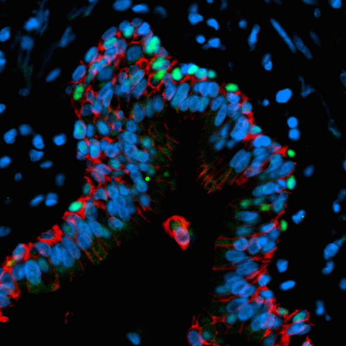
Figure 2. The panel on the left is a cross section through an organoid and on the right a cross section of a human airway way. By immunofluorescence we detected basal cells (green, P63 marker) in the organoids that were organized in a similar manner to the adult airway. The epithelium is labeled in red (beta-Catenin) and all the nuclei in blue (DAPI). Scale bar represents 50µm.

Starting with human pluripotent stem cells, we added ActivinA, which mimics Nodal signaling, a required pathway for differentiation of endoderm, to derive definite endoderm over the course of 4 days. Previous studies have shown that the definitive endoderm is capable of differentiation into foregut endoderm when TGFβ and BMP signaling are inhibited by the small molecule SB431542 (SB) and Noggin, respectively 9. The foregut gives rise to a variety of organs including lung, thyroid, liver, and pancreas. We found that adding SB and Noggin for four days caused the cultures to express lung and thyroid markers. Although the cells acquire the appropriate fate markers, they failed to self-organize and remained as a monolayer. Drawing from previous research, we added a Wnt agonist and FGF4 to cue the formation of 3D clusters in the dish. Activating Wnt and FGF signaling causes the cells to undergo “morphogenesis” in the culture dish, resulting in self-organizing cell clusters called spheroids that detach from the monolayer and float in the media 10,11. Treating endoderm with a combination of TGFβ and BMP inhibitors plus WNT/FGF stimulation led to the formation of self-organizing 3D spheroids that had the appropriate foregut endoderm fate.

Lastly, in order to generate lung specific tissue, we induced Hedgehog (HH) signaling using a Smoothen agonist, SAG. All together, definitive endoderm was given the following instructive cues: (1) TGFβ and BMP inhibitors to derive foregut endoderm, (2) factors inducing 3D spheroid tissue formation (WNT and FGF4), and (3) SAG, a small molecule stimulating HH signaling which enhanced lung lineage induction. These factors were added daily over the course of 6 days and the self-organizing spheroids that floated in the media were collected starting on the 4th day of treatment. These foregut spheroids possessed both epithelium and mesenchyme and expressed foregut and lung-specific markers including SOX2 and NKX2.1 respectively. The floating spheroids were collected and placed in a 3D extracellular matrix, Matrigel. Next, foregut spheroids were expanded into lung organoids through the addition of FGF10 to the media that overlaid the Matrigel droplet. FGF10 is a critical growth factor during lung development and adult homeostasis and we found that it is essential to maintain a healthy epithelium in long-term cultures. Every two weeks the lung organoids were placed in a fresh droplet of Matrigel.

After 65 days in culture (D65), the HLOs had proximal airway-like structures containing cells expressing markers of specific cell types found in this region of the lung including basal, ciliated and club cells. These proximal airway-like structures were surrounded by mesenchyme containing fibroblasts, myofibroblasts, and smooth muscle. In addition to these proximal-like structures, the D65 organoids expressed distal cell markers of both alveoli progenitors and mature alveoli cell types that had similar morphology to the adult alveoli cells, Type I and Type II alveolar cells.

Collectively, we observed that HLOs cultured over 65 days possessed some mature cellular features; however, some cellular features appeared to underdeveloped, leading us to hypothesize that HLOs were more similar to fetal tissue than adult. To address this, we used global transcriptional profiles obtained using RNA-sequencing, and performed an unbiased comparison of HLOs to fetal and adult lung. We found that the derived lung organoids closely resemble fetal lung. It is possible that HLOs remain fetal since they are grown in dish and lack several components of the human lung including blood vessels, nerves, and immune cells. Ongoing studies are aimed at understanding additional cues and cellular inputs to mature the tissue.

In summary, HLOs possess both developmental progenitors and differentiated cells along with structures that resemble the native human lung. Human lung organoids will be an important tool to study human lung development, adult homeostasis, and disease pathogenesis and we are excited for the many new avenues of research this system opens up.


Kadzik, R., & Morrisey, E. (2012). Directing Lung Endoderm Differentiation in Pluripotent Stem Cells Cell Stem Cell, 10 (4), 355-361 DOI: 10.1016/j.stem.2012.03.013

Longmire, T., Ikonomou, L., Hawkins, F., Christodoulou, C., Cao, Y., Jean, J., Kwok, L., Mou, H., Rajagopal, J., Shen, S., Dowton, A., Serra, M., Weiss, D., Green, M., Snoeck, H., Ramirez, M., & Kotton, D. (2012). Efficient Derivation of Purified Lung and Thyroid Progenitors from Embryonic Stem Cells Cell Stem Cell, 10 (4), 398-411 DOI: 10.1016/j.stem.2012.01.019

Mou, H., Zhao, R., Sherwood, R., Ahfeldt, T., Lapey, A., Wain, J., Sicilian, L., Izvolsky, K., Lau, F., Musunuru, K., Cowan, C., & Rajagopal, J. (2012). Generation of Multipotent Lung and Airway Progenitors from Mouse ESCs and Patient-Specific Cystic Fibrosis iPSCs Cell Stem Cell, 10 (4), 385-397 DOI: 10.1016/j.stem.2012.01.018

Wong, A., Bear, C., Chin, S., Pasceri, P., Thompson, T., Huan, L., Ratjen, F., Ellis, J., & Rossant, J. (2012). Directed differentiation of human pluripotent stem cells into mature airway epithelia expressing functional CFTR protein Nature Biotechnology, 30 (9), 876-882 DOI: 10.1038/nbt.2328

Ghaedi M, Calle EA, Mendez JJ, Gard AL, Balestrini J, Booth A, Bove PF, Gui L, White ES, & Niklason LE (2013). Human iPS cell-derived alveolar epithelium repopulates lung extracellular matrix. The Journal of clinical investigation, 123 (11), 4950-4962 PMID: 24135142

Huang, S., Islam, M., O’Neill, J., Hu, Z., Yang, Y., Chen, Y., Mumau, M., Green, M., Vunjak-Novakovic, G., Bhattacharya, J., & Snoeck, H. (2013). Efficient generation of lung and airway epithelial cells from human pluripotent stem cells Nature Biotechnology, 32 (1), 84-91 DOI: 10.1038/nbt.2754

Firth, A., Dargitz, C., Qualls, S., Menon, T., Wright, R., Singer, O., Gage, F., Khanna, A., & Verma, I. (2014). Generation of multiciliated cells in functional airway epithelia from human induced pluripotent stem cells Proceedings of the National Academy of Sciences, 111 (17) DOI: 10.1073/pnas.1403470111

<Dye, B., Hill, D., Ferguson, M., Tsai, Y., Nagy, M., Dyal, R., Wells, J., Mayhew, C., Nattiv, R., Klein, O., White, E., Deutsch, G., & Spence, J. (2015). In vitro generation of human pluripotent stem cell derived lung organoids eLife, 4 DOI: 10.7554/eLife.05098

Green, M., Chen, A., Nostro, M., d’Souza, S., Schaniel, C., Lemischka, I., Gouon-Evans, V., Keller, G., & Snoeck, H. (2011). Generation of anterior foregut endoderm from human embryonic and induced pluripotent stem cells Nature Biotechnology, 29 (3), 267-272 DOI: 10.1038/nbt.1788

Spence, J., Mayhew, C., Rankin, S., Kuhar, M., Vallance, J., Tolle, K., Hoskins, E., Kalinichenko, V., Wells, S., Zorn, A., Shroyer, N., & Wells, J. (2010). Directed differentiation of human pluripotent stem cells into intestinal tissue in vitro Nature, 470 (7332), 105-109 DOI: 10.1038/nature09691

McCracken, K., Catá, E., Crawford, C., Sinagoga, K., Schumacher, M., Rockich, B., Tsai, Y., Mayhew, C., Spence, J., Zavros, Y., & Wells, J. (2014). Modelling human development and disease in pluripotent stem-cell-derived gastric organoids Nature, 516 (7531), 400-404 DOI: 10.1038/nature13863

Thumbs up (3 votes)
Loading...

Tags: ,
Categories: Research

Meeting Report: Genome Editors and CRISPRs meeting, Edinburgh 23rd April 2015

Posted by , on 5 May 2015

Around 200 scientists from several Edinburgh research institutes gathered in central Edinburgh to discuss the current state and uses of zinc finger nucleases, TALENs (TAL effector nucleases) and CRISPR/Cas9 technology. The one-day meeting covered applications ranging from creating transgenic zebrafish lines to carrying out disease-relevant genetic modification of human cells. The meeting was lively and brought together researchers from disparate fields within animal, developmental, cell and biomedical biology. The breadth of research covered by the meeting greatly improved my understanding of the wide range of possible uses of the CRISPR (clustered regularly interspaced short palindromic repeats) system.

(more…)

Thumbs up (2 votes)
Loading...

Tags: ,
Categories: Events

The BSDB introduces the Cheryll Tickle Award

Posted by , on 2 May 2015

BSDBlogoThis is a news item which was first posted on the bsdb.org site. Please, note that not all items will be duplicated on The Node. To ensure you stay informed, please, take a minute to subscribe for email notifications on the bsdb.org site: simply enter your email address in the 3rd item of the side bar. Be ensured that the amount of emails sent to you will usually not exceed one per week or fortnight.

—————————–—————————–

Background & History

The BSDB have taken the decision to award a new annual medal, the Cheryll Tickle Medal, which will be awarded to a mid-career, female scientist for her outstanding achievements in the field of Developmental Biology. The first medal will be awarded at the 2016 BSDB Spring Meeting, where the recipient will present the Cheryll Tickle Award Lecture. BSDB members are invited to nominate suitable candidates.

————————————-

cheryll_tickleThe award is named after Cheryll Tickle (CBE FRS FRSE Hon FSB), an extremely eminent cell and developmental biologist who used the developing limb bud to explore pattern formation in embryogenesis. After her undergraduate studies at Cambridge and PhD work at Glasgow, she worked as a postdoctoral researcher at Yale University, then as a postdoc in the group of Lewis Wolpert at Middlesex Hospital (later merged into UCL) where she studied the morphogen model of digit patterning. This laid the foundation for her subsequent work on the elusive limb polarising factor, mechanisms of limb outgrowth, FGF signalling, HOX gene regulation and snake limblessness.

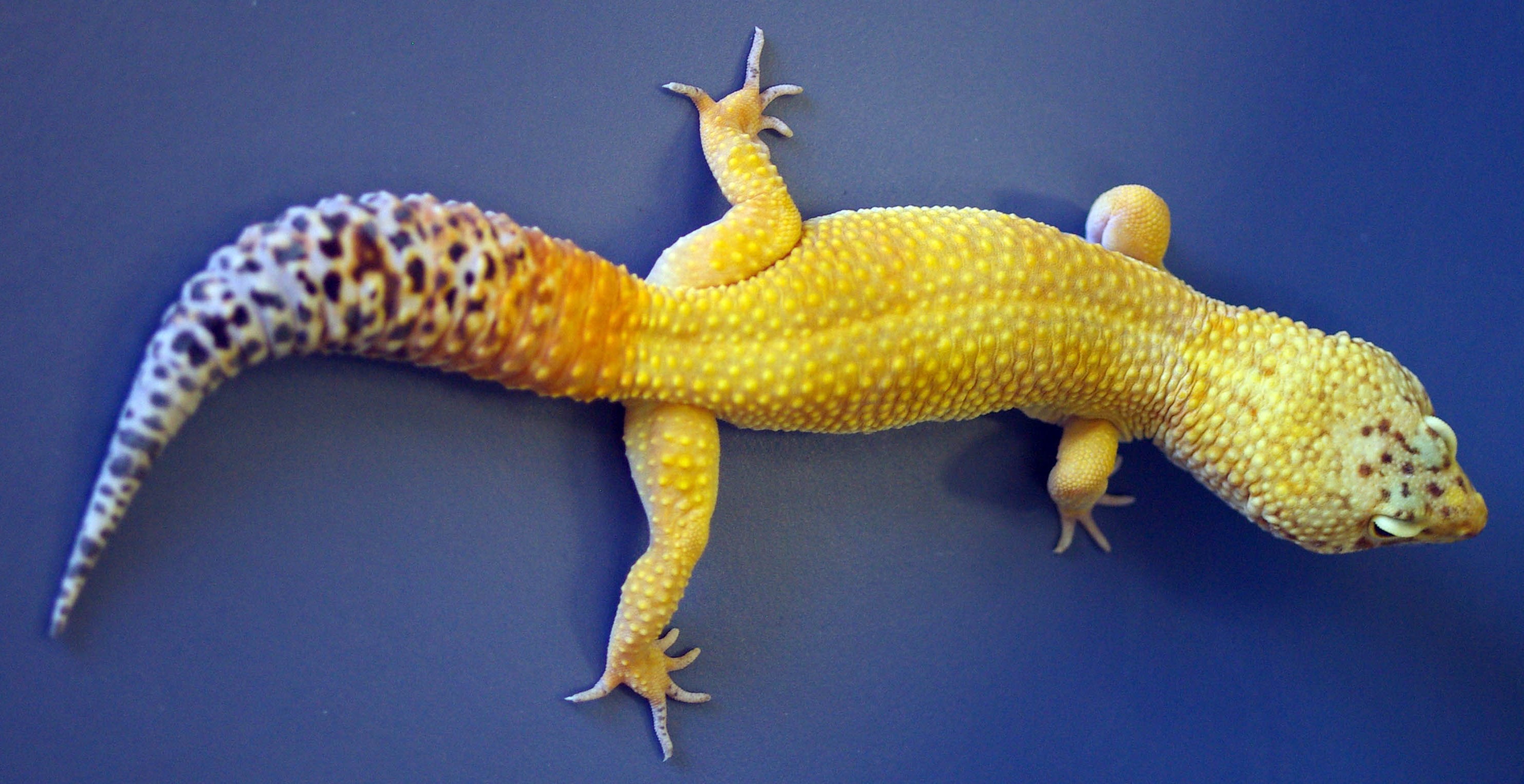
limbWhile at Middlesex/UCL, she moved up the ranks from lecturer, to reader and eventually to Professor, and shortly after she was elected a Fellow of the Royal Society, an acolade which was awarded the same year she moved to Dundee (1998). Cheryll was the first ever Waddington medal winner (1998) and became the first female Royal Society Foulerton Fellow (2000). Currently Professor Emeritus at the sticklebackUniversity of Bath, she continues to explore diverse limb projects such as the loss of the pelvic fin in natural populations of sticklebacks as well as ectopic bone formation in wounded war veterans.


Nominations for the Cheryll Tickle Medal

(this information will be permanently displayed on the BSDB web site)

Nominees should be outstanding female developmental biologists who have started their own research group in the UK within the last 15 years, with allowances for career breaks. Nominees should have made significant contributions to UK Developmental Biology and stand out as a role model for early career female researchers.


The following nomination procedure has been agreed by the Committee:

  1. Formal nominations should be made to the BSDB Secretary (secretary@bsdb.org) by the closing deadline which is 1st of July each year, but can be received at any time.
  2. Nominations should be submitted as a statement of support (maximum 1 page A4) from a Proposer and Seconder (both BSDB members), explaining why the candidate is suitable for the Medal and providing a short summary of their major contributions to Developmental Biology.
  3. Nominations must be accompanied by a CV of max. 2 pages.
  4. All nominations received will be considered, and voted upon, by the Committee.

The winner will be invited to present the Cheryll Tickle Award Lecture at the following BSDB Spring Meeting, where the medal will be presented, usually by the BSDB Chair.

Thumbs up (No Ratings Yet)
Loading...

Tags: ,
Categories: News, Societies