The community site for and by
developmental and stem cell biologists

Neuroblastoma may arise from problems with embryonic nerve development

Posted by , on 1 May 2015

Neuroblastoma is a tumour derived from the peripheral nervous system and is the most common cancer diagnosed within the first year of life. Although is a fairly rare disease, it does account for 15% of all pediatric cancer deaths. However, neuroblastoma is quite unique in that some, particularly very young, patients spontaneously regress requiring only clinical observation. Despite significant advances in the field, the underlying cause or driving mutations behind neuroblastoma have yet to be understood. Interestingly while most other cancers have significant genetic mutations underlying their disease, neuroblastoma cells on the whole appear to have their genomes largely intact, suggesting an alternative cause.

Given that the majority of neuroblastoma cases present during infancy, this implies that there is a unique window of susceptibility during development of the nervous system. During embryonic development, cells must maintain a fine balance between division, to properly generate enough cells, and the process of differentiation required for cells to perform specified functions within the embryo. Neuroblastoma arises from neural crest cells that go on to form the peripheral nervous system. Our work suggests that neuroblastoma may arise during a critical period of development because immature developing cells are incorrectly pushed towards division rather than differentiation.

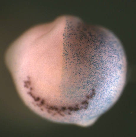
Using a widely used experimental system, tadpoles of the frog Xenopus laevis that develop outside the mother and so are almost uniquely accessible to observation and experimental manipulation of early embryonic stages, we have studied early development of peripheral nervous system. In work funded by the Neuroblastoma Society and recently published in the Journal “Disease Models and Mechanisms”, we identified a cell population of noradrinergic (NA) neurons in the developing tadpoles that are analogous to the immature nerves from which neuroblastoma is thought to derive. We then looked at genes turned on and off in these cells as the embryos progress through development and found that many genes such as Phox2a, Hand2 and Tyrosine Hydroxylase known to be expressed in NA cells in the tadpole are also found at high levels in NB. Most interestingly, we identified one key transcriptional regulator, Ascl1, that is normally only transiently expressed in normal development of NA cells but is found expressed in in both NB primary tumours and almost all the different types of NB cells we looked at. Ascl1, is a member of the basic helix-loop-helix transcription factor family, and we and others have found that is plays an important role in the switch between cell division and differentiation in a variety of neurons in the developing central and peripheral nervous systems. The fact that Ascl1 is expressed in NB cells could mean that it is aberrantly reactivated or that neuroblastoma results from a developmental stage at before Ascl1 downregulation has occurred. As well as offering a more complete understanding of the developmental stage of the neural crest from which NB derives, this also led us to explore whether the presence of Ascl1 may be of functional significance in development of neuroblastoma.

Building on our previous work in the central nervous system, we knew that the Ascl1 protein can be phosphorylated on multiple serine-proline (S)P sites and this phosphorylation limits its ability to drive neurogenesis; conversely a phosphomutant form of Ascl1 is much more effective at driving differentiation. We observed similar phospho-regulation of Ascl1 in the formation of NA neurons of the peripheral nervous system. Serine-proline sites are potential targets for cyclin-dependent kinases, as well as Map kinases, GSK3beta and other proline-directed kinases. Mechanistically, multi-site phosphorylation of Ascl1 on these sites limits its association with promoters and enhancers of downstream targets, and this prevents activation of these multiple targets that are needed for the differentiation process. Indeed, this regulation may be more wide-spread among proneural transcription factors as we have also seen similar multi-site phospho-regulation of a related proneural transcription factor Neurogenin2.


fig


Cdk-mediated phosphorylation of Ascl1 resulting in an inhibition of its ability to drive neuronal differentiation might provide a direct link between the cellular environment in rapidly proliferating cells, and the failure to activate genes that are absolutely required for differentiation. Supporting this hypothesis, when we overexpressed Cyclin A with Cdk2 NA neuron differentiation driven by wild-type Ascl1 was inhibited, but not that driven by expression of phosphomutant Ascl1. We saw a similar phenomenon when overexpressing the N-Myc protein in the embryo; ie that N-Myc inhibits the neurogenic activity of wild-type but not phosphomutant Ascl1 It is of note that NB as whole appears to be a CDK driven disease and that the MYCN gene is very commonly amplified in poor prognosis Neuroblastoma. This suggests that in both Xenopus development and neuroblastoma cells, Ascl1 phosphorylation in response to high levels of Cdks and/or N-Myc results in suppression of NA neuronal differentiation. However, if dephosphorylation of Ascl1 is sufficient to induce NA cell differentiation in Xenopus embryos, the same may be true in neuroblastoma where we find phosphorylated Ascl1 endogenously expressed. Forcing differentiation of NB cells would be expected to confer a much more favourable prognosis than that expected when NB cells are rapidly dividing; preventing phosphorylation of Ascl1 by Cdk inhibitors, possibly in combination with other inhibitors of proline-directed kinases, is a very real potential new treatment approach for NB.

From this work, we would highlight two important conclusions: that neuroblastoma may arise during the phase in sympathetic development when Ascl1 is transiently expressed, and that Ascl1 phosphoregulation plays an important role in control of NA neuron differentiation in normal development, a regulation that may be disrupted in neuroblastoma. From a wider perspective, our findings indicate that neuroblastoma may arise from an abnormality of arrested neuronal differentiation, and so can be viewed as a disease of development. Therefore, understanding the mechanisms underlying the normal development of the peripheral nervous system and how this differs from NB cell behavior will provide critical incite into both what goes wrong during the initial events of neuroblastoma formation, and also to develop future therapies to guide neuroblastoma cells back down their normal path of differentiation.

Wylie, L., Hardwick, L., Papkovskaia, T., Thiele, C., & Philpott, A. (2015). Ascl1 phospho-status regulates neuronal differentiation in a Xenopus developmental model of neuroblastoma Disease Models & Mechanisms, 8 (5), 429-441 DOI: 10.1242/dmm.018630

Thumbs up (3 votes)
Loading...

Tags: , , ,
Categories: Research

Navigate the archive

Use our Advanced Search tool to search and filter posts by date, category, tags and authors.

This month on the Node- April 2015

Posted by , on 1 May 2015

April has been a busy month on the Node! Here are some of the highlights:


Question of the Month- CRISPR technology:

This month a group in China reported genome editing in human embryos. What are the technical and ethical issues of using CRISPR? Share your thoughts here!


BSDB logoBSDB meeting:

April saw the joint Spring meeting of the British Societies for Cell Biology and Developmental Biology . Néstor wrote a meeting report with his perspective on this conference, while we interviewed the winner of the Beddington Medal for best PhD dissertation, John Robert Davis. You can check the full list of winners here. Also look out for the next instalment of our poster winners interview chain, which will be posted on the Node in the coming weeks!


Funding situation:

Scientists are being asked to provide their comments on the current funding situation in both the UK and the USA. Thomas posted his response to the Nurse review on science funding, while Vaibhav addressed the NIH request for information.


Also on the Node:

– Jill and Yoan wrote about their recent paper in eLife examining how branching patterns are established in moss, and what this tells us about the evolution of branching.

– What can an internet cat teaches us about rare diseases? Two postdocs launched a crowdfunding project to sequence the genome of LilBUB!

– Kate posted about her collaborative visit to Cambridge, sponsored by a Development travelling fellowship, to work with Dr. Andrew Gillis on skate axial patterning.

– Qiling highlighted some of the artistic creations of developmental biologists at the NIMR in London, part of their NIMR canvas project to mark the end of this illustrious institute.

– And the latest contribution to our model organisms series is by Sounak, a PhD student in the Aboobaker lab at the University of Oxford. Here is his ‘A day in the life of a planarian lab‘!


TF Criswell square


Happy Reading!

Thumbs up (No Ratings Yet)
Loading...

Categories: Highlights

Towards a mechanistic understanding of branching innovations in plant evolution.

Posted by , on 29 April 2015

Jill Harrison and Yoan Coudert.


The conquest of land by plants was one of the most significant events in our planet’s history, and was underpinned by a series of innovations in plant architecture. Amongst these, the innovation of branching stands out in allowing plants to colonize new volumes of space in the subaerial environment.

Unlike most plants, living bryophyte representatives of the earliest land plants have a biphasic life cycle with multicellular forms in both the haploid (gametophyte) and diploid (sporophyte) life cycle stages. The dominant photosynthetic phase of the life cycle is the gametophyte, and the sporophyte typically comprises a single ephemeral stem capped in a spore-bearing reproductive structure1.

Sporophytic branching forms are thought to have evolved once, contributing to the radiation of our dominant vascular plant flora (c. 260,000 species). In contrast, distinct gametophytic branching forms have evolved in each bryophyte lineage (c. 16,000 species)2.

Mosses are the most speciose bryophyte lineage (c. 10,000)2,3. Although all mosses are relatively small, having leaves that are a single cell thick, their branching habits are diverse and contribute to their ecology4 (Figure 1).


Harrison Fig.1

Figure 1: The diversity of branching forms in mosses. (A-E) Photographs of herbarium specimens of (A) Braithwaitea sulcata, (B) Hypopterygium arbuscula, (C) Cyatophorum bulbosum, (D) Ancistroides genuflexa and (E) Hymenodontopsis stresemannii showing variation in the vertical and radial distribution of lateral branches on the leafy gametophyte. The distribution of the slender leafless sporophytic stems also varies between species. In the species with erect gametophytic forms (A-C), sporophytes are preferentially localised at the top of the shoot, whereas in a species with a pendant form (D), the sporophytes are dispersed. (E) has sporophytes with a lateral and basal position. Dr Yoan Coudert is collaborating with colleagues at the Royal Botanic Garden, Edinburgh and the Natural History Museum in London to characterise evolutionary trajectories between these and other forms using a character mapping approach. Photos by Dr Yoan Coudert, with thanks to NHM for access to specimens. (click to see a bigger image)


There is also an interplay between the gametophytic branching habit and the arrangement of sporophytes on the stem, such that some forms have a single sporophyte at the tip, some forms have a cluster of sporophytes towards the top of the shoot, and others have sporophytes that are dispersed over the plant.

The functional basis and significance of these differences in architecture is not yet known.

Our recent work on the basis of branching patterns in the model moss, Physcomitrella patens, provides a starting point to identify the genetic mechanisms that underpinned the radiation of branching forms in mosses5,6. As there were no previous reports showing how branches arise in Physcomitrella, we started the project by characterising initiation. Using SEM and histology, we found that branches arise spontaneously from the epidermis with a patterned distribution6. Data from flowering plants7, other mosses8, other labs9 and other unpublished projects in our lab led us to believe that a hormonal interplay between auxin, cytokinin and strigolactone could contribute to branching patterns. We used a combination of computational modelling, genetics and pharmacology to show that the integrated action of these three plant hormones determines the distribution of branches up the gametophytic shoot6.

By varying the scope of contributions of each of hormone, we now aim to reproduce the diversity of branching forms in mosses in silico, and will use modelling to generate predictions that allow us to identify the basis of variation between species in future functional work.

The distribution of branches around the shoot is a key component of moss architecture that we have not yet taken into account (Figure 2), and several studies have indicated that the epidermis of Physcomitrella may be the primary site of auxin response5,10,11.


Harrison Fig. 2

Figure 2: Gametophytic branching distributions (A) as represented in Coudert et al. (2015), and (B-D) incorporating radial position. (A) Leaves were removed in a numbered series from gametophytic shoots, and if a branch was revealed, the position was recorded with dark green shading. (B) Movie of a rotating kitchen roll holder with green lines representing leaves ascending the shoot with a 137˚ divergence angle, and blue triangles representing a recorded branch distribution. (C) Photograph showing a cut that allowed us to unravel the kitchen roll holder to see (D) the radial distribution of branched represented in 2D. Photos by Dr Jill Harrison and hands from Dr Yoan Coudert. (click to see a bigger image. You can watch the movie below)


As branch initiation is an epidermal phenomenon, we will adapt our 2D modelling approach to analyse 3D branching architectures including radial patterning. We aim to analyse the level and distribution of each plant hormone in relation to the branching distribution with new fluorescent reporter systems in the future.

The work opens the door to mechanistic understanding of the transitions in form that happened during the evolution of branching- one of the defining features of our dominant land plant flora.


Further reading:

1 Langdale & Harrison (2008). ‘Developmental changes during the evolution of plant form‘ in Evolving Pathways: Key Themes in Evolutionary Developmental Biology (ed A. Minelli and G. Fusco) p.299-315.

2 Shaw, A., Szovenyi, P., & Shaw, B. (2011). Bryophyte diversity and evolution: Windows into the early evolution of land plants American Journal of Botany, 98 (3), 352-369 DOI: 10.3732/ajb.1000316

3 Laenen, B., Shaw, B., Schneider, H., Goffinet, B., Paradis, E., Désamoré, A., Heinrichs, J., Villarreal, J., Gradstein, S., McDaniel, S., Long, D., Forrest, L., Hollingsworth, M., Crandall-Stotler, B., Davis, E., Engel, J., Von Konrat, M., Cooper, E., Patiño, J., Cox, C., Vanderpoorten, A., & Shaw, A. (2014). Extant diversity of bryophytes emerged from successive post-Mesozoic diversification bursts Nature Communications, 5 DOI: 10.1038/ncomms6134

4 Farge-England, C. (1996). Growth Form, Branching Pattern, and Perichaetial Position in Mosses: Cladocarpy and Pleurocarpy Redefined The Bryologist, 99 (2) DOI: 10.2307/3244546

5 Bennett, T., Liu, M., Aoyama, T., Bierfreund, N., Braun, M., Coudert, Y., Dennis, R., O’Connor, D., Wang, X., White, C., Decker, E., Reski, R., & Harrison, C. (2014). Plasma Membrane-Targeted PIN Proteins Drive Shoot Development in a Moss Current Biology, 24 (23), 2776-2785 DOI: 10.1016/j.cub.2014.09.054

6 Coudert, Y., Palubicki, W., Ljung, K., Novak, O., Leyser, O., & Harrison, C. (2015). Three ancient hormonal cues co-ordinate shoot branching in a moss eLife, 4 DOI: 10.7554/eLife.06808

7 Domagalska, M., & Leyser, O. (2011). Signal integration in the control of shoot branching Nature Reviews Molecular Cell Biology, 12 (4), 211-221 DOI: 10.1038/nrm3088

8 von Maltzahn, K. (1959). Interaction between Kinetin and Indoleacetic Acid in the Control of Bud Reactivation in Splachnum ampullaceum (L.) Hedw. Nature, 183 (4653), 60-61 DOI: 10.1038/183060a0

9 Proust, H., Hoffmann, B., Xie, X., Yoneyama, K., Schaefer, D., Yoneyama, K., Nogue, F., & Rameau, C. (2011). Strigolactones regulate protonema branching and act as a quorum sensing-like signal in the moss Physcomitrella patens Development, 138 (8), 1531-1539 DOI: 10.1242/dev.058495

10 Bierfreund, N., Reski, R., & Decker, E. (2003). Use of an inducible reporter gene system for the analysis of auxin distribution in the moss Physcomitrella patens Plant Cell Reports, 21 (12), 1143-1152 DOI: 10.1007/s00299-003-0646-1

11 Jang, G., Yi, K., Pires, N., Menand, B., & Dolan, L. (2011). RSL genes are sufficient for rhizoid system development in early diverging land plants Development, 138 (11), 2273-2281 DOI: 10.1242/dev.060582


Thumbs up (4 votes)
Loading...

Tags: , ,
Categories: Research

BSDB/BSCB Spring meeting 2015, meeting report

Posted by , on 28 April 2015

A couple of weeks ago, the BSDB and the BSCB held their annual joint spring meeting at the University of Warwick. The spring meetings are their major annual event and a must-go for all cell and developmental biologists in the UK (and beyond). Now, before I go any further, here’s a disclaimer: I love this meeting. I cut my proverbial conference teeth at this meeting and I was lucky enough to attend it every year of my PhD. Maybe it is because of the excellent talks, maybe it is the collegiality and sense of community in the air – perhaps it is the fact that the Warwick campus has no other stimulus to distract you from the science… Whatever the reason, it manages to get you excited about science again, and this year it did not disappoint.

Sunday started with the graduate symposium and a careers workshop. Unfortunately I could not attend to these, but you can get some of the tips and keys of the workshop in this Storify from the Node. After dinner, Brigid Hogan and Jennifer Lippincott-Schwartz delivered two memorable plenary lectures that would make the best of Sunday primetime entertainment pale in comparison. Brigid Hogan first showed how lung cells are not always what they seem to be. Combining conditional alleles for lineage tracing and cell ablation with injury models, her lab has found that type 1 and type 2 cells can transdifferentiate into each other to repair the damaged tissue (some of this work has been recently published in Nature Communications). As if that was not enough, Jennifer Lippincott-Schwartz took the stage and, after making all jaws drop with some movies that would become the talk of the conference, she proceeded to give a beautiful lesson on fat metabolism and to show how fatty acids are transported inside the cell. LIVE. You can find all the details on the paper itself. This one would be the first of quite a few talks of metabolism that, as a few people mentioned, highlighted how little we actually know about it and how much more we should study it. By the way, here’s my suggestion to the organizers: Can we have a popcorn stand at the BSDB/BSCB 2016 meeting? All those movies, seriously…

Popcorn

Before I go on, the meeting saw a decent amount of twitter activity under #cbdb15, be sure to check out the twitter feed for more links, talk summaries and science banter!

Over the past few years, the meeting no longer has ‘BSDB’ and ‘BSCB’ sessions. Instead, they all fall into categories of common interest for everybody. I have a soft spot for anything that deals with collective cell behaviors, cell interactions, cooperation and competition, etc, so I mostly went to those sessions (and will comment on them below). That is why on Monday I was rather excited to see Roberto Mayor talk on collective cell migration and contact inhibition of locomotion. They have found how differences in the type of cadherin expressed by pre-migratory and migratory neural crest cells are responsible for their ability to move away from each other and to repolarize after collision in vitro. On a similar note, Paul Martin told us everything from the molecular basis of re-epithelialization, to prognosis markers for chronic wound patients, to the similarities between the immune response to wounds and tumors, all while peppering his talk with jokes mostly aimed at his former PhD student and 2014 Beddington Medal awardee, Will Razzell. If you want my opinion, his explanation of how tension and adhesion are regulated in epithelia via Eph/Ephrin signaling to allow cell movement during would closure was fantastic!

Speaking of wounds, Enrique Amaya talked on Tuesday about wounding and metabolism on this lecture on the role of reactive oxigen species (ROS) during regeneration and development. Interestingly enough, ROS, which are known to cause cellular damage and aging, are necessary for both regeneration and embryonic development in Xenopus. He ended his talk asking whether fertilization is actually an injury – one with life-long consequences, if you ask me! Laura Wagstaff, from the Piddini lab, told us about cell competition and the fascinating relationship between cell density, p53 levels and cell survival. José Gutierrez Marcos explained how plants can inherit stress tolerance through changes in DNA methylation. Jose Silva talked about MBD3, a member of the Nucleosome Remodeling and Deacetylase (NuRD) complex, which is essential for reprogramming into induced pluripotent stem cells (iPSCs) in certain contexts.

Later on, Kiyokazu Agata explained how planarians can make heads from tails when they regenerate after being cut into pieces. Antagonistic ERK and β-catenin activities establish an anteroposterior gradient that allows the polarization of the body pieces to reconstitute an entire body with the right orientation (see paper here). Not only is this a fascinating process, but one of the genes involved, a fibroblast growth factor receptor-like protein, has an awesome name too: nou-darake – brains everywhere!! Later on, Andrea Brand talked about how nutritional stimuli can activate dormant neural stem cells in Drosophila via the Insulin/Insulin-like factor pathway and how gap junctions at the blood-brain barrier (no, flies do not have blood, but the equivalent structure) are essential in this process. Andrew Johnson proposed that two different modes of germ cell specification in vertebrates – either at very early stages or later on in development – determine their evolutionary plasticity (and triggered a good number of questions from a skeptical audience!). Towards the end of Tuesday, Tristan Rodriguez beautifully brought together metabolism and cell competition, and presented how metabolic differences between cells can be the common outcome for a number of other differences observed between cells in competitive environments.

Monday and Wednesday were also days for medals. Kairbaan Hodivala-Dilke received the Hooke Medal from the BSCB while Lewis Wolpert received the Waddington Medal from the BSDB. Two scientists at very different stages of their careers, which was reflected on their talks. While Kairbaan Hodivala-Dilke talked about her bold approach to targeting angiogenesis during cancer progression, Lewis Wolpert gave a very personal career retrospective, where he expressed his views on the current state of science, gave us some quotes and suggested some big biological questions that remain unsolved – inspiration for the next grant proposal anyone?!

Another great piece of news this year was that the bar was to remain open until 3am after the conference dinner (!!!) Finally a worthwhile networking session!! Everyone rejoiced, except those scheduled to talk on the Wednesday morning session… Kudos to them, who had to face the hardest of audiences. Matthieu Piel, in a beautiful talk, showed how cells migrate very differently when constrained than when moving over a flat surface and how they can alter their cytoskeleton to squeeze the nucleus through very narrow spaces. We also saw some more awards: the BSCB’s brand new Women in Cell Biology (WICB) Medal going to Victoria Cowling for her work on the regulation of the mRNA methyl cap and the BSDB’s Beddington Medal to John R Davis (@drjrdavis) for his PhD work on contact inhibition of locomotion – don’t forget to read the recent interview of John R Davis with the Node! You can also catch a list of all the awards given at the conference (including poster awards!) here.

Finally, after the traditional hugging and hand-shaking, everyone rushed back home, hopefully with a good feeling and looking forward to #cbdb16!

And that pretty much sums it up… Really quick, before I sign off, here are my top 3 ‘wish I’d been there’ talks:

– Intestinal sex and contraception, by Irene Miguel-Aliaga
– The cellular and molecular basis for planarian regeneration, by Peter Reddien
– Embryonic transcription is orchestrated by maternal regulatory space, by Gert Veenstra

If anybody is willing to share their notes on these I would be forever grateful!

For any comments or conversation, I tweet @elneztor and you can find my e-mail on my profile at the top.

Thumbs up (3 votes)
Loading...

Tags: , , , ,
Categories: Events

In Development this week (Vol. 142, Issue 9)

Posted by , on 28 April 2015

Here are the highlights from the current issue of Development:


The light at the end of the tubule

FigureTubular structures, such as kidney tubules or blood vessels, carry out crucial functions in organisms. Their morphogenesis requires an orchestrated sequence of cellular rearrangements, the disruption of which leads to tubule dysfunction, as observed in polycystic kidney disease. While the initiation of lumen formation is well understood, less is known about the process of lumen expansion. Using time-lapse analysis of the Ciona intestinalis notochord, a simple model of tubulogenesis in which a lumen forms between two cells connected by an apical ring of cell-cell junctions, Di Jiang and co-workers (p. 1639) show that lumen growth is a non-linear process: it exhibits a lag phase, where the lumen continues to grow but the tight junction ring does not expand. These peculiar kinetics are regulated by the contractility and the dynamics of actomyosin filaments at the lateral cell-cell junctions. Mechanistically, TGFβ negatively regulates the enlargement of the tight junction ring through inhibition of the RhoA-ROCK pathway, which reduces actomyosin contractility. After the lag phase, the osmotic pressure created in the lumen by the apically localised anion transporter Slc26 allows the enlargement of the junctional ring to resume. This study reveals unexpected and complex lumen expansion kinetics, furthering our knowledge of tubule morphogenesis.


Tuning into cilium-mediated Shh transduction

FigureThe primary cilium is an antenna-like structure present at the surface of most cells and necessary for normal development. In particular, it is required for Shh signalling, a crucial developmental pathway. However, the molecular mechanisms underlying the connection between the cilium and Shh transduction remain elusive. To address whether the ciliary localisation of Gli2, a transcriptional effector of the Shh pathway, is required for its activation, Aimin Liu and colleagues (p.1651) generated a knock-in mouse strain in which Gli2ΔCLR, a non-ciliary variant of Gli2 that retains transcriptional activity and responds to its inhibitor Sufuin vitro, is expressed in a similar pattern to endogenous Gli2. Shh signalling is compromised in Gli2ΔCLR mutants and is not restored by the pharmacological activation of Smo, the co-receptor that transduces the Shh signal, in vitro or by the depletion of Ptc1, the Shh receptor that inhibits Smo, in vivo. Furthermore, the authors show that Gli2ΔCLR activates Shh targets at the same level as endogenous Gli2 in the absence of Sufu, indicating that the impaired Shh signalling observed in Gli2ΔCLR mutants results from an impaired ciliary-dependent release from Sufu inhibition. In summary, these findings reveal that the ciliary localisation of Gli2 is required for its activation, highlighting the importance of the primary cilium as a signalling compartment.


Shaping the cerebellum

FigureThe cerebellum, a posterior part of the brain crucial for motor coordination, is composed of folia – functionally distinct units that each receive specific combinations of inputs from the rest of the nervous system. Generation of folia, separated by fissures with anchoring centres at their base, requires the proliferation of granule cell progenitors (gcps) and their differentiation into granule cells (gcs). The basic pattern of folia (relative size, number) is conserved across species. To investigate how gcp behaviour regulates folium geometry, Alexandra Joyner and co-workers (p. 1661) performed a clonal analysis and evaluated the geometry of gc clones, their cell number and cell dispersion with respect to folium size and timing of generation. Gcp division and dispersion were preferentially oriented along the anterior-posterior (AP) axis, which correlates with the preferential AP growth of the cerebellum. Furthermore, gcps do not cross anchoring centres and show different behaviour (geometry and cell number) if they reside in short folia versus long folia. Lastly, by analysing clone geometry in En1+/−;En2−/− mice with shorter folia, the authors show that gcp behaviour does underlie folia size. This study uncovers how the behaviour of neuronal progenitors shapes cerebellar foliation, providing crucial insight into the possible mechanisms that lead to the emergence of a folded neocortex in gyrencephalic mammals.


Ignoring RA to maintain the germline stem cell pool

FigureIn tissues, niche-derived signals promote stem cell self-renewal and the spatially restricted environment shields stem cells from differentiating signals, thus maintaining the stem cell pool. In an open niche environment, such as the seminiferous tubules, where both self-renewal and differentiating signals are ubiquitously distributed, it is unclear how stem cells are maintained in an undifferentiated state. In this study, Shosei Yoshida and colleagues (p. 1582) use lineage-tracing analysis to show that retinoic acid (RA) induces the differentiation of a subpopulation of germ cells marked by NGN3, while another subpopulation (GFRα1+) is maintained. This differential response to RA is explained by the fact that the expression of RARγ, the RA receptor, is restricted to the NGN3+ cell population, thus conferring its differentiation capacity. Interestingly, forced expression of RARγ in GFRα1+cells provided them with the competence to differentiate. This study reveals that the selective response of different germ stem cell populations to RA preserves an undifferentiated stem cell pool, allowing spermatogenesis to persist throughout life.


PLUS:


Animal models for studying neural crest development: is the mouse different?

BronnerF1croppedThe neural crest is a uniquely vertebrate cell type and has been well studied in a number of model systems. Here, Bariga, Trainor, Bronner and Mayor discuss species-specific differences in neural crest development, urging the community to consider these differences and highlighting the need for further research in complementary systems. See the Spotlight on p. 1555


Sensory hair cell development and regeneration: similarities and differences

DEV1561Sensory hair cells are mechanoreceptors of the auditory and vestibular systems and are crucial for hearing and balance. Auditory hair cells in adult mammals are unable to regenerate whereas hair cells in the chick cochlea and the zebrafish lateral line are, prompting studies into the factors that regulate hair cell development and regeneration in various species. Here, Cheng and co-workers review these studies. See the Review on p. 1561


Intrinsic and extrinsic mechanisms regulating satellite cell function

DEV1572Muscle stem cells, termed satellite cells, are crucial for skeletal muscle growth and regeneration. Here, Rudnicki and colleagues review recent discoveries of the intrinsic and extrinsic factors that regulate satellite cell behaviour in regenerating and degenerating muscles. See the Review on p. 1572


Thumbs up (No Ratings Yet)
Loading...

Categories: Research

Animal models for studying neural crest development: is the mouse different?

Posted by , on 28 April 2015

In the latest issue of Development, Elias Barriga, Paul Trainor, Marianne Bronner and Roberto Mayor have contributed a Spotlight article that discusses conserved and non-conserved aspects of neural crest development across vertebrates, and highlights potential concerns or caveats regarding the use of the mouse for the analysis of early neural crest development. The piece raises a number of interesting issues, both from a technical point of view and from a broader evolutionary perspective.

As the article is relatively long, we have not reproduced it in full here (the abstract is copied below), but encourage interested readers to go to the Development website to access the full article for free. We also encourage you to leave any feedback you may have on the article in the comments section here.


Animal models for studying neural crest development: is the mouse different?

Elias H. Barriga, Paul A. Trainor, Marianne Bronner, and Roberto Mayor

The neural crest is a uniquely vertebrate cell type and has been well studied in a number of model systems. Zebrafish, Xenopus and chick embryos largely show consistent requirements for specific genes in early steps of neural crest development. By contrast, knockouts of homologous genes in the mouse often do not exhibit comparable early neural crest phenotypes. In this Spotlight article, we discuss these species-specific differences, suggest possible explanations for the divergent phenotypes in mouse and urge the community to consider these issues and the need for further research in complementary systems.

Thumbs up (5 votes)
Loading...

Tags:
Categories: Discussion

Discovery of an unexpected function of a protein linked to neurodegenerative diseases

Posted by , on 28 April 2015

A study done on fruit flies and published in Nature Communications reveals that the protein dDsk2, in addition to degrading proteins, also plays a key role in regulating gene expression.

This protein is also present in humans and is known to be mutated in several neurodegenerative diseases, including Alzheimer’s. But the mechanism by which these mutations contribute to the development of disease remains unclear.

IRB Barcelona is to start a study to examine the relationship between dDsk2 mutations and neurodegenerative diseases.


Until today, the proteins known as ubiquitin receptors have been associated mainly with protein degradation, a basic cell cleaning process. A new function now described for the protein dDsk2 by the team headed by Ferran Azorín, group leader at the Institute for Research in Biomedicine (IRB Barcelona) and CSIC research professor, links ubiquitin receptors for the first time with the regulation of gene expression. This discovery, published today in Nature Communications, opens up a double scenario, one focused on basic epigenomic research and the other biomedical, because of the link between dDsk2 and neurodegenerative diseases.


Double role of ubiquitin

In humans, there are about 100 proteins associated with ubiquitination, the process by which a protein labelled with ubiquitin is removed from the cell by specific cell machinery known as the proteosome. Ubiquitin receptors are involved in the detection of ubiquitination.

Ferran Azorín, head of the “Chromatin structure and function” group says, “although previous data pointed to the possibility of ubiquitin receptors also contribute to cell processes, data were scarce and a direct role in gene regulation had not been demonstrated.”

“Ubiquitination related to transcription proteins and to DNA repair had previously been described. But this is the first time that a protein, dDsk2, that recognises the ubiquitination of a histone, a protein that forms part of chromatin, has been identified.” Chromatin is a complex formed by DNA and histones —proteins tightly bound to DNA— packaging it into chromosomes and determining gene expression, a process known as epigenetics.

Recent years have brought about the discovery of the fundamental contribution of epigenetics to the development of disease. “We have now opened a new perspective for ubiquitin receptors and we should further this research”, explains Roman Kessler, a Swiss “la Caixa” PhD fellow at IRB Barcelona and first co-author of the paper. In the study, the researchers also reveal the molecular mechanism through which the protein dDsk2 binds to chromatin proteins, thus participating indirectly in the regulation of transcription.


The protein in neurodegenerative diseases

Subjects with Alzheimer’s disease and other neurodegenerative pathologies such as Huntington’s, have a mutation in the protein ubiquilin, the homologue of dDsk2 in humans. “The role of these mutations in the onset and development of disease is still unknown,” says Johan Tisserand, postdoctoral research and co-author of the study who is continuing with the project. “Now that we have discovered this new function, we aim to study whether it affects degradation or transcription, although probably both processes are altered. Our goal is to work towards unravelling these effects,” concludes Ferran Azorín. The new studies will be performed on Drosophila melanogaster and in cells in vitro.


Reference article:

Roman Kessler, Johan Tisserand, Joan Font-Burgada, Oscar Reina, Laura Coch, Camille Stephan-Otto Attolini, Ivan Garcia-Bassets and Fernando Azorín (2015) The ubiquitin receptor dDsk2 regulates H2Bub1 and RNApol II pausing at dHP1c-complex target genes  Nature Communications DOI: 10.1038/ncomms8049


This article was first published on the 28th of April 2015 in the news section of the IRB Barcelona website

Thumbs up (No Ratings Yet)
Loading...

Categories: Research

The science of the cat in your computer: our journey into crowdfunded sequencing of LilBUB

Posted by , on 27 April 2015

About a year ago – when spending some quality afterwork time on the Internet – me and my benchmate Dario stumbled upon LilBUB. If you’re an internet cat afficionado you’ve probably seen LilBUB around. She’s extremely cute and lovable, and she’s got something of a celebrity status. But, as developmental biologists, we were also intrigued by her in a different way. Which is why, two weeks ago, we launched a crowdfunding campaign to sequence the genome of LilBUB. This is the story of our campaign so far.


sofa XRAY-002-labeled hand2


Remember: there’s only one BUB.

LilBUB is not only terribly cute, but she was also born with a strange combination of phenotypic traits. She’s polydactyl (one extra digit on every paw), she’s really small, has a short jaw, possibly some sort of skull bossing, and so-called Erlenmeyer-flask bones. All of the latter traits hail from a very strong bone overgrowth diagnosed as osteopetrosis type IV, but the vets were unsure in their diagnosis. Finally, the bone overgrowth led to her jaws mineralizing too quickly, preventing almost all of her teeth to emerge. Also, since her tongue developed to normal length, it’s hanging out most of the time.


From human to cat genetics

Dario and I are postdocs at the Max-Planck Insitute for Molecular Genetics in Berlin. The focus of our work is to uncover the developmental pathomechanism underlying disease-causing mutations, which have been identified by our human geneticist colleagues. We primarily study mutations affecting limb patterning and bone biology. So we couldn’t help but notice the parallels between the patient-cases that we usually deal with and the phenotype of LilBUB.

Surprisingly though, while we could assign individual traits of LilBUB to one human disorder or another, her combination of traits (with polydactyly AND osteopetrosis) is unique and unlike any case we know. We immediately started hypothesizing what kind of mutation could cause her special looks. Did she have a new form of osteopetrosis? Or was she suffering from two rare diseases? We were super-curious really wanted to find out. So we wrote an email to BUB’s owner, asking if he would allow us to sequence LilBUB’s genome. The reply came within five minutes – he was totally up for it!


The power of the crowd

Having decided to sequence LilBUB we quickly realised that this should not be a traditional project, done somewhere in an isolated laboratory and then communicated only in a scientific journal.

  1. First, LilBUB is at home on the Internet. She has thousands of followers – it should only be fair that they might want to share LilBUB’s scientific journey with us.
  2. Second, LilBUB has a set of traits that resemble rare developmental diseases. Because they only affect very few people, such diseases often don’t get enough attention in the media, even though they can be devastating. Discussing our search for LilBUB’s mutation would make it possible to create awareness for these diseases.
  3. Finally, we were unlikely to get traditional funding: BUB’s an isolated case and an orphan, limiting the experimental approaches we could take.

Thus, we came up with the idea to crowdfund the project, to create some sort of hybrid of classical lab work, outreach and citizen science – and whatever else it would evolve to be. This way we could make thousands of people around the world co-owners of the project, and share our quest with them.


Preparing for battle

Months of preparation followed. First, we joined forces with Uschi, a former collaborator of ours, who had worked on polydactyly during her PhD. She was incredibly enthusiastic about the project, but equally importantly she also brought experience in science communication to the team. As a trio we then set up a project website and social media accounts, scripted a crowdfunding video, and devised a strategic plan for experiments and outreach. Most importantly, following months of brainstorming we came up a name: from a combination of LilBUB + genome, we created the LilBUBome!


And so, two weeks ago, we launched the crowdfunding campaign for the LilBUBome, as well as an accompanying blog, facebook and twitter account. The project launched simultaneaously with it’s sister project, the 99 cat lives initiative by Prof. Leslie Lyons from the University of Missouri. Leslie is an expert in cat genomics, and the 99Lives intends to sequence 99 cats from all over the world to figure out the genetic basis of feline biology and disease. It’s a great combination, when we get to sequence LilBUB we’ll have Leslie’s expertise and plenty of other data to use as a reference, and two projects hopefully reach more people than only one. There could have been no better time fort he LilBUBome than now.


First conclusions

After 2 weeks of fundraising this is what we know: It’s exciting. It’s fun. We love the interactions with our backers and followers. But in one respect it’s just like most other lab-related work: we don’t know whether it’ll work. If we fail to raise the money until May 24th, we get nothing! So, if you’re as curious as we are about the causes behind LilBUB’s magical looks, please spread the word, like, share and maybe even directly support us!

Daniel

Thumbs up (5 votes)
Loading...

Tags: , , , ,
Categories: Funding, News, Outreach

A response to NIH-RFI on funding policies and sustainability of biomedical research

Posted by , on 24 April 2015

Currently NIH has put out a request for information (RFI) to solicit input from the scientific community regarding “Optimizing Funding Policies and Other Strategies to Improve the Impact and Sustainability of Biomedical Research”(NOT-OD-15-084). The deadline to submit a response is May 17th 2015. More information can be found at Rock Talk blog post. Response need to be submitted at this website. 

NIH intends to maximize the impact of taxpayer investment in biomedical research by A] maximizing the productivity and creativity of the research workforce it funds,  B] ensure funding for a broad and diverse group of investigators studying a wide range of important questions. 

A response (unabridged version) submitted to this RFI is opened here. With the hope and intention of stimulating the scientific community to submit a response of their own as well as criticize, debate and use as resource, the points in this response.


Thumbs up (No Ratings Yet)
Loading...

Categories: Discussion, Funding

Young Embryologist’s Network Boston – Looking for speakers

Posted by , on 23 April 2015

The Young Embryologist’s Network is an organization in the UK – you may have seen the recent call for abstracts for the UK annual meeting – that is expanding to Boston! The aim of the first few Boston meetings is to bring together anyone with an interest in embryology and development to hear undergraduates, graduate students and postdocs give 20 minute talks about their work. Food and drink will be provided (for the May and June meetings, by the Dept of Systems Biology at Harvard Med School) for discussions and mingling afterwards.

Please get in touch (gary.mcdowell@tufts.edu) if you’d like to give a talk (events will be in May, June and resume after the summer), and please come along to listen! Please see the poster for more information and contact details.

YENjpeg1

Thumbs up (2 votes)
Loading...

Categories: Events