The community site for and by
developmental and stem cell biologists

I left my frog in San Francisco:

The 15th International Xenopus Conference

Asilomar CA, 2014

Posted by , on 22 September 2014

Every two years, the international Xenopus community gathers to meet. Two years ago I wrote about the meeting in Toulon, France and this time we crossed the pond to Asilomar, CA, just south of San Francisco. Cat Vicente recently wrote about Asilomar in the context of conference venues whilst attending the Genetics Society of America Xenopus conference for the Node.

The conference gives a chance for researchers in various fields, but with the common model system of Xenopus, to discuss their work. The meeting this year took a new direction in not having themed sessions, but rather mixing talks from very different fields into each session. This had the dual purpose of keeping everyone’s attention by switching between different subjects rapidly, but also ensuring people were more likely to stay around for talks outside their field. There was also a strong focus on getting junior faculty and postdocs to speak rather than just the usual senior researchers that are well-known. For these reasons, I personally found this the most engaging conference I have been to.

Keynote talks

There were special lectures given by John Gurdon (Gurdon Institute, University of Cambridge) and Marc Kirschner (Department of Systems Biology, Harvard Medical School), both senior figures in the Xenopus community. John’s talk in particular gave an historical perspective on the beginnings of Xenopus as a model organism, including the transport of frogs from South Africa to the UK by Lancelot Hogben. Apparently, in the early days he was forced to keep his frog colony in the crypt of a church and managed to balance the books by charging hospitals to use frogs as pregnancy tests: when injected with the urine of pregnant women, the hormones present would induce the release of eggs from the female frogs. And from such humble beginnings, the research community has grown, with of course achievements such as John’s Nobel Prize. John speculated on the possible future for research in the frog and the aspiration to be able to track the behavior of individual proteins, within a cell, in real time.

2014-08-25 13.18.52

John Gurdon’s special lecture: a historical perspective on the field of Xenopus research.

The keynote talk was given by Rebecca Heald of University of California, Berkeley. Rebecca’s lab focuses on questions to do with size and scaling of cells. In particular, looking at frogs there are basic questions about different species and different stages of development. Xenopus laevis eggs are nearly twice as big as Xenopus tropicalis and yet they are both single cells and, upon fertilization, undergo cleavage in the same way. How does this relate to the DNA content of the cells and how is this remarkable feat of engineering, in an cell so large, achieved? Also, as frog embryos develop, cells continuously divide whilst the embryo as a whole does not grow. What are the implications of maintaining a similar mechanism of cell division, starting in a cell as large as 1 mm in diameter and ending up at a fraction of the size? Work in frog egg and embryo extract is a significant part of the Xenopus field but not often universally used by frog researchers and it was an overview of some of the amazing work that can be achieved with the simple yet versatile extract system.

P8260087

Rebecca Heald’s tribute to the “Founding Froggers”. Photo courtesy of Cat Vicente.

The community

The frog community also has numerous resources that we simply couldn’t do without: you can check out all things frog at Xenbase and there are three resource centres around the world: the National Xenopus Resource (NXR) in Wood’s Hole, MA; the European Xenopus Resource Centre (EXRC) at the University of Portsmouth, UK; and the National BioResource Project (NBRP) in Hiroshima, Japan which have stocks of wild type and transgenic frogs as well as components such as DNA clones and antibodies. All of these groups presented updates on work they are doing to support the community, such as the genome-editing workshop planned for November 2014 at Wood’s Hole.

Two special workshops were also arranged. One for trainee researchers looking for career advice both in academic and non-academic areas; and one for junior faculty looking for advice on getting tenure. I attended the PhD/postdoc session (as did the Node’s Cat Vicente – as a speaker for non-academic careers) and although the outlook is, as ever, sobering, it was still good to hear some advice specific to people who had worked on frog and some key things to keep in mind. Especially that it’s never too early to be thinking about the next step.

These are both key examples of why I’ve enjoyed my scientific training in the Xenopus community. I started off after finishing my Master’s in Chemistry and moved into a frog lab, and have worked in 3 frog labs/projects since. The sense of community and support provided by the Xenopus field is really quite unique and many who have passed through frog meetings have commented on this. There are some key figures driving the support of more junior faculty and trainees, which is sadly not the norm across all fields of academia.

Looking forward

Work in the model organism Xenopus seems to be really taking off because of all the different techniques that are available to frog researchers. Genetics and genome editing, with TALENs and the CRISP/Cas system, have expanded recently, particularly using the diploid genome of Xenopus tropicalis, but even the pseudoallotetraploid genome of Xenopus laevis, for so long considered a disadvantage, is by its nature of being so unique and outbred generating a lot of questions that other inbred model systems simply can’t answer. The use of Xenopus extracts for in vitro biochemical work was showcased in particular by Rebecca Heald’s lab and the questions that can be answered by the cell-in-a-test-tube system that Xenopus provides still leaves many frog researchers surprised. The rapid development and easy manipulation of frog embryos also allows many simple but elegant in vivo experiments. So, in reality, frogs can be used to answer a whole lot of questions rapidly (and cheaply). I will try to expand upon some of these themes in the near future; needless to say I don’t have time here except to mention what an adaptable model system is across many fields, from bioengineering to biochemistry to biophysics.

2014.08.27 R0002872-2

Frog friends! Photo courtesy of Atsushi Suzuki, from the National BioResource Project, Japan.

This was my third Xenopus conference; each time I’m unsure if I’ll be at the next one, so I always make sure to enjoy the company of my froggy friends; we always make sure to have a good time!

If you want to keep up with frog-related updates, you can follow various twitter accounts for Xenbase @Xenbase, the National Xenopus Resource at the Marine Biological Laboratory at Wood’s Hole @XenopusNXR and I also blog at bewareofthefrog.tumblr.com and @BiophysicalFrog.

Thumbs up (7 votes)
Loading...

Tags: , , ,
Categories: Events

Navigate the archive

Use our Advanced Search tool to search and filter posts by date, category, tags and authors.

International Research Conference on Science, Health and Medicine 2014 (IRCSHM 2014)

Posted by , on 22 September 2014

[Apologies for cross-postings!]

Call for Papers – IRCSHM 2014

October 15-16,

Dubai, UAE

Dear Colleague,

The unique idea behind International Research Conference on Science, Health and Medicine 2014 (IRCSHM 2014) is to provide an opportunity for leading academicians, scientists, researchers, doctors, medical practitioners, paramedical specialists and industry professionals from around the world to network and have scientific discussion on the latest advancements in the closely interlinked domains in science, medicine and health and it’s research benefits for each other’s domain progress.

IRCSHM 2014 will address multiple topics and issues of interest in the areas of science, medicine and health by practical exposure in the form of specialized sessions, poster presentations, plenary sessions and renowned speeches from the leading practitioners reinforcing the upcoming challenges to be faced and their potential solutions. .

We are emailing you this Call for Papers, hoping that you will consider submitting a paper for IRCSHM 2014 which will be held in Corp. Executive Al Khoory Hotel, Dubai, UAE during October 15-16, 2014. For further details, please visit www.ircshm.com.

The types of articles that will be considered for publication include case studies, reviews, as well as theoretical and empirical research. All submitted manuscripts are peer reviewed, and decisions about a manuscript are based only on the importance, originality, clarity, and contribution of the submission to knowledge in the conference scope.

According to this initiative selected invited extended version of best quality papers presented in IRCSHM 2014 will be recommended for publication consideration with Indexed Journals according to the participating Journal publishing policies and requirements.

  1. International Journal of Emerging Technology and Advanced Engineering (IJETAE)(Special Issue) (ISSN 2250–2459(Online)), IJETAE is indexed in many professional databases, IJETAE Impact Factor of 1.932 as per International Society for Research Activity for the year April 2013.


  1. International Journal of Recent Development in Engineering and Technology (IJRDET) (Special Issue) (ISSN 2347 – 6435 (Online)), IJRDET is indexed in many professional databases.


  1. Medical Science (MS)(Special Issue) (ISSN 2321 – 7359; EISSN 2321 – 7367), MS is indexed in many professional databases.


  1. Bioengineering and Bioscience (Special Issue) (ISSN: 2332-0028 (Online)), is is indexed in CABI, Index Copernicus, EBSCO A-Z, J-Gate, JournalTOC, Google Scholar, WorldCat many other professional databases.


The paper submission extended deadline is: 25th September 2014.  You can submit your research paper via email to ircshm@gmail.com.

The topics of interest for submission include, but are not limited to:

Biology • Aerobiology • Astrobiology • Bioengineering • Bioinformatics • Biomedical research • Biotechnology • Synthetic Biology • Botany • Cell biology • Conservation biology • Cryobiology • Environmental biology • Genetics • Hematology • Integrative biology • Marine biology • Microbiology • Molecular biology • Neurobiology • Paleontology • Pathobiology or pathology • Physiology • Phytopathology • Psychobiology • Sociobiology • Structural biology Life Sciences • Affective neuroscience • Biocomputers • Biocontrol • Biodynamics • Bioinformatics • Biomaterials • Biomedical science • Biomonitoring • Developmental biology • Environmental science • Ethology • Evolutionary biology • Evolutionary genetics • Food science • Genomics • Health Sciences • Immunogenetics • Immunology • Immunotherapy • Kinesiology • Nanomaterial Science • Sports science • Structural biology • Systems biology • Zoology
Physics • Biophysics • Classical physics • Acoustics • Fluid dynamics • Geophysics • Quantum physics • Thermodynamics • Theoretical physics • Mathematical Methods • Classical Mechanics • Quantum Mechanics • Classical Electrodynamics • Solid State • Electronics • Atomic Spectroscopy • Nuclear Physics Chemistry • Analytical chemistry • Biochemistry • Inorganic chemistry • Materials chemistry • Neurochemistry • Nuclear chemistry • Organic chemistry • Physical chemistry • Chemical thermodynamics • Chemical kinetics • Electrochemistry • Statistical mechanics • Spectroscopy • Astrochemistry • Theoretical chemistry • Organic Spectroscopy • Organic Synthesis
Medical Science • Anatomy • Anesthesia • Neuroethology • Neuroscience • Oncology • Optogenetics • Optometry • Parasitology • Pathology • Pharmacogenomics • Pharmacology • Physiology • Proteomics • Psychology • Neurology and Neurological Sciences • Neurosurgery • Obstetrics and Gynecology • Cancer Biology (IDP) • Ophthalmology • Cardiothoracic Surgery • Orthopaedic Surgery • Otolaryngology • Pathology • Dermatology • Pediatrics • Psychiatry • Genetics • Urology Paramedical Science • Medical Laboratory Technology • Physiotherapy • Physical Therapy and Rehabilitation • Electro-Cardiogram Technology • Medical Radiographic (X-Ray) Technology • Dental Alternative Science • First Aid • Family Welfare & Population Education • Hygiene & Sanitation • Home Nursing • Health Care Administration • Medical Records Management • Dialysis Technology • Dental Mechanics • Ophthalmic Technology • Audiology & Speech Therapy • Pharmaceutical sciences • Family and Community Medicine
Animal Sciences • Veterinary nursing • Animal health • Animal anatomy • Animal physiology • Animal pathology • Animal pharmacology • Animal toxicology • Animal pharmacy • Animal nutrition • Animal welfare • Veterinary public health • Overseas veterinary development • Animal Health & Welfare • Animal Breeding Health Science • Evidence Based Practice Resources • Healthcare Quality • Family Medicine • Forensic Science • Multimedia Resources • Nursing • Pharmacology • Public Health • Psychiatry and Mental Health • Rehabilitation Therapy • Resources for Health Sciences Research • Natural, Environmental and Health Sciences
Food Science & Nutrition • Chemistry of food • Food microbiology, safety, and risk assessment • Metabolic, molecular, and genetic mechanisms in nutrition • Safety and security analysis of global food supplies • Food preservation, storage, and hurdle technology • Food toxicology • Engineering of food processing technologies • Handling and packaging of foods • Quality assurance of food products • Food oral processing • Health and nutritional implications of food • Bioavailability and disease prevention • Nutritional methodologies, behaviors, and modeling • Sensory and consumer science • Community and international nutrition • Enology and fermentation technology • Food and dietary supplement • Health claims • Interdisciplinary research spanning food science and nutrition Agriculture • Agriculture research on plant production, utilization, biomass, and environment • Horticulture • Fisheries science • Forestry • Sericulture • Agriculture Engineering • Agricultural Marketing, Banking & Co-operation • Agricultural & Food Systems • Crop Production • Crop establishment and variety choice • Livestock Production • Management & Economics • Soil Use & Management • Plant diversity • Agronomy • Animal Production • Agronomy of Combinable Crops • Cereal agronomy • Business Management • Financial Management • Organic Farming • Agricultural & Rural Policy • Grassland Management and Ecology • Farm Business Administration • Nature Conservation • Land Management • Cereal Management and Marketing • Agronomy of Root Crops • Animal Welfare • Dairy Production • Equine Management


Please try to forward this message to as many potentially interested people as possible. For further updates, please visit www.ircshm.com or email to ircshm@gmail.com.

Best Regards,

Organising Secretary,

IRCSHM 2014.

Thumbs up (1 votes)
Loading...

Categories: Events

The Node at the EMBO conference on interdisciplinary plant development

Posted by , on 18 September 2014

The Node is on the road again, but this time not very far! We are going to attend the EMBO conference on interdisciplinary plant development, which starts this Sunday (21st September) at the Sainsbury Laboratory here in Cambridge (UK). We are planning on tweeting with the hashtag  #EMBOplantdev, and will try to include some photos of their beautiful building and the botanical gardens around it! If you are attending the meeting, our community manager will be there, and we look forward to meeting you! We are also looking for someone to blog about the meeting, so if you are interested drop us an email!


Node plant EMBO

Thumbs up (1 votes)
Loading...

Tags: ,
Categories: Events

Mid-career/Senior Group Leader Position in Stem Cell Biology, Institut Pasteur

Posted by , on 17 September 2014

Closing Date: 15 March 2021

The Institut Pasteur (Paris, France) announces an international call for an outstanding mid-career/senior candidate wishing to establish a new research group in the field of “Stem Cells and Development”. The recruitment is part of the Revive Laboratory of Excellence(LabEx – http://www.pasteur.fr/revive) programme on “Stem Cells and Regenerative Biology and Medicine”. Candidates will be integrated into the cutting edge interdisciplinary environment provided by the Department of Developmental & Stem Cell Biology.

The successful candidate will develop a program specializing in the fields of stem cells, genetics, regeneration, translational research or ageing. He/She will be integrated in an internationally renowned Institute combining fundamental and translational research, in an attractive location in central Paris, in close proximity to other major research centres.

Applications will be evaluated on the basis of scientific excellence. A highly attractive package to match the experience of the candidate will be provided.

Candidates should send their formal applications by E-mail to the Director of Scientific Evaluation, Prof. Alain Israël, at the Institut Pasteur g5revive@pasteur.fr.

Application deadline: December 21, 2014

Short-listed candidates will be invited for interview.

Further information on the Institute and on-campus facilities can be found at http://www.pasteur.fr.

Applicants should provide the following (in order) in a single pdf file:

1. A brief introductory letter

2. A Curriculum Vitae, 10 most important publications and a full publication list

3. A description of past and present research activities (up to 2 pages with 1.5 spacing; Times 11 or Arial 10 font size).

4. The proposed research project (up to 4 pages with 1.5 spacing; Times 11 or Arial 10 font size).

Thumbs up (No Ratings Yet)
Loading...

Categories: Jobs

BSMB/BSDB Joint meeting – The Musculoskeletal System: from development to disease

Posted by , on 11 September 2014

BSMB/BSDB Joint meeting – The Musculoskeletal System: from development to disease

1st -3rd September 2014, University of East Anglia

The first joint meeting of the BSMB and BSDB was, in our opinion, a great success. The meeting was held over three days and was packed with brilliant science from areas of musculoskeletal research from the talks, posters and general discussions. The conference was held in the newly built Julian Study Centre at the University of East Anglia, which boasts well-equipped lecture theatres and small meeting rooms, with inviting break out spaces.

The opening keynote speaker, Tom Rando, provided a brilliant start to the conference with a fascinating account of his group’s work into a primed state of quiescent stem cells, which turned out to be one of the highlights of the conference for me (DB), as it’s closely related to my research. This was followed by an exciting afternoon of presentations within the fields of signalling and development. Standout talks from this session included Gabrielle Kardon on the development of the diaphragm, Malcolm Logan on muscle and tendon formation in the limb and Christine Hartmann on bone development. One great feature of the conference was the opportunities offered to PhD students and post-docs to present their work should they be selected from submitted abstracts. I (GFM) had my abstract selected and was able to present my work during this session along with Anne-Gaelle Borycki, Dylan Sweetman, Susanne Dietrich, Clare Thompson and Sue Kimber. The first day ended with an evening poster session and reception.

The atmosphere at the poster session was exciting and stimulating, fuelled by a couple of glasses of free wine and nibbles, conversations around posters were bustling.  This was my (DB) first opportunity to present some of my PhD work in the form of a poster and the sessions allowed for brilliant discussions of my (DB) work with other students working in the field, but also with experienced researchers. It felt great to be able to discuss and get feedback on my work from my peers.

The second day consisted of two sessions, the first focusing on ‘Mechanobiology and Anatomy’. The session started with a fascinating talk from Eli Zelzer, with his group’s novel take on the mechanics of fracture repair. Other great talks included Mario Giorgi on the role of fetal movement in joint morphogenesis and Chrissy Hammond on jaw development in zebrafish. After a quick coffee break, I (GFM) was hugely impressed with talks by Ronen Schweitzer on tendon growth in the mouse limb and Andy Pitsillides on his group’s work on limb growth and joint formation.

The afternoon session was on ‘Human Genetics and Pathology’ with talks by Mike Briggs, Qing-Jun Meng, Madelaine Durbeej, Linda Troeberg and Veronique Lefebvre. The talks offered a great insight into the current research into various dystrophies and we think this was another ‘plus point’ for the meeting as it allowed the combination of developmental biologists and matrix biologists to share ideas with each other, thus initiating stimulating discussions and possible collaborations.

The conference dinner was held at St. Andrews Hall in the centre of Norwich, a grade 1 listed building dating back to the 14th Century. This provided a great venue and atmosphere for more relaxed discussions of the days events. The three-course meal included salmon, pan-fried lamb and lemon tart (plus unlimited wine!).

The final day played host to the final session on ‘Transcriptional and Epigenetic Regulation’ with great talks by Cay Kielty on genotypes of fibrillinopathies and Simon Tew on gene regulation in chondrocytes. The BSMB also awarded the Young Investigator Award to Blandine Poulet and she gave a lovely talk on her work on modelling osteoarthritis. The final keynote speaker, David Glass, in my opinion (GFM) gave the most fascinating and entertaining talk on his group’s work in developing an antibody treatment for patients with muscular atrophies. It was a great talk to end a fantastic conference. The final session of the day concluded with prizes awarded for PhD students and post-docs for best talk and best posters judged by the invited keynote speakers – this was a great incentive for those who presented their posters and a great way to network.

We would like to give our thanks and congratulations to everyone involved in organising the conference (Ulrike Mayer, Andrea Munsterberg, Graham Riley, Ian Clarke and Tonia Vincent. It was a privilege to be involved in the first joint meeting of the BSMB and BSDB, which we thought was a great success between the two societies and their members, and hope that there will be future joint meetings again. We would also like to thank the BSDB/Company of Biologists for providing travel awards for us to attend.


Danielle Blackwell (PhD student) and Gi Fay Mok (Post Doc), Münsterberg lab, UEA

Thumbs up (3 votes)
Loading...

Tags: , , , ,
Categories: Events, Societies

What is your favourite conference venue?

Posted by , on 10 September 2014


Last month I was lucky enough to attend the Xenopus meeting, which took place at the Asilomar conference grounds, close to Monterey, California. This meeting was fantastic, combining great science with a great venue. As there is already someone lined up to write about the scientific aspects of the meeting, I thought I would focus here instead on the venue.

Asilomar is one of the best conference venues I have visited so far. The grounds consist of cabin-style buildings (with fireplaces!) in a pleasant wood, which include accommodation, conference room and cafeteria. Just across the road is the beach and the beautiful californian coastline, there is a swimming pool, and we are surrounded by nature: deer and squirrels in the woods, and whales that can be seen from the coast! Though its natural beauty is definitely a winning feature, what I particularly liked about Asilomar conference grounds was its relative isolation. Conferences are as much about the talks as the interesting discussions that take place in between. When a conference takes place in a big city, attendees tend to stay in different hotels and go their own way after the afternoon sessions, in search of restaurants for dinner. This reduces quite significantly the possibility of chance encounters and hence the opportunity to meet new people and discuss science! This was not a problem in Asilomar, as all the attendees stayed at the grounds and ate all their meals there. And as it was not in a famous world city, there was less of a chance that anyone would skip a session to visit a nearby world attraction! The serene and relaxing location, where you could hear the sea in the nearby beach, kept you could focus on the science and on discussions with the other attendees. At the same time, Asilomar is close enough to Monterey, so that those who wanted to see the local sights (such as the famous Aquarium!) could do so in the free afternoon. And particularly important if you live in the UK, there was that beautiful (and reliable!) californian sun.

This is, of course, my personal perspective, and you may prefer a conference that takes place among the excitement of a big city! What do you think makes the perfect conference venue? Have you been to any conference locations that you thought were particularly good? Let me know what you think by leaving a comment below or on twitter using the hashtag #BestMeetingVenue


Asilomar combined

Thumbs up (4 votes)
Loading...

Tags:
Categories: Discussion, Lab Life

postdoctoral position — neural crest migration

Posted by , on 9 September 2014

Closing Date: 15 March 2021

A postdoctoral position is available in the laboratory of Dr. Shuyi Nie at Georgia Institute of Technology. The lab employs multiple approaches including developmental biology, cell biology, imaging, and biochemistry to study the mechanisms of neural crest cell migration during embryonic development. Potential projects are investigating the regulation of cranial neural crest cell migration by actin cytoskeletal regulators and the roles of transcriptional factors in cardiac neural crest migration, etc.  The lab is well funded with extramural grants and Gatech start-up.

We are looking for highly motivated researchers with training in development biology, molecular biology, or cell biology. Experience in frog or chick embryology, genomics or confocal microscopy are preferred but not required.

For more information, contact Shuyi Nie (shuyi.nie@biology.gatech.edu). Please send a brief statement of scientific/research interests, your curriculum vitae, and a list of 3 references.

Thumbs up (No Ratings Yet)
Loading...

Tags:
Categories: Jobs

The origin of blood

Posted by , on 4 September 2014

DEV110841 3319..3323

As for the origin of species, the question of the origin of blood during development has unleashed a lot of passion among the scientific community. As a matter of fact, the failure to derive blood stem cells (haematopoietic stem cells, HSCs) from pluripotent stem cells (stem cells that can generate any type of cells) has generated a lot of frustration for many scientists and has emphasized the need to understand how HSCs first arise during development.

Various researchers have shown that, in the mouse embryo, HSCs emerge in the dorsal aorta in a step-wise manner: endothelial cells lining the dorsal aorta first commit to the haematopoietic lineage (they start to express the surface marker CD41), and further transition to haematopoietic stem cell fate with the expression of the surface marker CD45. Each of these transitions is tightly regulated by precise stage-specific molecular mechanisms and these been under intense scientific scrutiny.

In a recent report published in Development, Liakhovitskaia and colleagues show that the transcription factor Runx1 is not necessary for the first haematopoietic commitment to CD41+ stage but is essential for the commitment to the CD45+ HSC stage.

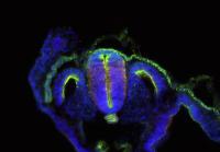
In the picture you can observe the dorsal aorta of a normal mouse embryo at day 10.5 of development. Blue corresponds to the DNA contained in the cell nuclei. The lining of the aorta is composed of endothelial cells marked by CD31 (green) from which some express the first blood commitment marker CD41 (red). These CD31+CD41+ cells are thought to be the immediate ancestors of HSCs.

Interestingly, the authors further observe that similar CD41+ (red) cells are present in the dorsal aorta of embryos from which the protein Runx1 has been deleted, showing that Runx1 deficiency does not prevent the formation of CD41+ cells. However, Runx1 deficient embryos fail to develop CD45+ HSCs, showing that Runx1 deficiency prevents the progression towards HSC lineage.

Altogether, this report provides a better understanding of Runx1 dependent checkpoints during HSCs development. This small step is another forward step towards the understanding the origin of blood and hopefully towards the generation of HSCs from pluripotent stem cells.


Picture credit:

Liakhovitskaia, A., Rybtsov, S., Smith, T., Batsivari, A., Rybtsova, N., Rode, C., de Bruijn, M., Buchholz, F., Gordon-Keylock, S., Zhao, S., and Alexander Medvinsky (2014). Runx1 is required for progression of CD41+ embryonic precursors into HSCs but not prior to this. Development 141, 3319-23. doi: 10.1242/dev.110841

Thumbs up (4 votes)
Loading...

Tags: , ,
Categories: Research

Designing ChIP primers

Posted by , on 4 September 2014

Hello everyone,

Recently I got assigned with the task of designing good primers for ChIP. My supervisor advised me to use the Primer BLAST tool from NCBI together with AmplifiX to get some computer-generated primers and at the same time test some I designed myself. Problem is we were discussing yesterday and eventually we came up with a question: should the primers include the binding sites for the transcription factor we are interested in or there is no problem if they don’t as long as the binding site is sufficiently close to the region amplified by the primers?

In the meantime I already looked for some info online and the idea I got is that it’s not mandatory that the designed primers include the binding site region. However there is not much more info on the subject (at least that I could find…)

So I would like to ask to people that are more familiriazed with this technique: in your opinion what is the best?

Thank you so much in advance!

Cheers! ;)

Thumbs up (1 votes)
Loading...

Categories: Discussion, Lab Life, Research

Researchers grow ‘seed’ of spinal cord tissue in a dish

Posted by , on 3 September 2014

Neuromesodermal progenitors (green) generated in a culture dish form normal precursors of muscle (pink)

Medical Research Council (MRC) scientists have for the first time managed to turn stem cells into the specialised cells that go on to form spinal cord, muscle and bone tissue in the growing embryo. Their discovery could lead to a new way of studying degenerative conditions such as spinal muscular atrophy, which affects the nerve cells in the spinal column, and may pave the way for future treatments for this and other neuromuscular conditions.

During normal embryo development the spinal cord, muscle and skeleton all form from a group of cells called NMPs (neuro-mesodermal progenitors). This process is driven by a series of carefully timed chemical signals, which instruct NMPs to turn into the different cell types in the growing embryo.


By carefully studying and then mimicking this process in a petri dish, researchers at the MRC National Institute for Medical Research and the MRC Centre for Regenerative Medicine, at the University of Edinburgh, were able to coax mouse and human embryonic stem cells into becoming NMPs and then spinal cord cells.

Dr James Briscoe, who co-led the research from the MRC National Institute for Medical Research, said: “There have been some great advances in the field of stem cell research in recent years, with scientists being able to grow liver, heart and even some brain tissue in the lab. The spinal cord, however, has remained elusive because the NMP cells have largely been overlooked – even though they were first discovered more than 100 years ago.

“The real breakthrough for us was realising that we had to coax the stem cells into this intermediate ‘stepping stone’ cell type before turning them into spinal cord and muscle cells. We can’t yet produce the tissues themselves, but this a really big step. It’s like being able to make the bricks and raw materials but not yet build the house.”

Researchers have previously been able to grow some types of nerve, muscle and bone cells in the lab by converting them directly from stem cells. But this is the first time the intermediate NMP cell type, which acts like a ‘stepping stone’, has been created from stem cells. The advantage provided by guiding cells through the routes used in normal development is that the resulting cells may bear closer resemblance to those that occur naturally in the body. This may help any future therapy utilising these cells by providing positional cues to allow them to better integrate with the surrounding tissue.

Neuromesodermal progenitors (green) generated in a culture dish form normal precursors of the spinal cord (pink)

In the near-term being able to grow NMP cells in the lab will allow researchers to learn more about normal human development in a part of the embryo that is otherwise difficult to study. In future the method could also be refined to allow scientists to grow tissue from patients with diseases that affect the spinal cord, muscles, or the motor neurones that connect muscles to the brain and spinal cord. This would provide a powerful new tool to study in a dish how these diseases progress and take hold in the body.

Prof Val Wilson, the co-leader of the research from the MRC Centre for Regenerative Medicine, at the University of Edinburgh, added: “NMPs are important because they’re the source of the spinal cord and most of the bones and muscles in our body. But they have been like Cinderella cells. Although recognised for more than a century in the embryo, they’ve tended to be ignored by scientists trying to make these cell types in a dish. We hope this work will bring them out of obscurity and highlight their importance.”

Dr Rob Buckle, Head of Regenerative Medicine at the MRC, said: “This study is a fantastic example of how combining different branches of science can lead to new discoveries. While there have been many important advances in reprogramming stem cells, it’s important that we explore all the possible routes to generating the specific cell types best suited to clinical development. Incorporating detailed knowledge of early developmental processes is likely to play an important role in providing the fine tuning required to achieve this.”

This article was first published on the 26th of August 2014 in the news section of the MRC website.

The paper, entitled ‘In vitro generation of neuromesodermal progenitors reveals distinct roles for Wnt signalling in the specification of spinal cord and paraxial mesoderm identity’, by Gouti et al, is published in the journal PLOS Biology. Further information available from the MRC press office: press.office@headoffice.mrc.ac.uk

Thumbs up (1 votes)
Loading...

Tags: , ,
Categories: Research