The community site for and by
developmental and stem cell biologists

A simple step to reverse ageing

Posted by , on 3 May 2014

april 2014

How great would it be if we knew how to reverse ageing and turn old organs into young ones? Actually, this might not be as crazy as it sounds. As a matter of fact, a team of scientists managed to regenerate the thymus in old mice and observe what closely resembles the juvenile thymus!

The thymus is a key organ of the immune system as it is where T cells, major actors of one’s immunity, develop and mature. In normal healthy people, the thymus degenerates with age (a process called thymic involution) and this results in a decline of the immune system function. Since our immune system is what protects us against diseases, it is evident that being able to restore the function of the thymus in elder people would be very beneficial.

Interestingly, in this study recently published in Development, Bredenkamp and colleagues achieved thymus regeneration in old mice. They observed a juvenile-like thymus after using genetic engineering to force the expression of the protein foxn1 in the aged thymus.

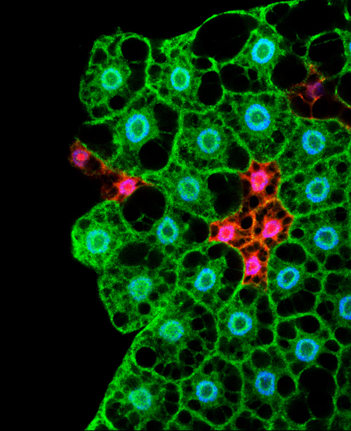
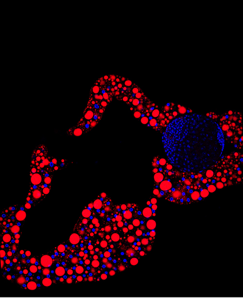
In this picture, you can observe the cortical thymic epithelial cell marker CDR1 in green and the medullary thymic epithelial cell marker keratin 14 (K14) in red. The cortex and the medulla are distinct regions of the thymus and they deteriorate during thymic involution. This results in reduced distinction between cortex and medulla as observed in the bottom picture taken in 24 months old mice. However, when foxn1 is over-expressed in the same 24 months old mice, you can observe that the clear distinction between cortex (green) and medulla (red) is restored, indicative of thymus regeneration.

In addition to the restoration of thymic architecture, the authors show that the regenerated organ has an increased T cell output and a gene expression profile similar to the juvenile thymus.

Most amazingly, apart from being able to “reverse ageing” in the thymus, they show that this regenerative process relies on the over-expression of a single factor (foxn1), making it a lot simpler than one might have anticipated! Thus, this study brings a new provocative concept that will most likely have a broad impact for regenerative biology.


Picture credit:

Bredenkamp, N., Nowell, C., & Blackburn, C. (2014). Regeneration of the aged thymus by a single transcription factor Development, 141 (8), 1627-1637 DOI: 10.1242/dev.103614

Thumbs up (1 votes)
Loading...

Tags: , ,
Categories: Research

Navigate the archive

Use our Advanced Search tool to search and filter posts by date, category, tags and authors.

6th Young Embryologist Annual Meeting

Posted by , on 2 May 2014

ImageJ=1.48a

6th Young Embryologist Annual Meeting
Friday
27th June 2014
JZ Young LT, Anatomy Building, UniversityYEN2014_small College London

Last call for abstracts

The 6th Young Embryologist Meeting aims to bring together developmental biologists from across the UK and abroad to discuss their work at a leading UK institution by presenting a talk or a poster. This year, we are pleased to include The Sammy Lee Memorial Lecture presented by Professor Bill Harris (University of Cambridge). We will also have a Q&A session about ‘Careers in Science’. In addition, prizes will be awarded to the best talks and posters, including a medal in memory of Sammy Lee

This unique meeting is run by PhD students and Post Docs at UCL, KCL, NIMR and  Imperial and aims to create a more interactive research community. Everyone with an interest in developmental biology is welcome to attend this free meeting!
 
We hope the meeting will provide a platform for young researchers (preference is given to PhD and Post Docs) to present their work in a series of 20 minute talks and in the poster session. We will favour diversity of topics and models in the field of embryology.
 
Our main goal is to generate a space where students, postdocs and PIs can discuss a wide variety of topics in developmental biology in a relaxed setting. Ultimately, we would like to strengthen  the Young Embryologist Network to enhance the research environment for embryologist, so please come!
 
Registration is free and can be done here: http://yen2014.eventbrite.co.uk
If you are interested in giving a 15 minute talk or presenting a poster please submit your abstract by clicking here
Deadline for submission is May 19th 2014
 
More information can be found on our website: http://www.youngembryologist.org/
If you want to get more out of your research, meet fellow researchers and get new ideas then join the Young Embryologist Network (YEN) by emailing: youngembryologistnetwork [at] gmail.com
Thumbs up (1 votes)
Loading...

Categories: Events

This month on the Node- April 2014

Posted by , on 30 April 2014

Our jobs page has been particularly busy this month, with a range of job positions from research technicians to senior lectureships! Here are some of the other highlights:


Research:

parhyale squared– Nikos wrote about what his work in Parhyale, recently published in Science, can tell us about the evolution of regeneration.

– The undergraduates at Reed College posted their second contribution, with a journal club discussion of a 2011 paper by Sasai and colleagues on eye morphogenesis in a dish.

– And Caroline re-posted a press release on a recent Development paper in which regeneration of the aged murine thymus was achieved by expression of a single transcription factor


Outreach: cellular architectures

– Andreas was challenged to project cells onto buildings– cells that had been grown in micropatterns matching the buildings’ architecture. Read his post for more about this exciting outreach project in Paris!

– This month we announced the winners of our outreach metaphor competition. Congratulations to Ewart and Roel from the Hubrecht Institute!


Also on the Node:

– Lilian wrote about her favourite gene names. Share your favourite gene name by leaving a comment!

– We interviewed William Razzell, winner of this year’s Beddington medal, awarded by the British Society for Developmental Biology to the best PhD thesis. William did his PhD with Paul Martin at the University of Bristol, and worked on wound healing in Drosophila.

– Mariana wrote about her collaborative visit to Seville, where she produced the transgenic fish lines that will help her establish her new lab in Costa Rica.

– Megan reported from a state meeting of the Australia and New Zealand Society for Cell and Developmental Biology in New South Wales, Australia.

– And at a recent meeting we asked the Node readers to tells us what is the most exciting scientific advance of the last few years, and what is the best scientific advice they ever received. We collated their answers in this short video.


Happy reading!

Thumbs up (1 votes)
Loading...

Categories: Highlights

Research Assistant (St Johnston group)- genome engineering

Posted by , on 29 April 2014

Closing Date: 15 March 2021

Salary: up to £27,004 depending on experience.

Applications are invited for a Wellcome Trust funded Research Assistant position to join an international team in the Gurdon Institute, University of Cambridge. We are looking for a highly motivated and well-organised person who is willing to learn new techniques to join a project using CrispR-mediated homologous recombination to perform targeted genetic engineering in Drosophila . The project is a collaboration between groups in Cambridge, Yale and Oxford and is funded by a Wellcome Trust Strategic Award for 30 months. Applicants should have a degree in a relevant area of biology. Experience in molecular biology or Drosophila genetics would be an advantage.

The Gurdon Institute is a world-renowned centre in the fields of developmental, cell, and cancer biology, located in the heart of the historic city of Cambridge, and part of the University’s School of Biological Sciences. Founded in 1991, its purpose is to provide the best possible environment for research, and to foster interactions and collaborations between scientists with diverse but complementary interests. It is generously supported by core funding from the Wellcome Trust and Cancer Research UK, and benefits from state-of-the art facilities in a friendly, modern, purpose-built environment (see www.gurdon.cam.ac.uk).

To apply online or view further information please visit: http://www.jobs.cam.ac.uk/job/3745

Applications should be submitted via the online application facility.

The University values diversity and is committed to equality of opportunity.

The University has a responsibility to ensure that all employees are eligible to live and work in the UK.

Thumbs up (No Ratings Yet)
Loading...

Categories: Jobs

Research Assistant (St Johnston group)- imaging

Posted by , on 29 April 2014

Closing Date: 15 March 2021

Salary: up to £27,004 depending on experience.

Applications are invited for a Wellcome Trust funded Research Assistant position to join an international team in the Gurdon Institute, University of Cambridge. We are looking for a highly motivated and well-organised person who is willing to learn new techniques to join a project investigating polarised secretion in epithelia and how this is controlled by polarity factors. The project is a collaboration between groups in Cambridge, Yale and Oxford and will involve using new STED and PALM super-resolution microscopes to image polarized secretion in living tissues, and is funded by a Wellcome Trust Strategic Award for 30 months. Applicants should have a degree in a relevant area of biology. Experience in microscopy, molecular biology or Drosophila genetics would be an advantage.

The Gurdon Institute is a world-renowned centre in the fields of developmental, cell, and cancer biology, located in the heart of the historic city of Cambridge, and part of the University’s School of Biological Sciences. Founded in 1991, its purpose is to provide the best possible environment for research, and to foster interactions and collaborations between scientists with diverse but complementary interests. It is generously supported by core funding from the Wellcome Trust and Cancer Research UK, and benefits from state-of-the art facilities in a friendly, modern, purpose-built environment (see www.gurdon.cam.ac.uk).

To apply online or view further information please visit: http://www.jobs.cam.ac.uk/job/3743

Applications should be submitted via the online application facility.

The University values diversity and is committed to equality of opportunity.

The University has a responsibility to ensure that all employees are eligible to live and work in the UK.

Thumbs up (No Ratings Yet)
Loading...

Categories: Jobs

Exciting advances and scientific advice- the Node readers have their say!

Posted by , on 25 April 2014

Last month we attended the joint meeting of the British Society for Developmental Biology and the British Society for Cell Biology in Warwick. At the time we had the opportunity to chat with Node readers, and gather their thoughts on two different questions:
 

– What is the most exciting scientific advance of the last few years?

– What is the best scientific advice that you have ever received?


We collated their answers in the short video below:


Which scientific advance would you highlight? And have you received advice that you would like to share? Share your thoughts by leaving a comment below!

Thumbs up (1 votes)
Loading...

Categories: Discussion, Video

Research Assistant (Hendrich Lab)

Posted by , on 24 April 2014

Closing Date: 15 March 2021

Department/Location: Wellcome Trust – Medical Research Council Cambridge Stem Cell Institute, University of Cambridge, Tennis Court Road, Cambridge, CB2 1QR

Salary: £24,289-£27,318

Reference: PS03240

Closing date: 22 May 2014

Fixed-term: The funds for this post are available until 30 November 2016 in the first instance.

The Wellcome Trust – Medical Research Council Stem Cell Institute draws together outstanding researchers from 25 stem cell laboratories in Cambridge to form a world-leading centre for stem cell biology and medicine. Scientists in the Institute collaborate to generate new knowledge and understanding of the biology of stem cells and provide the foundation for new medical treatments.

Applications are invited for the position of research assistant in Dr. B. Hendrich’s research group.

You must be committed to a career in science and have previous experience with multidisciplinary projects. A wide range of techniques will be used and therefore you should have considerable laboratory experience. Practical experience in biochemistry (western blots, immunoprecipitations), molecular biology (real time PCR, RT-PCR, transfections, gene cloning), and mammalian stem cell culture is essential. Experience in human ES cell or iPS cell culture would be advantageous.

The ideal candidate will have experience in molecular and cell biology and be familiar with the principles of good laboratory practice. Good interpersonal/communication and note-keeping skills are essential, as is the ability to work independently as well as within a laboratory team, as required. You may be required to give oral presentations of your research work to other lab members and prepare written reports for your supervisor. You should also be willing to present your work in oral or poster form at international meetings. The post will require frequent weekend work and a flexible approach to working hours. Careful observation and accurate record keeping are essential.

You should have been awarded a BSc degree or equivalent and have several years basic laboratory experience.

The position will be under the direct supervision of Dr. Brian Hendrich, and is funded by the European Commission 7thFramework Programme Project “4DCellFate.”

Once an offer of employment has been accepted, the successful candidate will be required to undergo a health assessment.

To apply online for this vacancy and to view further information about the role, please visit: http://www.jobs.cam.ac.uk/job/3792. This will take you to the role on the University’s Job Opportunities pages. There you will need to click on the ‘Apply online’ button and register an account with the University’s Web Recruitment System (if you have not already) and log in before completing the online application form.

Please upload your current CV and cover letter with your application by 22nd May 2014.

Informal enquiries are also welcome via email: cscrjobs@cscr.cam.ac.uk.

Interviews will be held week commencing 2nd June 2014. If you have not been invited for interview by 30th May 2014, you have not been successful on this occasion.

Please quote reference PS03240 on your application and in any correspondence about this vacancy.

The University values diversity and is committed to equality of opportunity.

The University has a responsibility to ensure that all employees are eligible to live and work in the UK.

Thumbs up (No Ratings Yet)
Loading...

Categories: Jobs

Lecturer position in Developmental Biology/Reproduction in New Zealand

Posted by , on 23 April 2014

Closing Date: 15 March 2021

Applications are now invited for a lecturer in the Department of Anatomy, University of Otago, New Zealand.

University_of_Otago__Clocktower

Job details: https://otago.taleo.net/careersection/2/jobdetail.ftl?lang=en&job=1400800


Department of Anatomy: http://anatomy.otago.ac.nz/


I’m happy to answer any questions you have about working at Otago.

Dr Megan Wilson

meganj.wilson@otago.ac.nz

@DrMegsW

Developmental Biology laboratory, Department of Anatomy.

Thumbs up (No Ratings Yet)
Loading...

Categories: Jobs

In Development this week (Vol. 141, Issue 9)

Posted by , on 22 April 2014

Here are the highlights from the new issue of Development:


Hemogenic endothelium flexes some muscle

FigureMesoangioblasts (MABs) are progenitor cells of embryonic derivation with mesodermal potential. They have been successfully used to restore skeletal muscle loss in dystrophic mice, but despite the clinical potential of these cells, their origin and role during development has not been defined. Now, on p. 1821, Silvia Brunelli and colleagues identify embryonic MABs that originate from the hemogenic endothelium during the early stages of mouse embryogenesis. The authors use a lineage tracing approach based on VE-cadherin expression to show that the MABs originate from endothelial cells (ECs) in the yolk sac and the placental tissues from approximately embryonic day (E) 8.5 until E10.5, and that these cells contribute to multiple mesodermal lineages during development, including skeletal muscle. The authors further show that this VE-Cadherin-positive extra-embryonic endothelium also generates the first wave of hematopoietic cells that colonise the embryonic mesenchyme. This study demonstrates for the first time that the embryonic hemogenic endothelium can generate extra-vascular mesodermal tissue in vivo.


Motor efferent axons lead the way

FigureThe assembly of the peripheral nervous system occurs in a precise order: motor efferent axons (MEs) emerge first, followed by somatosensory afferent axons (SAs), and then by sympathetic efferent axons (SEs). While this order is clearly defined, it is not clear whether the pioneering axons provide instructive cues for the trailing axons to follow, and thus whether the network represents a true hierarchy. In this issue (p. 1875), Till Marquardt and colleagues take an evolutionary approach to address this issue, and find that peripheral nerve assembly is governed by a stringent hierarchy of axon-dependent interactions. Using elegant in vivo genetic analyses to manipulate sensory and motor axons networks in three different vertebrate organisms – fish, chick and mouse – the authors show that MEs act as pioneer axons, laying down tracks that are followed by SAs, which in turn act as pioneers for SEs. The authors argue that this hierarchy mirrors the phylogenetic emergence of peripheral nerve types during vertebrate evolution.


On growth and gradients

FigureHow does a developing tissue know how much to grow and when to stop? On p. 1884, Marcos Gonzalez-Gaitan and colleagues address this question using theDrosophila eye as a model. This study follows their earlier work proposing a temporal model for growth control in the wing, whereby cells divide when the levels of Decapentaplegic (Dpp) signalling increase by a defined percentage. In the eye, spatial growth patterns are very different from those in the wing, and growth is partially dependent on a Dpp gradient, the source of which – the morphogenetic furrow – moves as development progresses. The authors find that, as in the wing, the signal gradient scales with tissue size – which grows and then shrinks with the progression of the furrow. They then show that their temporal model is quantitatively consistent with observed patterns of proliferation in wild-type and in various mutant conditions. Intriguingly, they also show that the Dpp-independent component of growth control can be explained by a temporal model – implying a similar cellular response to a different signalling gradient. Thus, a model of tissue growth that involves cells dividing in response to defined increases in signalling levels may be applicable across multiple tissues and multiple signalling inputs.


Developing concepts of wound healing

FigureWound repair is a fundamental process that is required for tissue homeostasis and regeneration following damage. Most studies of wound healing have focussed on changes in the leading edge of wounded cells, but here William Razzell, Will Wood and Paul Martin show that morphogenetic cell shape changes that occur multiple cell rows back from the wound are important for efficient wound re-epithelialisation (p. 1814). Using laser-induced wounding of the Drosophila embryo epidermis as a model, the researchers first show that multiple rows of cells around the wound stretch towards the closing tissue. They further reveal dramatic shrinking of the cell-cell junctions that are perpendicular to the pulling force of the wound. This shrinking, which is driven by pulses of myosin that are directed to the cell junctions, leads to cell intercalations. Importantly, these morphogenetic changes, which resemble those observed during the developmental event of germband extension, are essential for wound closure; blocking myosin activity in cells behind the leading edge results in delayed wound contraction. This work highlights an important role for cells surrounding the wound in its closure, and suggests that the cellular morphogenetic mechanisms used during development are recapitulated during wound healing.


miR-8 enables correct synaptogenesis

FigureSynaptogenesis is a complex process that involves the coordinated assembly of pre- and postsynaptic compartments. Various extracellular pathways and cues have been shown to regulate synapse formation but here, on p. 1864, David Van Vactor and colleagues show that the microRNA miR-8 controls synapse structure by repressing the actin regulator Enabled (Ena) and hence modulating synapse morphogenesis at the Drosophila neuromuscular junction (NMJ). The authors previously identified miR-8 as a potent regulator of NMJ architecture and presynaptic morphogenesis, and now find that Ena is direct target of miR-8 that is crucial for mediating its activity in synapse formation. Ena is enriched in the postsynaptic peribouton area surrounding the presynaptic compartment, and this localisation appears to depend on conserved actin-binding domains in the C-terminus of Ena. Further studies suggest that miR-8 controls NMJ architecture by inhibiting Ena expression and, hence, limiting the levels of postsynaptic Ena-dependent actin assembly, which in turn can regulate the expansion of presynaptic arbours. Together, these studies uncover a novel morphogenetic mechanism that coordinates the remodelling of pre- and post-synaptic compartments.


Plus…


Actomyosin networks and tissue morphogenesis

1789- actomyosinTissue morphogenesis is driven by coordinated cellular deformations and recent studies have shown that these changes in cell shape are powered by intracellular contractile networks comprising actin filaments, actin cross-linkers and myosin motors. In their Development at a Glance poster article, Munjal and Lecuit provide an overview of the mechanics, principles and regulation of actomyosin-driven cellular tension driving tissue morphogenesis. See the article on p. 1789


Bioengineering approaches to guide stem cell-based organogenesis

1794- bioengineeringBioengineering approaches promise to bridge the gap between stem cell-driven tissue formation in culture and morphogenesis in vivo, offering mechanistic insight into organogenesis and unveiling powerful new models for drug discovery, as well as strategies for tissue regeneration in the clinic. Here, Lutolf and colleagues draw on several examples of stem cell-derived organoids to illustrate how bioengineering can contribute to tissue formation ex vivo. See the Review article on p. 1794


Genomic imprinting in development, growth, behavior and stem cells

1805- imprintingGenes that are subject to genomic imprinting in mammals are preferentially expressed from a single parental allele. These imprinted genes can directly regulate fetal growth, and recent work has also demonstrated intricate roles for imprinted genes in the brain and in induced pluripotent stem cells and adult stem cells. As Bartolomei and colleagues review here, these findings highlight the complex nature and developmental importance of imprinted genes. See the Review on p. 1805


Thumbs up (No Ratings Yet)
Loading...

Categories: Research

Flippase recognition target: orientation matters, so why care?

Posted by , on 16 April 2014

FRT sites are used often (at least in Drosophila) for inducing deletions or “flipping out” of markers in transgenic constructs.

When there are two FRTs sequences in tandem, after inducing flippase the DNA sequence that is between these two sites will be deleted. If two FRT sites are facing each other (or looking away), the DNA that sits in between them can be inverted after induction of flippase.

FRTFig. 1: different behavior of flippase induced recombination of FRT sites. Left: sin the situation where two FRT sites are in different orientation, the DNA laying between them (the thick line with a red to yellow coloration) can be inverted. As the FRT sites are not deleted in the process, the inversion can happen many times. Right: If the FRT sites sit in tandem on the DNA (thick line), the DNA laying in between them can be deleted. Only one FRT site is left on the original DNA.

Figure 1 illustrates how important the orientation of the FRT sites is for experimental design. So there should be a convention on how to label the orientation of those elements  (as I did in the figure above, where the arrow shows from 5′ end to 3′ end of the element). Most scientist draw the FRT sites in a similar way on their plasmid maps. The big problem is, that this is not done in a consistent way.

The wikipedia article on FRT states the following:

5′GAAGTTCCTATTCtctagaaaGtATAGGAACTTC3′

This is a clear definition of orientation, and the article writers took this information out of a paper published in 1994 (Schlake and Bode, Biochemistry 33 (43): 12746–12751).

Others use a different annotation of  the FRT element, where exactly the other strand is the leading strand (for example in “Drosophila, a laboratory handbook”, Ashburner et al., second edition).

This can cause some problems, especially when sharing plasmids and flies between laboratories or even between people in the same lab. One has to be consistent, and not trust the graphic map of a plasmid, but its sequence.

I hope this blog post helps other scientists to prevent the bad luck I had with experimental design. Always check the sequence and be consistent :)

Thumbs up (11 votes)
Loading...

Categories: Discussion, Research, Resources