Of all the articles on helpimascientist.com, this one garnered the most views—several thousand within a few days after posting about it on Twitter. And I was reminded of the subject after several conversations this week with several new trainees, who wanted to discuss the topic. As always, I’m somewhat reticent to give very specific advice to others, but not because I don’t have definite opinions. I certainly know what being “successful” in science has required of me. What I don’t generally know is how to answer that question for others. Still, I made a stab at it a few years ago, and I stand by what I wrote. If nothing else, it’s a good starting point for open honest discussions.
I would now add that despite working hard at my job, I essentially never missed my son’s or daughter’s sporting events, concerts, etc. I even helped coach baseball and skiing, took up many new hobbies, and spent plenty of quality time with my wife and family. Achieving a balance is never easy and is more of a “lifetime-average” goal than day-to-day objective. But it’s possible if you work at it.
We are pleased to invite you to participate in the Salamander Meeting 2026, which will be held at the Universidad Nacional Autónoma de México (UNAM) in Mexico City from October 6 to 8, 2026.
We invite you to register and submit your abstract on the website.
This meeting will bring together researchers, postdoctoral fellows, and students working on salamander biology, with a particular focus on regeneration, development, and related areas, especially in Ambystoma mexicanum and other salamander models.
The scientific program will include 3 keynote lectures, 17 short talks, and a poster session, providing opportunities for discussion and exchange of ideas across the field.
As this edition will be held in Mexico, the meeting will also include a special cultural component, including a visit to Xochimilco, the axolotl’s native habitat, and a lecture on the broader historical and cultural context of the axolotl in Mexico.
During the meeting, the organizing committee will recognize Dr. Malcolm Maden for his significant scientific contributions to limb regeneration and for clarifying the role of retinoic acid in this process.
IMPORTANT POINTS TO CONSIDER
*The registration fee includes: a visit to Xochimilco, conference materials, coffee breaks, and 2 meals, as well as breakfasts for those staying at the host hotel.
*Accepted abstracts may be included in an e-book to be published in Frontiers in Cell and Developmental Biology (for authors who choose this option) and will be indexed with Crossref and CLOCKSS.
*The conference will include a special issue in Frontiers in Cell and Developmental Biology titled “Salamander Regeneration and Development: Cellular, Molecular, and Evolutionary Insights”; please also consider submitting your work.
We would be delighted to welcome you to Mexico City for this meeting.
As new journal impact factors are released this week, it’s worth considering what an impact factor really is and whether the academic community should put so much emphasis on a single metric.
In the latest issue of Development, Editor-in-Chief James Briscoe and I openly discuss our views on the impact factor and how the field of developmental biology is perceived through its lens. As part of a larger series drawing on real conversations we’ve had with our community, we highlight that the impact factor is not an accurate measure of Development’s real contribution to research and researchers. We detail how authors and readers can take practical steps towards ethical publishing practices that support our communities, as well as the field-specific, society or not-for-profit journals that serve them.
We hope this Editorial ignites discussion and empowers researchers to make change.
“We are all, in a sense, co-authors of the IF. It is not a number that descends from on high; it emerges from millions of individual citation decisions made by researchers like you. If we collectively cite the work that matters to us, in the journals that serve our field, the metrics will follow. We also have a collective responsibility to challenge how IF is used: in grant reviews, in hiring decisions, in promotion committees. If we believe that research should be judged on its own merit, we need to act on that belief, not just as editors, but as reviewers, as panel members, and as colleagues”
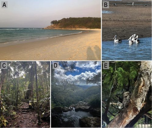
If you drive inland from Meanjin, Australia, also known as Brisbane City, you will find yourself surrounded by subtropical rainforests. Trees and skies are filled with bowerbirds, rainbow lorikeets, cockatoos, my personal favourite, whipbirds, or at night, you may even come across a tawny frogmouth. If you then drive along the coastline, you will meet pelicans, gulls, curlews, spoonbills, oystercatchers, and cormorants sweeping over the ocean surface. If you come to the city, though, and visit The University of Queensland, you may, quite unexpectedly, find the Japanese quail, Coturnix japonica.
Images from Brisbane surroundings, Yuggera, Turrubal, and Quandamooka lands. A) Cylinder Beach, located on Minjerribah Island, also known as North Stradbroke Island; B) Pelicans, spotted in the Moreton Bay region; C-D) Hiking through Springbrook National Park; E) A tawny frogmouth, spotted at The University of Queensland.
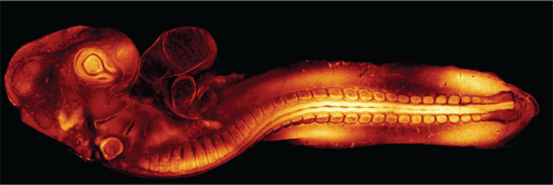
At the Institute for Molecular Bioscience (IMB) at The University of Queensland, you will find my research lab, led by Dr Melanie White . In our lab, we use the quail to study spinal cord development and how, when this process goes wrong, neural tube defects (NTDs) develop. As an imaging lab, we work towards beautiful, state-of-the-art imaging of live embryo development, a process that requires us to use fluorescently labelled, transgenic quails (Alvarez et al., 2024).
A Lifeact-eGFP transgenic quail embryo grown for ~72 hours and imaged using confocal microscopy (Alvarez et al., 2024).
Quails and human spine development
It may surprise you to learn that human spinal cord development closely resembles that of avian species (Dady et al., 2014). The neural tube begins to develop by week three in humans. A flat sheet of epithelial cells folds and thickens into a tube that will become the brain and spinal cord. The anterior region of this tube, the top, is formed by a process called primary neurulation, where cells fold up into a ‘V’ shape, then bend further to create a closed ‘O ‘- shaped tube. The posterior section, the bottom, of the neural tube is formed by secondary neurulation. In secondary neurulation, the flat sheet thickens before lumens form and fuse to make the hollow neural tube (Copp et al., 2003).
This is a generally agreed-upon set of processes, but it wasn’t until recently that a region bridging these two mechanisms was discovered, junctional neurulation. A region where the primary neurulation process tapers off, and the secondary neurulation process begins to take over (Dady et al., 2014; Wang et al., 2026). This overlapping region, termed junctional neurulation, is unique in that it has not yet been seen in any species outside of humans and avians, making birds arguably the best model for human spine development.
Why use the quail?
Using an in ovo model, a model that grows in an egg, has the ethical and practical benefits of development outside of a uterus. However, if all avian species develop their spines in this way, then why use the quail? Simply, they grow very fast. A quail will reach sexual maturity at seven weeks old. For creating a transgenic line, this is a huge advantage: you need to breed multiple generations of birds before you have a line suitable for studies, a process which is much faster with a quickly maturing species. It also allows us to work as fast as possible within the lab. When we grow embryos for our studies, we can grow them for as little as 24 hours before they are old enough to begin studying.
8am: Culturing a quail embryo
A culturing day starts early, and picking up a coffee on the way to the lab is a necessary first step. But then it is time to set up for the day. Agar and albumin (egg white) coated plates are warmed up, and quail eggs are retrieved from the incubator; the eggs having been incubated at 37°C for 24-48 hours beforehand. I often work with embryos after only 24 hours of incubation to catch the earliest stages of neural tube formation.
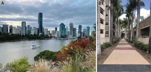
A Brisbane morning showing A) Brisbane City from Kangaroo Point; B) The walkway into IMB
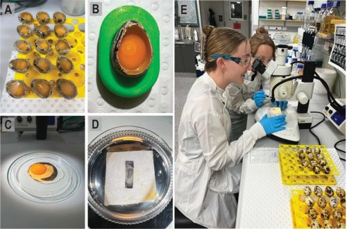
Very carefully, a window is cut into the delicate shell of each egg to check on the embryos, still smaller than a grain of rice and growing on the surface of each yolk. From here, we can grow them longer, leave them in the egg and provide drug treatment or perform in ovo electroporation, allowing them to grow in their eggs as long as possible; more often, we will culture them out of the egg.
Culturing out a quail embryo means pouring them out of their eggs. The albumin is entirely removed, and the yolk itself is patted dry with tissue. Only now can a culture paper be placed over the embryo; a small square of paper with a rectangle slightly bigger than the embryo itself is placed with the embryo in the space. Scissors are used to cut around the edges of the paper, and the embryo is lifted from the yolk, now suspended within that space. The embryo can then be placed on the warmed agar albumin plates, making sure to place it so that, when the heart begins to develop in another ~6-12 hours, there is room for it to fold and grow before it slowly starts to beat.
The embryo culturing process, summarised: A) Windowed quail eggs, covered in parafilm for moisture retention; B) A close-up windowed egg showing the tiny embryo balanced on top of the yolk; C) The same egg emptied into a culturing dish; D) The same embryo, now suspended on a culturing paper; E) Our students on a culturing day.
Embryos can then be returned to the warm 37°C incubator to grow for four to five days. But we rarely need an embryo to grow for this length of time. I can also choose to electroporate embryos at this stage or apply a drug treatment to observe if there is any effect on neural tube development.
1pm: Live Imaging
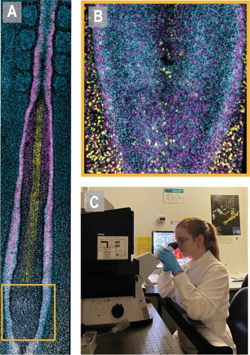
Embryos can be fixed and stained with antibodies for imaging or imaged live. Live imaging of our transgenic quails allows us to study development in real time, recording the symphony of folding and movement that ultimately produces the complete spinal cord and brain. At the IMB , we are lucky to have access to the advanced microscopy facility, with a range of state-of-the-art microscopes and a dedicated microscopy team always willing to troubleshoot any problem.
For live imaging, embryos are mounted on glass-bottom plates with an incredibly thin layer of that Agar-Albumin gel to keep them happy. If, for example, we were using our Lifeact-eGFP embryos in which the actin network is visible through a GFP tag, the embryos would be mounted and then set up on one of our confocal microscopes (Alvarez et al., 2024). After delicately balancing laser power so that it is strong enough to get a signal but not so strong that it bleaches or even damages the embryo itself, you will be able to see the fluorescent, complex, interconnected cabling that actin creates within a single embryo. This beautiful maze of actin will change, move, and grow before your very eyes. Not a bad sight for a Tuesday afternoon.
Live imaging of a single embryo can take anywhere from one hour to 12 hours, depending on which processes you are studying. Development is much slower than it looks when we speed up the final video, but every minute is worth it to unravel these systems. These stunning videos and images don’t just provide us with data; they also allow audiences to connect with our work in unique ways, as visual information drives curiosity. That connection to other scientists and the public drives our research, our passion for the unknown and for this model system.
The developing quail neural tube showing A) part of the quail neural tube imaged at 10x magnification and; B) a close-up of the junctional neural tube imaged at 40x magnification. These images are of a fixed quail embryo stained for various proteins of interest; C) Me, working with the confocal microscopes.
5pm: Pack-up
By 5 pm, I have worked a long but rewarding day. On some culturing days, you may do everything from egg to image; on others, you might just culture and fix your embryos to work on throughout the week. These big culturing days usually happen once a week, with the rest of the week spent processing your images and data from previous weeks, planning future experiments and prepping in the lab. We have the distinct advantage in the lab of everyone using the same techniques, so our lab “chores” are communal; replace what you use, and everything will be ready for the next person. When everything is restocked and packed away, it’s time to head home, where a cup of tea and a book are waiting for me
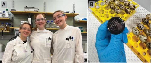
The Quail Morphogenesis lab; A) some of our student team, including myself (in the middle); B) our team logo on a pin.
Alvarez, Y. D., van der Spuy, M., Wang, J. X., Noordstra, I., Tan, S. Z., Carroll, M., Yap, A. S., Serralbo, O., & White, M. D. (2024). A Lifeact-EGFP quail for studying actin dynamics in vivo. J Cell Biol, 223(9). https://doi.org/10.1083/jcb.202404066
Copp, A. J., Greene, N. D., & Murdoch, J. N. (2003). The genetic basis of mammalian neurulation. Nat Rev Genet, 4(10), 784-793. https://doi.org/10.1038/nrg1181
Dady, A., Havis, E., Escriou, V., Catala, M., & Duband, J. L. (2014). Junctional neurulation: a unique developmental program shaping a discrete region of the spinal cord highly susceptible to neural tube defects. J Neurosci, 34(39), 13208-13221. https://doi.org/10.1523/jneurosci.1850-14.2014
Wang, J. X., Alvarez, Y. D., Tan, S. Z., Ranie, S. N., Stehbens, S. J., & White, M. D. (2026). Quantitative live imaging reveals PRICKLE1 controls junctional neural tube morphogenesis independent of Planar Cell Polarity. Nature Communications, 17(1), 3654. https://doi.org/10.1038/s41467-026-71242-0
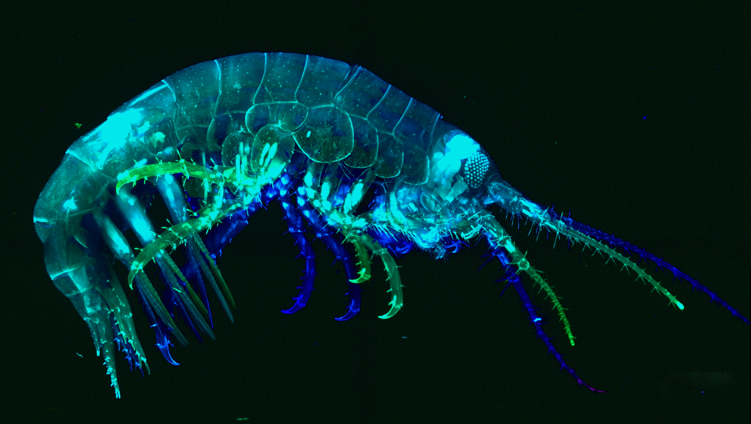
In January of 2025, advanced graduate students and postdoctoral fellows from across the Americas gathered in Chile to participate in the International Course on Developmental Biology, an EMBO Practical Course held at the Marine Biology Station of Quintay (CIMARQ). Over two weeks of intensive training, the students generated some beautiful images of development across different species. We’re excited to launch a competition to pick an image taken by the students to become the cover of an issue of Development, immortalised in a future issue of the journal – a testament to the bright future of developmental biology in Latin America. The next cohort of the International Course on Developmental Biology is currently accepting applications until 30 July 2026.
Please vote for your favourite image using the poll at the bottom of the page. The voting will close on Wednesday, 24 June 2026, at 13:00 BST (UTC + 1). One vote per person.
Browse through the gallery (click to view full image)
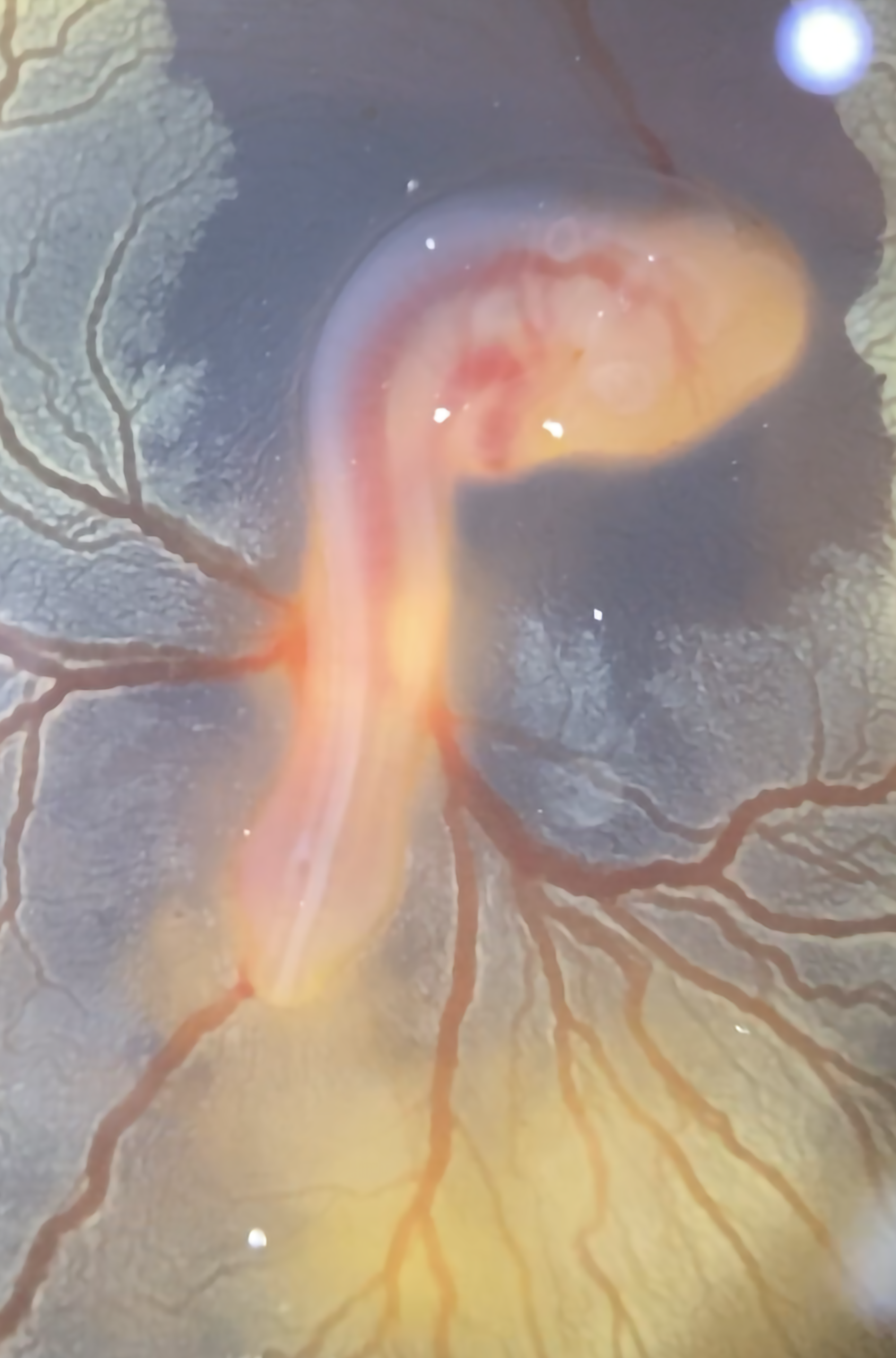
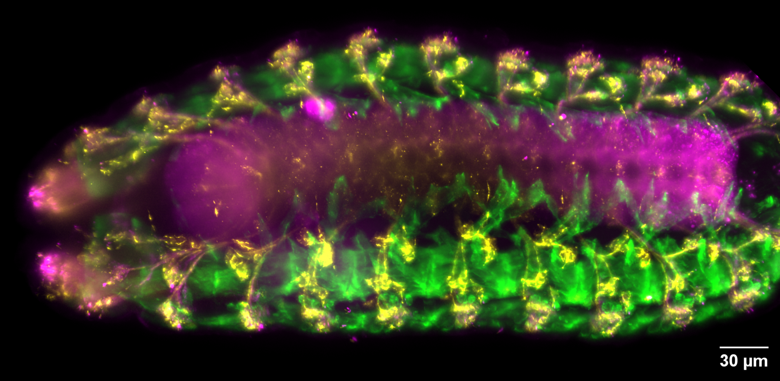
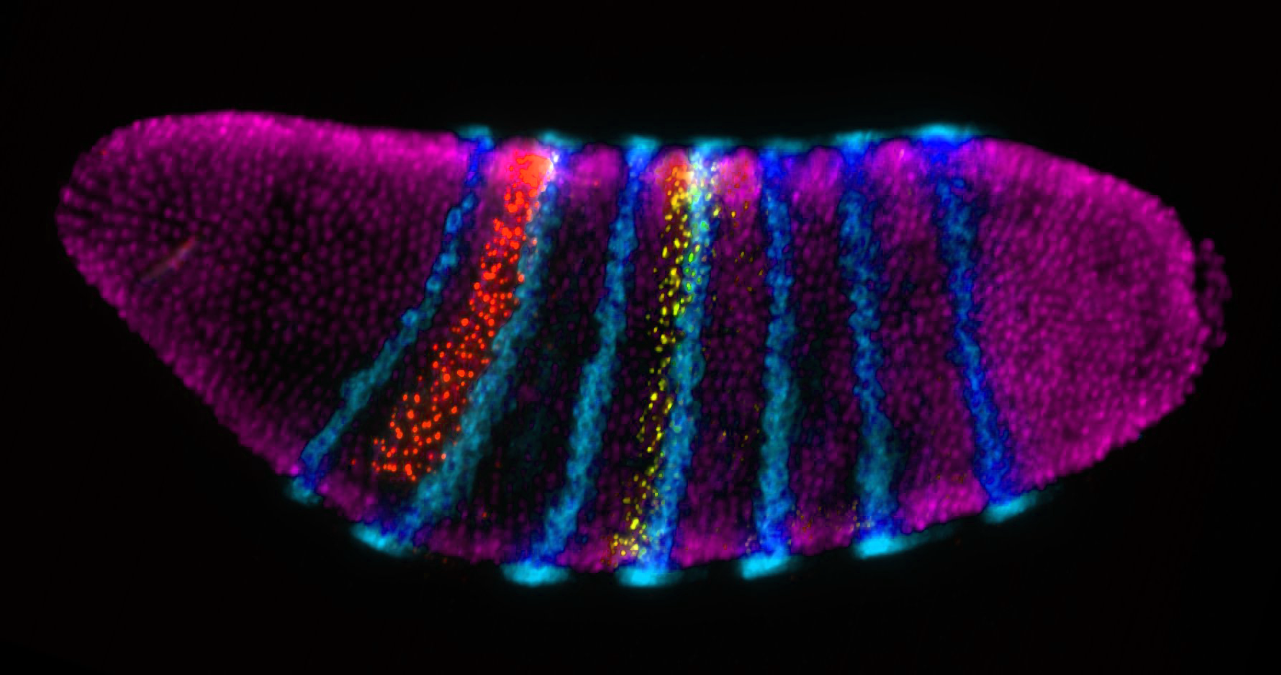
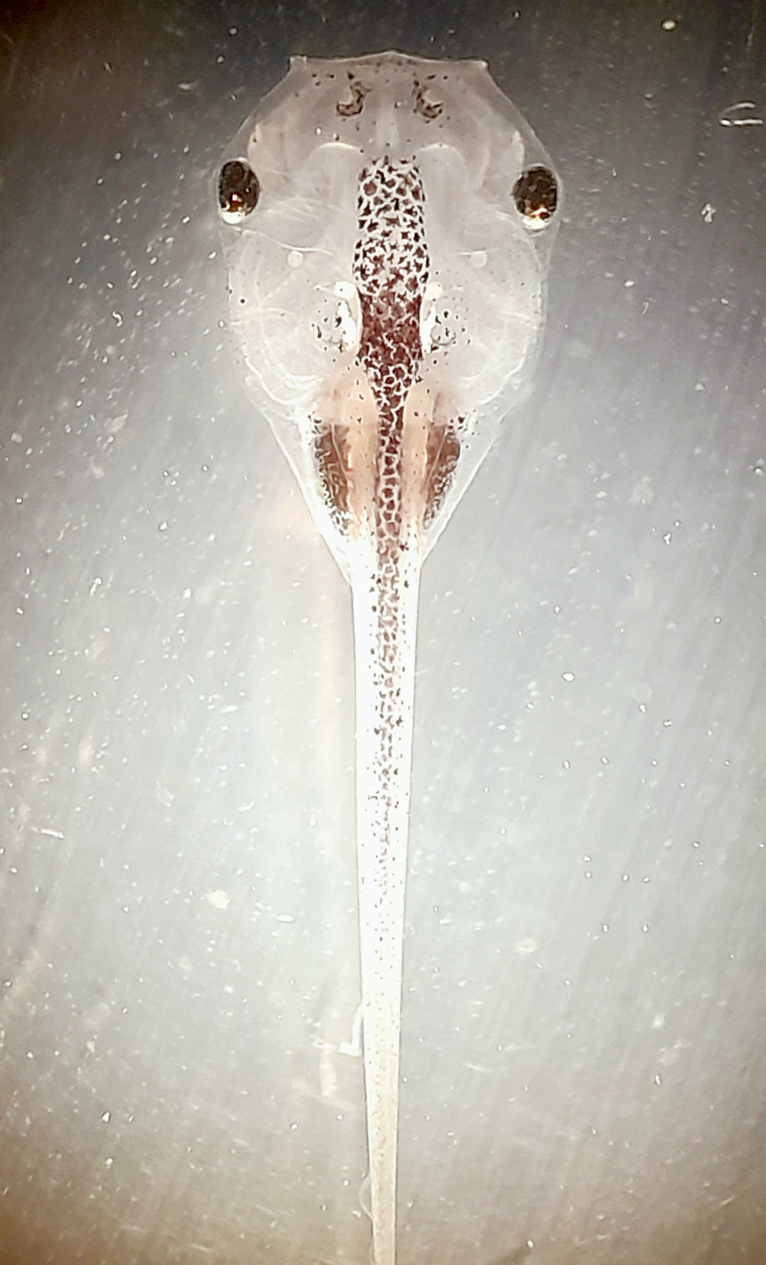
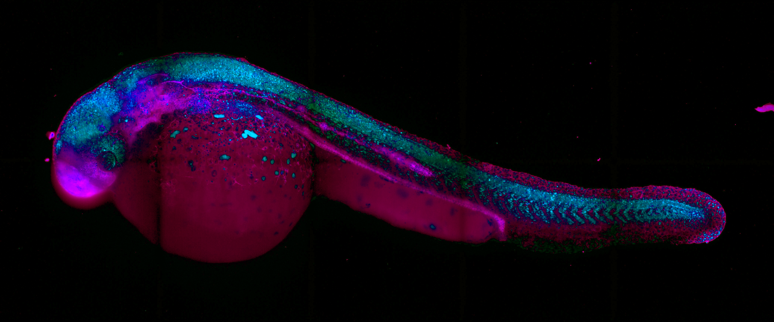
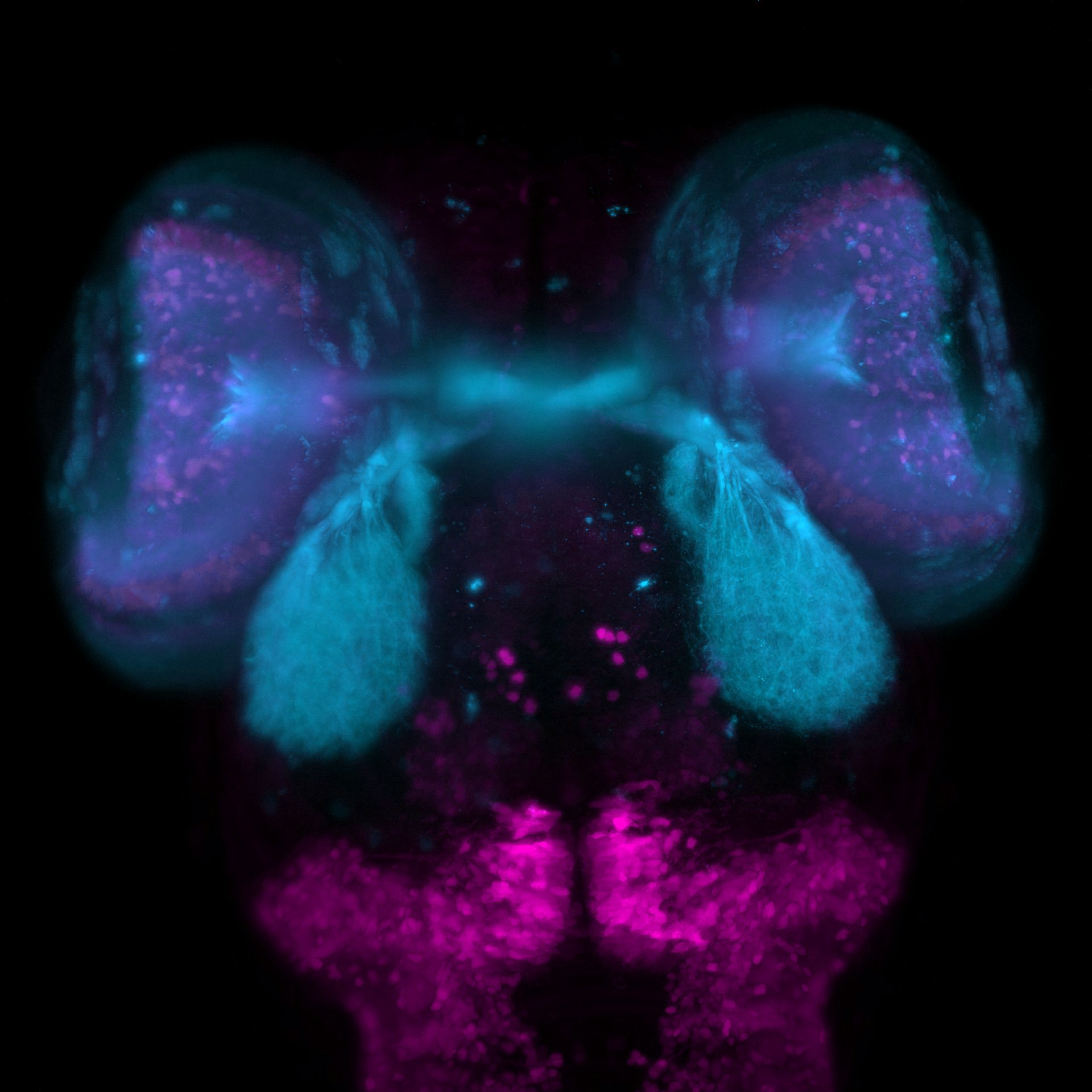
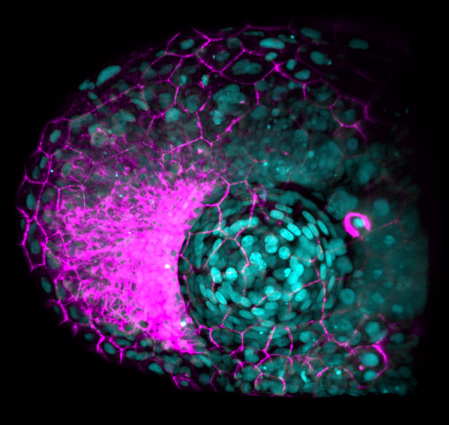
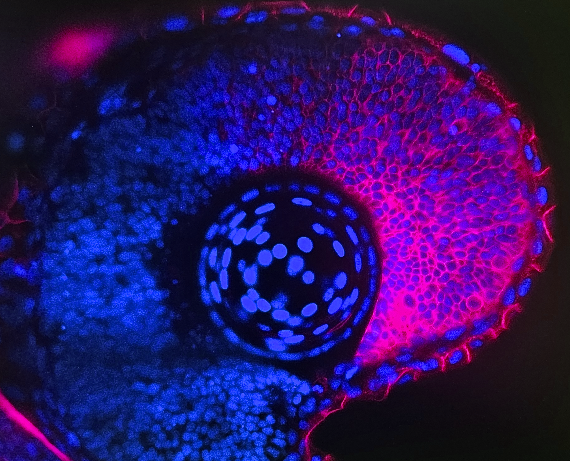
1. Amphipod. Amphipod collected in Quintay Bay by students of the 2025 course, and stained with phalloidin and DAPI.2. Chicken embryo. Live chicken embryo observed under a stereomicroscope.3. Late-stage Drosophila embryo. Confocal micrograph captures a late-stage Drosophila melanogaster embryo viewed from a ventrolateral or lateral perspective. It features triple-fluorescence labelling targeted at the developing musculoskeletal and nervous systems.4. Blastoderm-stage Drosophila embryo. Segmentation and homeotic (Hox) gene expressions in a blastoderm-stage Drosophila melanogaster embryo5. Tadpole Xenopus laevis. 6. Transgenic zebrafish embryo. Transgenic zebrafish embryo (Danio rerio), 24 to 48 hours post-fertilisation. Cyan: GFP; Magenta: DAPI7. Developing zebrafish brain. Micrograph of a developing zebrafish brain, showing cellular circuits and asymmetrical structures in cyan and magenta8. Zebrafish ocular structure. This image captures the developing ocular structure of a zebrafish embryo, visualised using high-resolution confocal/light-sheet fluorescence microscopy.9. Zebrafish embryo eye. Fluorescence microscopy image of a developing zebrafish embryo eye. Blue: DAPI, Magenta: retinal tissue
Our April webinar featured two of the papers named as finalists of Development’s 2025 Outstanding Paper Prize. Two of the papers were presented by Deputy Editor, Steve Wilson. Here, we share the talks from Xi Yang (Purdue University, USA) and Bénédicte Lefèvre (Institut Curie, France). Learn more about the finalists in our Editorial and read the full shortlist of nominated papers in Development’s subject collection.
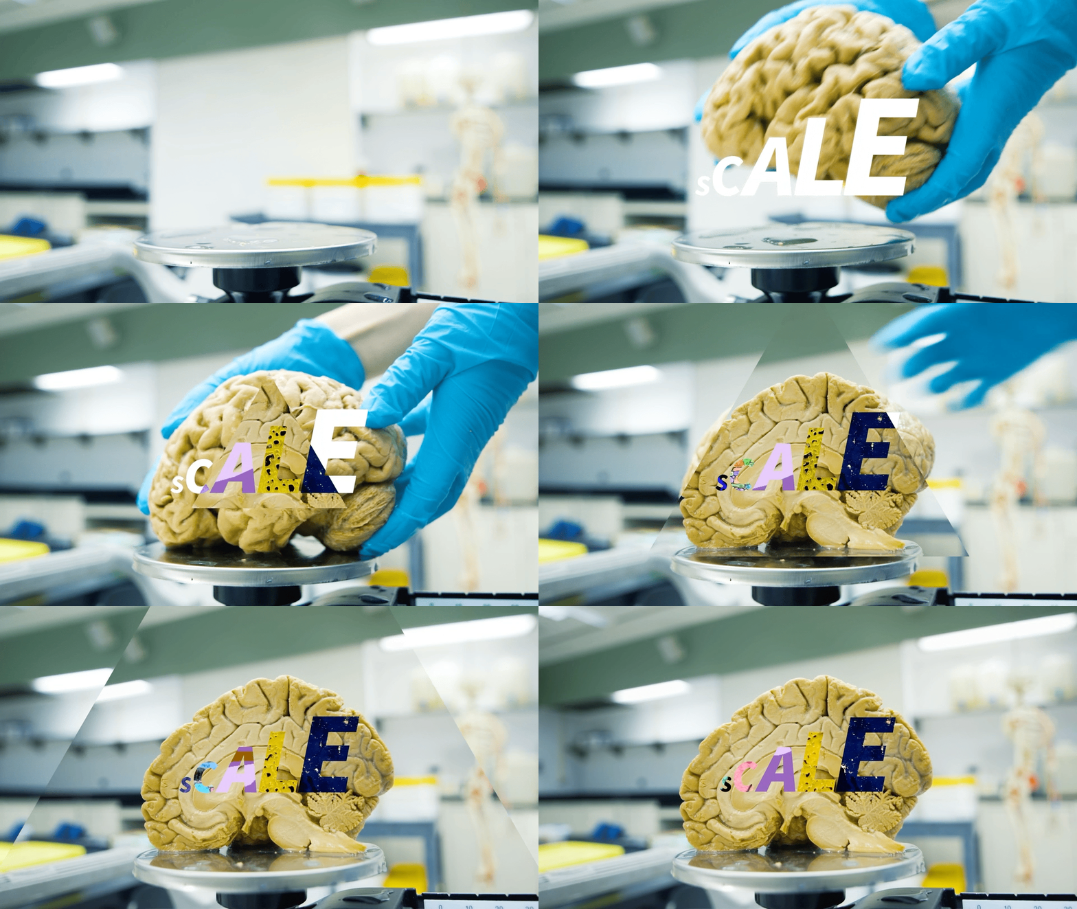
In this SciArt profile, we meet Ramin Rahni, a developmental plant biologist from New York, USA, who recently moved to London, UK, to focus on his creative work. In his artwork, Ramin works across graphic design, motion graphics/animation, music, and video art, integrating precision and noise in ways both dramatic and understated.
Can you tell us about your background and what you work on now?
I’m trained as a developmental plant biologist and did my PhD and postdoc at New York University, USA. I’m also a multidisciplinary artist and have always had creative projects going alongside my research, working across graphic design, motion graphics/animation, music, and video art. I just relocated from New York to London to focus more fully on creative work.
02. “postcard_016” from my video art series, post-CAD
Were you always going to be a scientist? And what about art – have you always enjoyed it?
As a child, I really wanted to be an artist. To try to and encourage me to get more into science, my parents would get me these science coloring books on biology, astronomy, etc. Those books really informed my love of the synthesis of art and science. When I got interested in science again, towards the end of my undergraduate studies, it was really through a love of the figures and the aesthetics of science graphics. The creative problem-solving and hands-on nature of benchwork and experimental biology were also always really appealing and rewarding to me. But a PhD and a postdoc later, my favorite part of research is still the visuals.
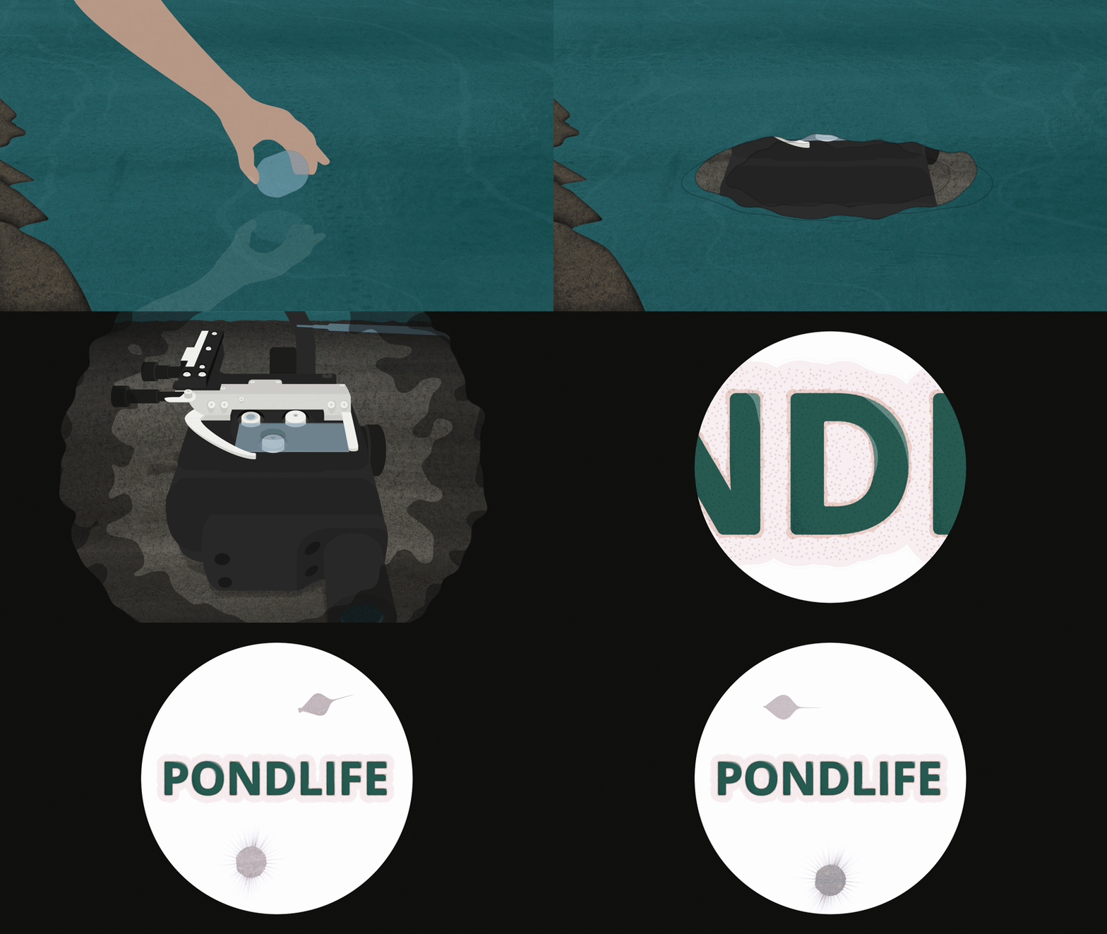
03. Animated title sequence for Pondlife, a web miniseries collaboration between Sally Warring and the American Museum of Natural History
What or who are your most important artistic influences?
I’m equally drawn to precision and noise. On the one hand, I love the clean line art of technical drawings, manuals and other documentation, scientific and medical textbook diagrams, and architectural drawings. At the same time, I love experimental media of all kinds: works by people at the margins of their fields, outsider art, non-linear storytelling, purely textural and abstract pieces, horror B-movies, and high-concept works that don’t take themselves too seriously. Trying to integrate those extremes, in ways both dramatic and understated, motivates a lot of my creative work. For example, in a recent project for Pioneer Works Science Studios, I got to combine a 2D line-art animation style with more abstract textures I made using my video synthesizer.
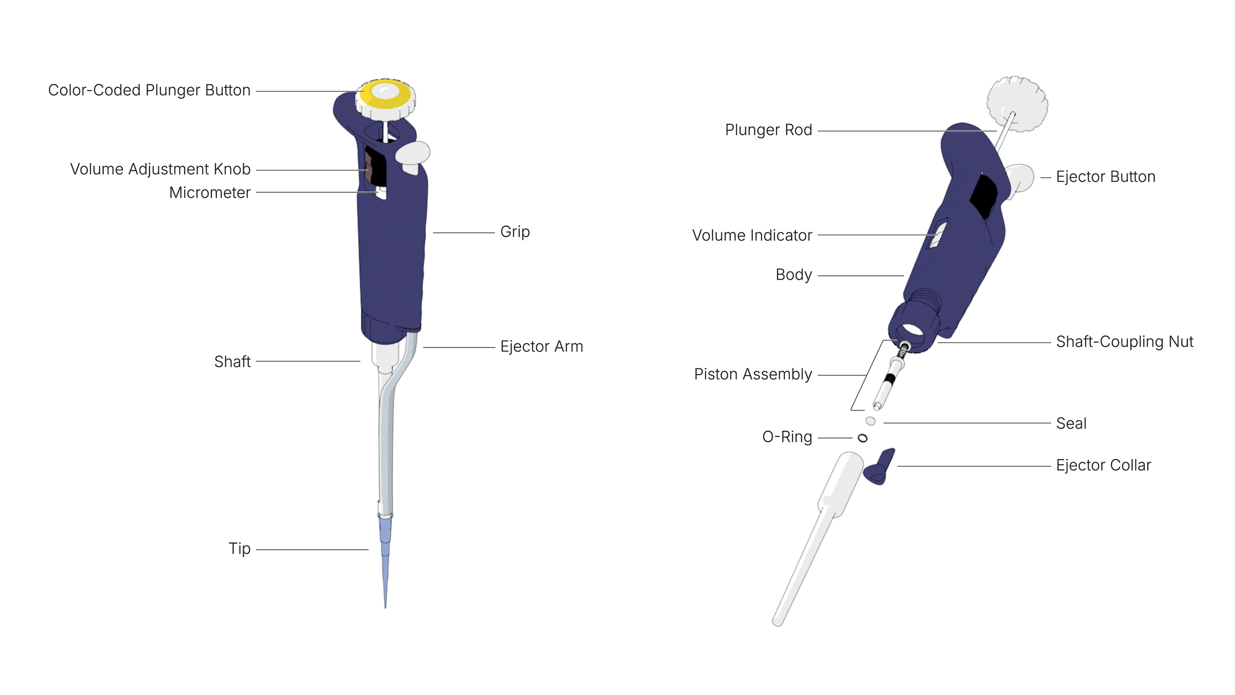
04. Annotated technical illustration of Gilson P200 pipette
I struggle with favorites, but here are a few works I’ve enjoyed recently: The Wax Child by Olga Ravn, the architecture of the Kyoto International Conference Center, the fresh-mint ice cream from Towpath in London (it tastes like actual mint!), and Good Morning Extra! by Water With Water.
For my graphic design projects, I primarily work in Figma, Illustrator, and InDesign. I use After Effects for motion graphics, 2D animation, and compositing. I also use 3D software for a lot of my static and animated work, but render everything using toon shaders to give it all a technical line-art feel. Lately, I’ve been experimenting with interactive animations using Rive and developing interactive biology learning materials. I make my video art using analog modular video synthesizers.
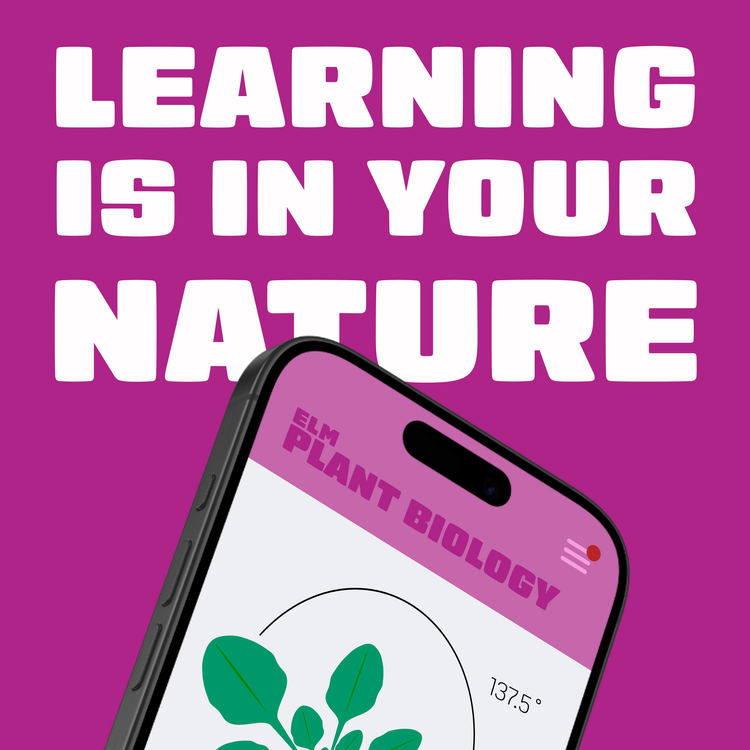
06. Brand application for ELM, an interactive life science learning app concept early in development
Does your science influence your art at all, or vice versa, or are they separate worlds?
They definitely mutually reinforce one another. The meticulous, organized, and cross-disciplinary nature of science lends itself well to the pixel-perfect and collaborative demands of graphic design. And being able to think in terms of systems, parts, and processes helps with the procedural nature of making art using analog video or other node-based workflows.
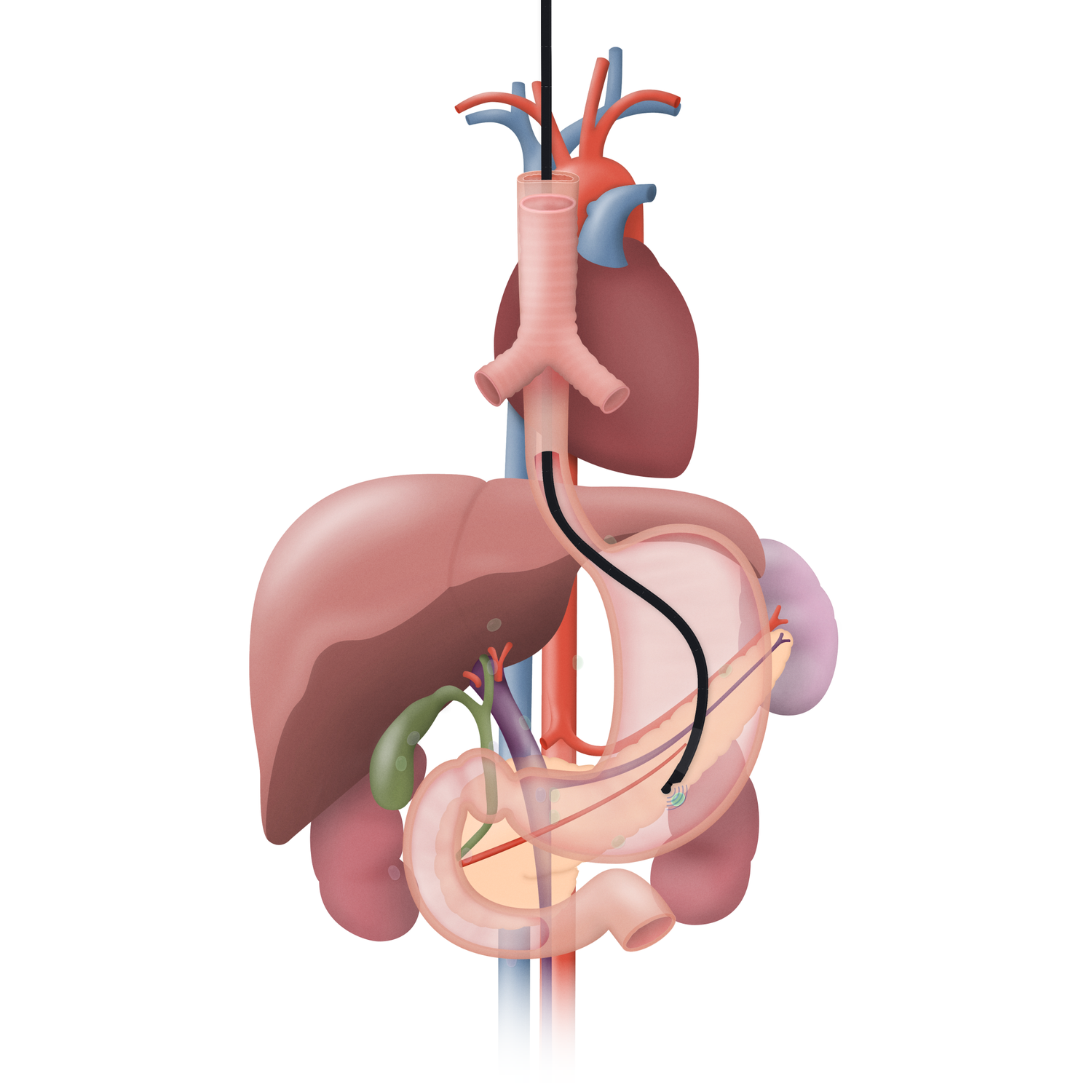
07. Still image from endoscopy animation for PENTAX Medical
What are you thinking of working on next?
On the personal side, I am working on an ongoing video art series called post-CAD that sits somewhere between postcards, graffiti, and thrift store re-paintings. Outside of my visual art practice, I play in the art rock duo Tar Of and the SWANA electronic duo Googoosh Dolls, and co-organize the annual technowruz series in NYC. We are about to mix a new Tar Of album so that’s next on my plate as well.
Professionally, I’m currently looking for opportunities where I can focus more fully on creative work informed by my science background, either in-house somewhere or within a studio or agency.
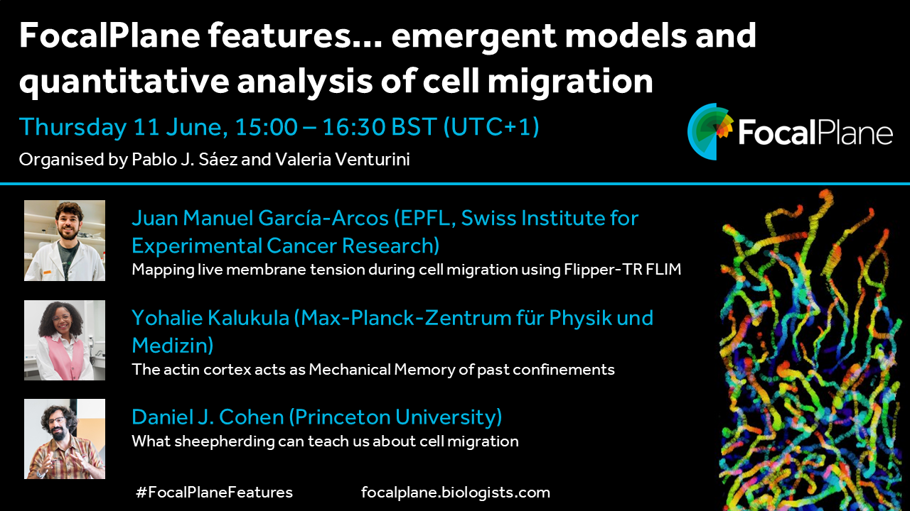
In this quarterly webinar series, hosted by Pablo J. Sáez and Valeria Venturini, we aim to showcase the latest research in the field, and we’d love to hear from researchers (especially early-career researchers) who would like to present their work. You can get in touch with us at focalplane@biologists.com or fill in our short application form.
Development, host of the Node, invites you to submit your latest research to our upcoming Special Issue – Plant and Algae Development. This issue will be coordinated by Academic Editor Dominique Bergmann (Stanford University, USA) and Guest Editor Susana Coelho (Max Planck Institute for Biology, Tübingen, Germany).
Developmental biology aims to understand how a single cell, the zygote, becomes a complex multicellular organism with specialised cell types organised into functional units. Derived from unicellular ancestors, algae, plants and animals use shared developmental principles, such as biophysical interactions, signalling, patterning and cell fate determination, to innovate and overcome obstacles for multicellular development, such as coordinating growth. The highly plastic development of photosynthetic organisms have long offered specific opportunities to answer questions regarding the origins of multicellularity, stem cell maintenance and regeneration, as well as environmental and microbial interactions and integration. In recent years, advances in genomics, imaging and synthetic biology are revealing how gene regulatory networks, mechanical biology and signalling interact to shape form and function in plants and algae. Furthermore, mathematical, computational and modelling approaches combined with traditional experimental biology have revealed, explained and predicted quantifiable properties behind phenotypes and across scales. We also see an exciting range of research organisms in use today, including species of algae, moss, ferns, flowering plants and others. In this special issue, we aim to showcase the contribution of plants and multicellular algae to our understanding of development, regeneration and evolution, highlighting quality research across the entire breadth of developmental biology.
The deadline for submitting research papers is 2 November 2026.
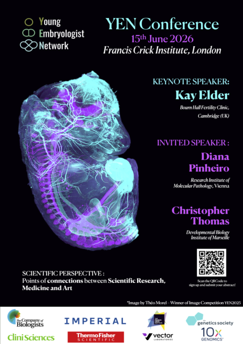
Note: it appears that some people are having issues with google forms, apologies if this has affected you. If you cannot use the form, please email your entry to us (see below for what is needed) and we will add it for you!
Looking forward to seeing all your images!
Many thanks,
Timothy
On behalf of YEN committee
Competition details:
Submit an original image you have generated related to developmental or stem biology. This may be a microscopy image, an image analysis output, or an artwork inspired by your scientific work.
Provide a title and a short description of the image, explaining the scientific context and techniques used (e.g. microscopy method or analysis strategy).
Provide your full name and affiliation. This information will be used for our internal records and future acknowledgement, but will not be displayed during voting.
You can view examples from the YEN 2025 Image Competition here.
Terms and Conditions
By sending us an image for the YEN Image Competition, you agree to the YEN Committee displaying the image publicly (including on our website and social media) and using it on YEN materials to promote future events. We will always credit you unless you request otherwise.
All submissions will undergo an initial review by the YEN Committee based on quality and visual impact. Applicants will be notified of the outcome of this selection through the email address provided in this form.