Howdy all,
As promised, here’s a post with book recommendations for developmental biologists.
~First, there are two books that together provide a simply exceptional entry into the field. They will be entertaining for developmental biologists looking for a broader view, but also represent good recommendations to friends and relatives hoping to grasp what the heck we are doing:
Coming to Life: How Genes Drive Development, by Christiane Nüsslein-Volhard. Written by one of our own Nobel Laureates, this book presents all you really need to know about developmental biology in a brisk and lively 150 pages, accompanied by exceptionally cogent illustrations. Undeniably my favorite in the genre.
Life Unfolding: How the Human Body Creates Itself, by Jaime Davies. This very readable, if somewhat dense, book focuses more on human development and gets into many of the gritty details passed over by Coming to Life.
~Two additional books that do the same thing, but from the vantage point of the early 1990s and early 1960s, respectively. Both are written by absolute giants in the field, people whose ideas very much molded our modern practice, These provide a very entertaining glance back in time, and both are geared to a popular audience.
The Triumph of the Embryo, by Lewis Wolpert
How Animals Develop: A Short Account of the Science of Embryology, by C.H. Waddington
~Next are two books that offer a glimpse into the minds of working developmental biologists.
Egg & Ego: An Almost True Story of Life in the Biology Lab, by Jonathan Slack focuses on his work that of others in the race to link growth factors to embryonic induction and the Spemann-Mangold Organizer in Xenopus. Find out which Xenopus hot-shot is “tall and bluff.”
The Dance of Life: The New Science of How a Single Cell Becomes a Human Being, by Magdalena Zernika-Goetz (with Roger Highfield) is a fun read that focuses on her recent work with early mammalian embryos.
~Here are two outstanding books chart the long, and dare I say it, glorious history of developmental biology:
Embryos Under the Microscope: The Diverging Meanings of Life, by Jane Maienschein unfurls the history of developmental biology with a view toward its impact on ethical and policy issues.
A History of Embryology, by Joseph Needham is a prolix tome that leaves no corner of European thought on embryos unturned from ancient times to the close of the 18th Century. He concludes with a plea for “a theoretical embryology suited in magnitude and spaciousness to the wealth of facts which contemporary investigators are accumulating day by day.” Guess we all gotta keep our day jobs.
~Two books for thinking about developmental biology as it relates to human form:
Extraordinary Bodies: Figuring Disability in American Culture and Literature, by Rosemarie Garland-Thompson is among the founding documents of disabilities studies and this meditation on human variation is essential reading for all developmental biologists.
Mutants: On Genetic Variety and the Human Body, by Armand Leroi is an exceptionally well-written book explaining developmental biology through discussion of human structural variation. The title has aged poorly, but the book is spectacular.
~Two great books about evolution that nonetheless brought developmental biology to the masses.
Your Inner Fish: A Journey into the 3.5 Billion-Year History of the Human Body, by Neil Shubin provides not just a great overview of developmental biology, but also nicely demonstrates how it informs our understanding of animal evolution.
Endless Forms Most Beautiful: The New Science of Evo Devo, by Sean Carroll is a fantastic synthesis developmental biology’s impact on our understanding of evolution.
~Two books on ancient pondering of the embryo:
The Human Embryo, Aristotle and the European Tradition, Ed. G.R. Dunstan. This collection of essays edited by a central figure in 20th Century medical ethics provides an excellent introduction to ancient European thought about embryos.
Imagining the Fetus: The Unborn in Myth, Religion, and Culture, Ed. Vanessa Sasson and Jane Marie. With a far wider lens than the title above, this collection of essays was the book that first set me on the path to thinking seriously about the non-European history of embryological thought.
~Miscellanies:
Black Apollo of Science: The Life of Earnest Everett Just, by Kenneth Manning is surely the most important (and best) biography of a developmental biologist. Charting Just’s experiences as a Black man in science in the early 20th Century, the book is heartbreaking in its humanity. But the science will be exhilarating to developmental biologists.
The Heritage of Experimental Embryology: Hans Spemann and the Organizer, by Viktor Hamburger is an excellent first-person account of the heady days of experimental embryology. We can thank his narrative of Hilda Mangold’s life in the Appendix of this book for her finding her rightful place in the minds of modern developmental biologists.
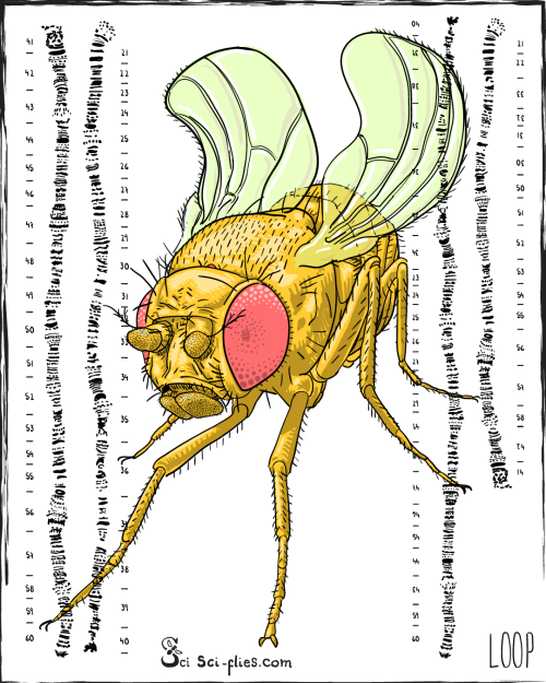
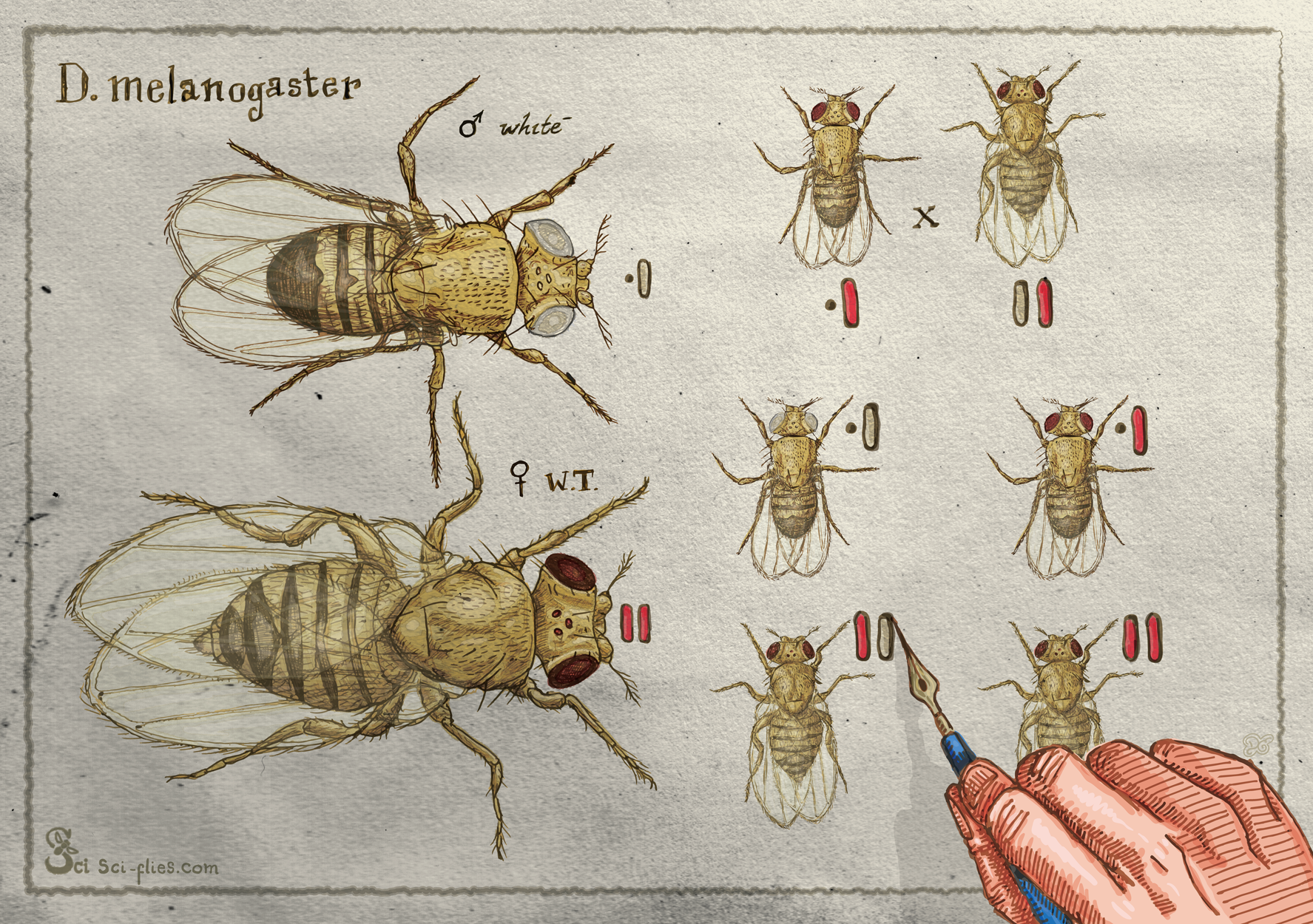
Lords of the Fly: Drosophila Genetics and the Experimental Life, by Robert Kohler provides a richly detailed history of the establishment and early evolution of Drosophila genetics as a craft. A fascinating read.
From Egg to Embryo: Regional Specification in Early Development (Second Edition), by Jonathan Slack is written for specialists but provides an outstanding overview of the state of the art in about 1990, by which time the major themes of modern developmental biology were squarely in focus. Provides and exceptional primer on the big picture of our field; great for new students.
On Growth and Form, by D’Arcy Wentworth Thompson is a book that Steven Jay Gould describes as the “greatest work of prose in twentieth-century science.” A heralded masterpiece, its quantitative approach was prescient to say the least!
The Dog Stars, by Peter Heller is a novel that has nothing whatsoever to do with developmental biology. But it reads like a mashup of A River Runs Through It and Mad Max and I really liked it and you might want to read it.
 (No Ratings Yet)
 Loading...

 (1 votes)
