The Turing Centre for Living Systems (CENTURI) wishes to attract talented PhD students to the Luminy campus. To do so, CENTURI will fund up to 10 PhD positions to start in 2021. PhD students will work in an interdisciplinary life science environment, and have backgrounds in any of the following fields: cell or developmental biology, immunology, neurobiology, biophysics, theoretical physics, computer science, bioinformatics, applied mathematics, engineering.
Candidates can either apply to one of the advertised CENTURI projects or submit their own project, providing that they meet the application criteria and that their application is supported by at least 2 host labs.
PhD students will be co-supervised by two or three supervisors from our community. Candidates can apply to a maximum of three projects.
Do not hesitate to contact the projects’ supervisors for more information.
Applications must be submitted via the project’s application form and must be written in english.
Deadline for application: February 16, 2022
Duration: 3 years
On-site or video interviews: April 26 to April 27, 2022
Expected profile – selection criteria
Candidates will be evaluated based on the following criteria:
Academic achievements
Past research experience (internships, master thesis)
Interest to work in a multidisciplinary research environment
Enthusiasm and communication skills
To apply please fill the form associated to each project. Applications must include the following documents, in English (compiled into a single PDF file):
CV
cover letter
transcript of your MSc’s grades (M1 and M2 if available)
2 letters of recommendation must also be sent by your references. Please note that an automatic email will be sent to them so that they can upload their recommendation letter. We invite you to contact them to make sure that they have received the notification email.
Who are we?
CENTURI brings together leading institutes in biology, physics, mathematics, computer science and engineering to decipher the complexity and dynamics of living systems. CENTURI offers an exceptional international environment for the development of interdisciplinary projects, in developmental biology, immunology and neurosciences.
CENTURI is mainly located on the Luminy campus of Aix-Marseille University and is affiliated to Aix- Marseille University, CNRS, INSERM and École Centrale Marseille.
At the end of the year people take stock and reminisce. This also applies to data visualization scientists, who like to review the best visualizations of 2021. I enjoy Maarten Lambrechts summary of all the “Best of 2021” posts [note added: by now this post is included in his list!].
While musing whether to keep or toss my Nature print issues (still undecided!), I browsed through all the science visualizations of 2021. Well, I mainly checked the biology and medicine figures as I really do not understand enough of physics to comment on their charts and plots. Some visualizations and themes really stood out to me – so I decided to summarize the top 10 science visualization trends of 2021 for you.
[Notes: (1) all photos show anonymous excerpts for educational purposes from Nature articles 2021. (2) because of technical difficulties this article was already posted on my personal site.]
Viva viridis!
The viridis color-scheme is now omnipresent. Viridis was developed by Stefan van der Walt and Nathaniel Smith in 2015 as the default sequential color map for matplotlib. Their goal was to create a color scheme in which color changes are perceptually uniform, and to replace the non-linear Jet/Rainbow-color schemes used previously for sequential data.
Viridis quickly gained popularity as we can see in the many examples below. By now viridis is however no longer only used for sequential data. Instead we also see it being applied to diverging and categorical data, which may be not exactly ideal at times. But for now, let’s celebrate the end of rainbow color schemes!
Charts with viridis color-scheme colors
Various charts with the viridis-color scheme.
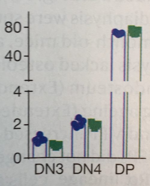
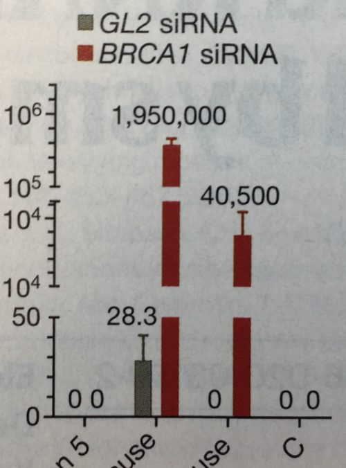
2. Still trending: axis breaks
Yes, axis breaks, necessary or not, are unfortunately still a thing in 2021. And so far, no end in sight. Every time I teach a Data Viz Course, I challenge the students that all axis breaks are avoidable – I have yet to see one where the break really was necessary. Send me yours!
Maybe necessary, unnecessary, and a really-really unnecessary axis break.
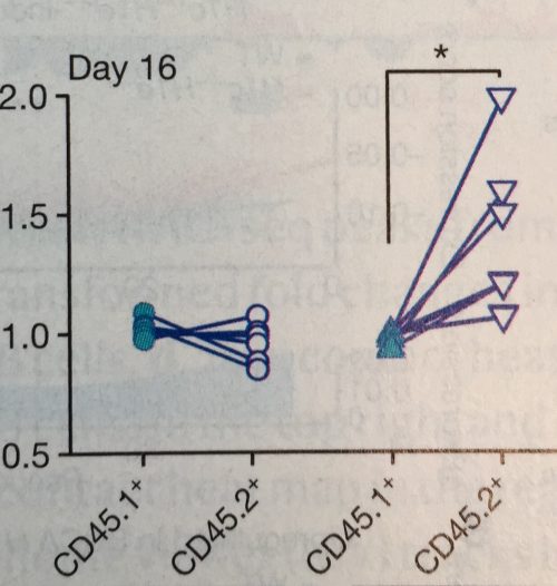
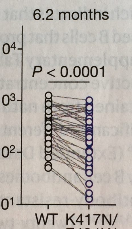
3. Hello slope-charts
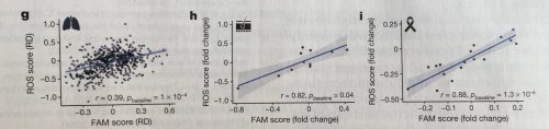
In 2016 the #BarBarChart initiative called for a ban of bar charts for data distributions. They were successful and I have not any in 2021. At that time, Tracey Weissgerber published a notable paper encouraging the use of a slope chart for dependent observations. It seems the science world listened again: numerous papers now use slope charts to illustrate dependent measurements of specimens, for example mice before and after treatment, responders and non-responders in cohorts etc.
Slope-charts for dependent measurements.
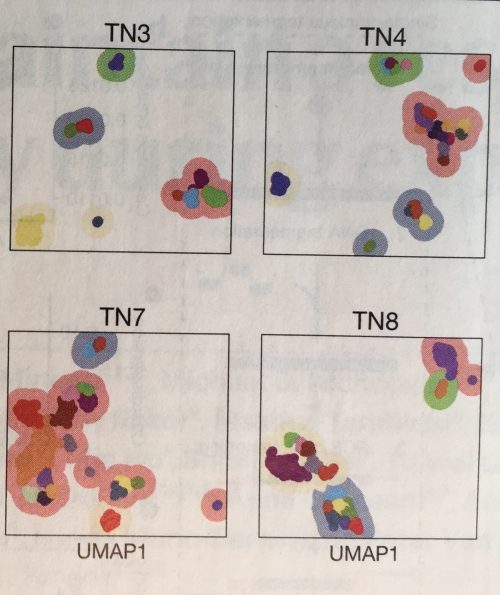
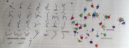
4. tSNE, UMAP, PCA
There don’t seem to be any papers that don’t have at least one dimension reduction plot. t-SNE, UMAP and PCA are omnipresent. These plots are however rarely explained in articles and figure legends, and I suspect even more rarely understood by audiences. In my courses participants are often not familiar with them – not surprising given that they are a recent addition in statistics and still heavily researched in the vis community! To educate your students refer them to these resources from Claus O. Wilke and StatQuest.
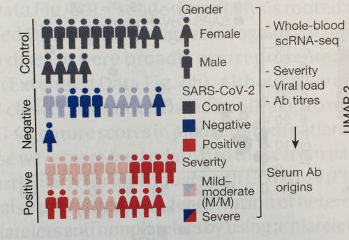
Eye-catching plots like tSNE, UMAP and PCA are useful for genomics, single cell sequencing and even letter-analysis in brain-robots!
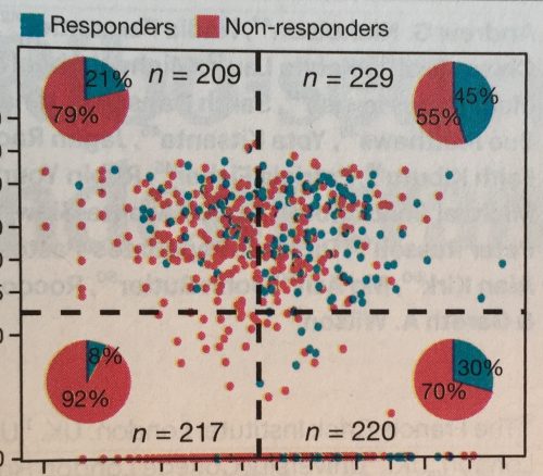
5. Mixing charts
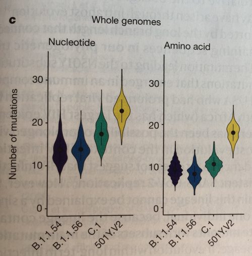
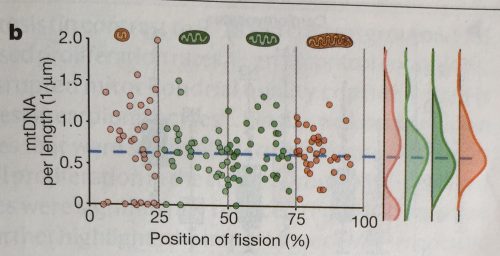
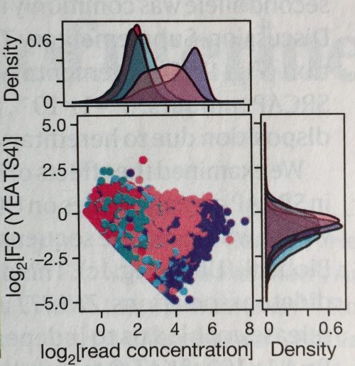
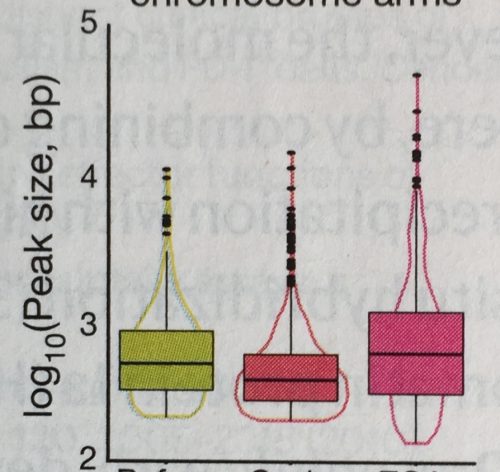
A big fashion trend of 2021 was mixing styles and patterns, and it seems this did spill over to the visualization world. Researchers have become quite experimental with mixing chart types (see also earlier post). I’ve seen a number of scatter plots showing the data distribution summarized along each axis: above for the x-axis and left for the y-axis. I’ve also seen pie charts summarizing the quadrants of a scatter-plot, charts mixing violin and box-plots, of course box-plots with data points, and many more.
Mixed-plots: it has become useful to merge plot types where needed.
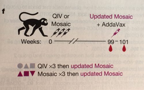
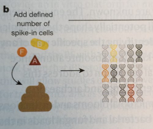
6. Pictograms
Often novel insights come from a new methodological approach. To familiarize audiences with a new method, papers now regularly include a sketch of the experimental procedure. And, good news, these have begun to look rather nice and clear, whether it’s a stool preparation method, an approach stem cell differentiation, or for a mouse neurobiology set-up.
Pictograms help explain experimental designs.
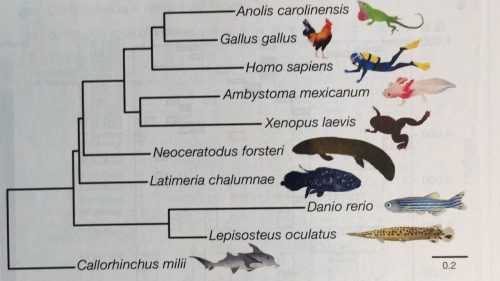
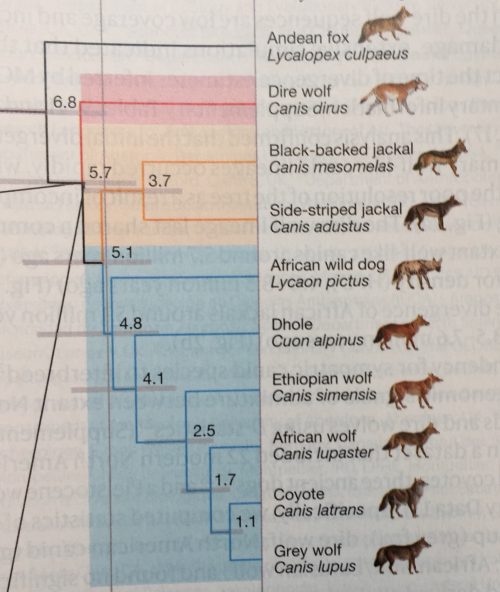
7. Pictograms for labeling
Pictograms are not only used for explaining procedures, but also often help to quickly orient audiences. Pictograms are for example used instead of text: I’ve seen pictograms as a title, pictogram to name a leaf in a phylogenetic tree, or even a pictogram chart. Overall pictograms seem to be most popular in mouse molecular genetics and evolutionary biology. Go figure.
Pictograms can make titles and labels understandable at first glance.
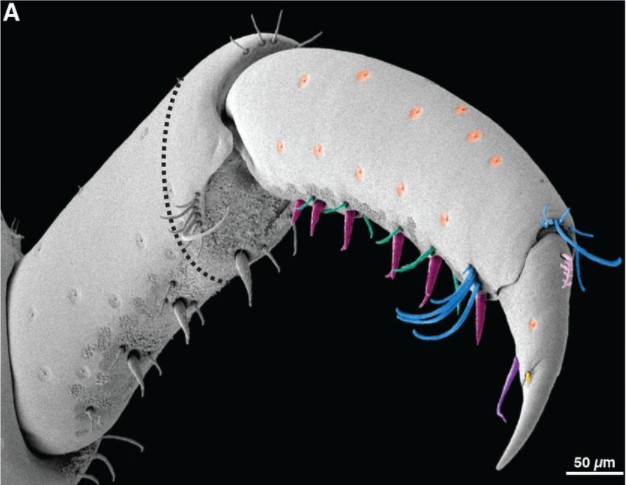
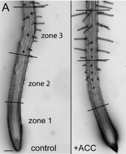
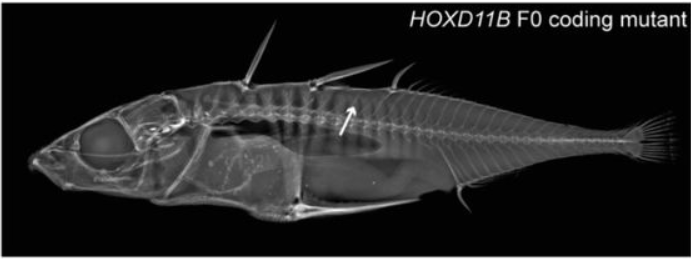
8. Images now come with scale bars!
Scale bars in images are apparently here to stay. A few years ago, I had noticed that scale information was quite often missing in scientific images, even in high profile journals. Eventually this observation prompted a collaborative project to systematically screen the quality of image data in publications.
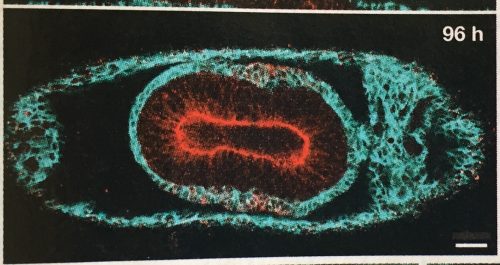
This year the tide has definitely shifted. Except for a few cases, scale bars are now visibly placed on all images; even in plant and histology images, two fields so far quite notorious for omitting scale information.
Now regularly with scale-bars: images!
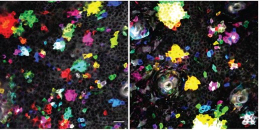
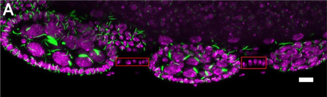
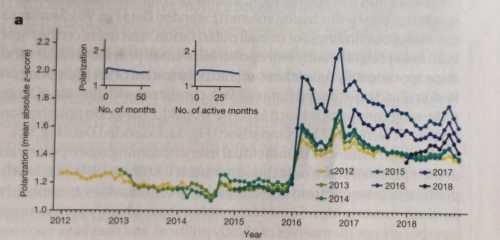
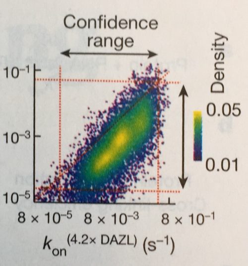
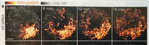
9. Colors in images: still problematic
Despite many recent efforts, most recently a technology feature in nature feat. your truly, no end is in sight for images that generously mix red and green, making it illegible for many color-blind people. In addition, often images that show false-color scales do not come with a legend explaining the color intensities.
A glimpse of hope: I’ve seen a color legend once this year (still without quantities in legend), which is already a 100% increase from previous years! And, rescue is near, with QUAREP we have a collaborative consortium addressing quality of light microscopy. Join our working group that develops guidelines for image visualization in publications!
Include legends for intensity values – ideally with quantifications.
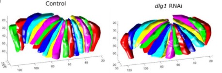
10. More space for figures!
Figures get more space in publications. This is quite easy in electronic publications, but also in print editions half or even full-page figures are becoming a sight. This makes it quite enjoyable to read some of the figures, which before were entirely illegible due to being squeezed into a small space.
More space for figures means less space for other sections of a paper. Often materials and methods are now entirely moved to the supplementary materials and citations are limited to a few only.
The figure below for example each took up 2/3 of a page – lovely!
Regionalising the nervous system: the steps to produce spinal cord.
The nervous system is one of the first structures that develops during embryogenesis. However, the process by which regional identity is established in this system remains unclear and is a long-standing unanswered question of developmental biologists. The Metzis lab aims to elucidate some of the molecular mechanisms defining regional identity in the nervous system during its initial development. To address these questions, the group uses embryonic stem cells (ESCs) as an in vitro model of neural development combined with high throughput sequencing approaches and comparisons to in vivo mouse models. Previous research has demonstrated that the generation of spinal cord requires the activity of the transcription factor CDX2 (1, 2), but it is still unknown exactly how this happens at the molecular level. My project focussed on identifying potential downstream targets of CDX2, using a combination of experimental and computational approaches.
The Hox family are a set of transcription factors that have fascinated developmental biologists for decades due to their role in patterning the body plan and their conservation across Bilateria (animals with left-right symmetry). Interestingly, it’s found that these genes have distinct rostro-caudal domains of expression in the developing spinal cord (3) and are expressed in a colinear fashion (4). Hox genes are regulated by CDX transcription factors (5, 6), so the main aim of my project was to predict which Hox genes are directly regulated by CDX2during the emergence of spinal cord. This could elucidate potential Hox genes that act in conjunction or as a result of Cdx2 upregulation to promote spinal cord identity.
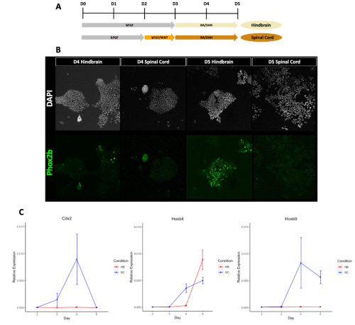
I initially approached this project by processing and analysing high throughput sequencing data from previous mRNA-seq experiments (7). This allowed me to identify gene expression patterns that occur during the differentiation of pluripotent ESCs into hindbrain versus spinal cord progenitors in vitro. I then evaluated these predictions by performing RT-qPCR to confirm that the published data from the mRNA-seq dataset matched the in vitro differentiations being conducted in the lab using the same protocol. I found that in general the predictions made were reproducible (Figure 1), so I finally went about verifying if CDX2 occupied the Hox genes that I had predicted, by analysing ChIP-seq datasets from previous studies (2, 8).
Figure 1:(A) Schematic of how mouse ESCs differentiate to neural progenitors in vitro with hindbrain (top; HB), or spinal cord (bottom; SC) identity. Both progenitors are produced with basic fibroblast growth factor (bFGF) treatment from day (D) 0 to D2. Hindbrain progenitors are treated with bFGF for another day and then Retinoic acid (RA) and Sonic Hedgehog (SHH) is added from D3 to D5 to promote the generation of motor neuron progenitors. Spinal cord progenitors are generated following a brief pulse of bFGF and active WNT signalling conditions from D2 to 3 (light orange shading; Adapted from Metzis et al., 2018). (B) Immunofluorescence imaging shows hindbrain progenitors express PHOX2B, indicating visceral motor neuron identity, whilst this identity is suppressed under spinal cord conditions. (C) RT-qPCR analysis of Cdx2, Hoxb4 and Hoxb9 expression at different days of the in vitro differentiation from ESCs, verifying predictions made from mRNA-seq data (Gouti et al. 2014).
It was exciting but challenging completing my own independent research project for the first time, and it forced me to step outside my comfort zone and grow quickly. A good example of this was using 384-well plates for the first time during qPCR. It was daunting to plan each plate, calculate the reagent needed and pipette each one out in a reproducible manner. Initially it took me a long time and I made mistakes; nevertheless, I soon got the hang of it and could do them with my eyes closed! Furthermore, having had no previous experience in coding, I initially struggled to successfully output any results. However, the dry lab aspect of this project taught me to be perseverant and explore your options, as there is often more than one way to get to the correct result. Over this summer I have also learnt to think more deeply about why I am doing a specific experiment, what it can teach me, and what my next steps will be after obtaining the results.
Although difficult, I found my project in the Metzis lab thoroughly enjoyable, and the experience gained was invaluable. I learnt to always approach intimidating tasks quickly and get stuck in, which was also really helped by the fact that the lab team were supportive and would guide me through new techniques. I learnt to always have a plan B – it is unlikely everything will go perfectly the first time, so preparing for that eventuality is necessary to not slow down your workflow too much.
This project has opened my eyes to what a professional research lab is like but also the questions and challenges present in the field of developmental biology. It has encouraged me to think more deeply about what we don’t already know about this field, and potentially some unanswered questions that I could look to answer in the future. I have had an amazing summer at the Metzis lab, and I very much look forward to getting back to the bench to explore those unanswered questions!
A picture of me and the lab team!
1. Metzis V, Steinhauser S, Pakanavicius E, Gouti M, Stamataki D, Ivanovitch K, et al. Nervous System Regionalization Entails Axial Allocation before Neural Differentiation. Cell. 2018;175(4):1105-18.e17.
2. Mazzoni EO, Mahony S, Peljto M, Patel T, Thornton SR, McCuine S, et al. Saltatory remodeling of Hox chromatin in response to rostrocaudal patterning signals. Nat Neurosci. 2013;16(9):1191-8.
3. Carpenter EM. Hox genes and spinal cord development. Dev Neurosci. 2002;24(1):24-34.
4. Wellik DM. Hox patterning of the vertebrate axial skeleton. Dev Dyn. 2007;236(9):2454-63.
5. Amin S, Neijts R, Simmini S, van Rooijen C, Tan SC, Kester L, et al. Cdx and T Brachyury Co-activate Growth Signaling in the Embryonic Axial Progenitor Niche. Cell Rep. 2016;17(12):3165-77.
6. Neijts R, Amin S, van Rooijen C, Deschamps J. Cdx is crucial for the timing mechanism driving colinear Hox activation and defines a trunk segment in the Hox cluster topology. Dev Biol. 2017;422(2):146-54.
7. Gouti M, Tsakiridis A, Wymeersch FJ, Huang Y, Kleinjung J, Wilson V, et al. In vitro generation of neuromesodermal progenitors reveals distinct roles for wnt signalling in the specification of spinal cord and paraxial mesoderm identity. PLoS Biol. 2014;12(8):e1001937.
8. Mahony S, Edwards MD, Mazzoni EO, Sherwood RI, Kakumanu A, Morrison CA, et al. An integrated model of multiple-condition ChIP-Seq data reveals predeterminants of Cdx2 binding. PLoS Comput Biol. 2014;10(3):e1003501.
Welcome to our monthly collection of preprints on developmental and stem cell biology (and related fields). The list includes a number of more unusual model organisms including squirrels, hippos, the crustacean Parhyale hawaiensis, deer mice, sticklebacks, mouse lemurs and Agaricomycetes
The preprints this month are hosted on bioRxiv and arXiv – use these links to get to the section you want.
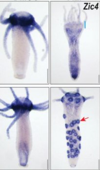
The transcription factor Zic4 acts as a transdifferentiation switch Matthias Christian Vogg, Jaroslav Ferenc, Wanda Christa Buzgariu, Chrystelle Perruchoud, Panagiotis Papasaikas, Paul Gerald Layague Sanchez, Clara Nuninger, Céline Delucinge-Vivier, Christine Rampon, Leonardo Beccari, Sophie Vriz, Stéphane Vincent, Brigitte Galliot, Charisios D. Tsiairis
Histone demethylome map reveals combinatorial gene regulatory functions in embryonic stem cells Pratibha Tripathi, Pushkar Dakle, Majid Mehravar, Varun K. Pandey, Michael J. Bullen, Zhongming Zhang, Dhaval Hathiwala, Marc Kerenyi, Andrew Woo, Alireza Ghamari, Alan B. Cantor, Lee H. Wong, Jonghwan Kim, Kimberly Glass, Guo-Cheng Yuan, Luca Pinello, Stuart H. Orkin, Partha Pratim Das
Effects of α-crystallin gene knockout on zebrafish lens development Mason Posner, Kelly Murray, Brandon Andrew, Stu Brdicka, Alexis Butterbaugh-Roberts, Kirstan Franklin, Adil Hussen, Taylor Kaye, Emmaline Kepp, Mathew McDonald, Tyler Snodgrass, Keith Zientek, Larry David
Kismet/CHD7/CHD8 affects gut biomechanics, the gut microbiome, and gut-brain axis in Drosophila melanogaster Angelo Niosi, Nguyên Henry Võ, Punithavathi Sundar, Chloe Welch, Aliyah Penn, Yelena Yuldasheva, Adam Alfareh, Kaitlin Rausch, Takhmina Rukhsar, Jeffery Cavanaugh, Prince Yadav, Stephanie Peterson, Raina Brown, Alain Hu, Any Ardon-Castro, Darren Nguyen, Robert Crawford, Wendy Lee, Mikkel Herholdt Jensen, Eliza J. Morris, Kimberly Mulligan
Cellular development and evolution of the mammalian cerebellum Mari Sepp, Kevin Leiss, Ioannis Sarropoulos, Florent Murat, Konstantin Okonechnikov, Piyush Joshi, Evgeny Leushkin, Noe Mbengue, Céline Schneider, Julia Schmidt, Nils Trost, Lisa Spänig, Peter Giere, Philipp Khaitovich, Steven Lisgo, Miklós Palkovits, Lena M. Kutscher, Simon Anders, Margarida Cardoso-Moreira, Stefan M. Pfister, Henrik Kaessmann
Gene loss during a transition to multicellularity Berenice Jiménez-Marín, Jessica B. Rakijas, Antariksh Tyagi, Aakash Pandey, Erik R. Hanschen, Jaden Anderson, Matthew G. Heffel, Thomas G. Platt, Bradley J. S. C. Olson
Tabula Microcebus: A transcriptomic cell atlas of mouse lemur, an emerging primate model organism The Tabula Microcebus Consortium, Camille Ezran, Shixuan Liu, Stephen Chang, Jingsi Ming, Olga Botvinnik, Lolita Penland, Alexander Tarashansky, Antoine de Morree, Kyle J. Travaglini, Kazuteru Hasegawa, Hosu Sin, Rene Sit, Jennifer Okamoto, Rahul Sinha, Yue Zhang, Caitlin J. Karanewsky, Jozeph L. Pendleton, Maurizio Morri, Martine Perret, Fabienne Aujard, Lubert Stryer, Steven Artandi, Margaret Fuller, Irving L. Weissman, Thomas A. Rando, James E. Ferrell Jr., Bo Wang, Iwijn De Vlaminck, Can Yang, Kerriann M. Casey, Megan A. Albertelli, Angela Oliveira Pisco, Jim Karkanias, Norma Neff, Angela Wu, Stephen R. Quake, Mark A. Krasnow
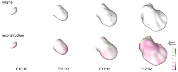
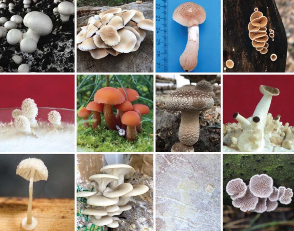
Agaricomycetes from Nagy, et al.
Lessons on fruiting body morphogenesis from genomes and transcriptomes of Agaricomycetes László G. Nagy, Peter Jan Vonk, Markus Künzler, Csenge Földi, Máté Virágh, Robin A. Ohm, Florian Hennicke, Balázs Bálint, Árpád Csernetics, Botond Hegedüs, Zhihao Hou, Xiao-Bin Liu, Shen Nan, Manish Pareek, Neha Sahu, Benedek Szathmári, Torda Varga, Hongli Wu, Xiao Yang, Zsolt Merényi
Improving Lab Culture through Self-Assessment: A Case Study Soleil Hernandez, Raymond Mumme, Laurence Court, Daniel El Basha, Skylar Gay, Barbara Marquez, Yao Xiao, Kai Huang, Hana Baroudi, Wenhua Cao, Carlos Cardenas, Raphael Douglas, Jack Duryea, Zaphanlene Kaffey, Deborah Mann, Kelly Nealon, Tucker Netheron, Callistus Nguyen, Kyuhak Oh, Adenike Olanrewaju, Carlos Sjogreen, DJ Rhee, Jinzhong Yang, Cenji Yu, Lifei Zhang, Yao Zhao, Hamid Ziyaee, Mary Gronberg
A remote lecture series roadmap to equity, diversity, and inclusion in STEM Evan A. Boyle, Gabriela Goldberg, Jonathan C. Schmok, Jillybeth Burgado, Fabiana Izidro Layng, Hannah A. Grunwald, Kylie M. Balotin, Michael S. Cuoco, Keng-Chi Chang, Gertrude Ecklu-Mensah, Aleena K. S. Arakaki, Noorsher Ahmed, Ximena Garcia Arceo, Pratibha Jagannatha, Jonathan Pekar, Mallika Iyer, DASL Alliance, Gene W. Yeo
The Importance of Mentors and How to Handle More Than One Mentor Andrea G. Marshall, Lillian J. Brady, Caroline B. Palavicino-Maggio, Kit Neirkirk, Zer Vue, Heather Beasley, Edgar Garza-Lopez, Sandra Murray, Denise Martinez, Haysetta Shuler, Elsie C. Spencer, Derrick Morton, Antentor Hinton Jr
Launched in 2020, FocalPlane is a curated and centralised platform for the microscopy community to share news and techniques, discuss issues relevant to the field and read about the latest research and events. We are now looking for an enthusiastic and motivated person with fresh ideas and a willingness to learn to join us to develop and maintain this site.
Core responsibilities of the position include:
• Creating and commissioning content for FocalPlane, including writing posts and soliciting content from the academic community, societies, companies and other organisations
• Providing user support and ensuring site functionality on a day-to-day basis
• Providing creative and practical input into the development of the site
• Maintaining and developing the site’s presence on social networking sites such as Facebook and Twitter
• Representing Journal of Cell Science and FocalPlane at international conferences
Essential skills:
• PhD in a relevant scientific field, ideally with experience of microscopy
• Willingness to grow and develop knowledge of microscopy
• Demonstrable ability to write for an online audience and/or produce social media content
• Clear understanding of the online environment as it applies to scientists
• Excellent interpersonal and communication skills
• Strong networking abilities online and in person
Desirable:
• Experience with additional media (e.g. video or podcasting)
• Experience with WordPress
• Contacts within the microscopy community
This is an exciting opportunity to develop a hub for the microscopy community – in a similar vein to the Company’s established community site for developmental biologists, the Node – and to engage with relevant people at all levels: academics, developers, facilities, institutes and companies. The Community Manager will work alongside an experienced in-house team, including the Executive Editor of Journal of Cell Science. Additional responsibilities may be provided for the right candidate.
The Company of Biologists exists to support biologists and inspire advances in biology. At the heart of what we do are our five specialist journals – Development, Journal of Cell Science, Journal of Experimental Biology, Disease Models & Mechanisms and Biology Open. All are edited by expert researchers in the field, and all articles are subjected to rigorous peer review. We believe that the profits from publishing the hard work of biologists should support scientific discovery and help develop future scientists. Our grants help support societies, meetings and individuals. Our workshops and meetings give the opportunity to network and collaborate.
The Company of Biologists is an inclusive organisation and we believe that everyone has a contribution to make. We want all our employees to feel included, valued and appreciated. We believe in equality of opportunity for all staff and encourage applications from all individuals regardless of sexual orientation, gender identity or expression, religion, ethnicity, age, neurodiversity, disability status and citizenship.
Applicants should send a CV along with a covering letter that summarises their relevant experience, and in particular any specific microscopy/image analysis skills, and includes links to any online activities, salary expectations, and details about why they are enthusiastic about this opportunity.
Applications and informal queries should be sent by email to recruitment@biologists.com by 31 January 2022. We may request written tests in advance of any interview.
Thanks to everyone who has contributed to the Node in 2021. We hope you have enjoyed interacting with the site over the past year. As a reminder, the Node is a community site so anyone with an interest in developmental biology or stem cells can, once registered, post on the website. This could be a research story, a commentary, an interview or a job or event listing. If you have any suggestions for new features or would like advice from the Node team you can contact us at thenode@biologists.com. But before we look ahead to 2022, we focus on 2021 in numbers:
Regeneration is the mechanism by which a species can restore damaged or missing cells, tissues, organs, or body parts. Different living organisms have vastly differing regenerative capabilities. Humans, while capable of regenerating some organs following damage or disease, most notably the liver, are very limited in their capacity. Other organisms, however, have remarkable regenerative capacities that facilitate re-growth of entire limbs, parts of nervous systems, or even whole-body regeneration like planarians.
Despite the relative prevalence of regeneration and the potential contributions of its research to modern medicine, the field is widely understudied, especially from a comparative evolutionary perspective (Lai and Aboobaker 2018 Dev Bio).
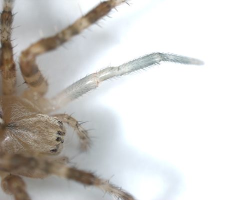
This summer, the BDSB Summer Studentship gave me the opportunity to undertake an exciting research project, further studying the regenerative abilities of the European Garden Spider, Araneus diadematus, within the McGregor Laboratory at Oxford Brookes, under the supervision of Research Fellow and spider expert, Anna Schönauer.
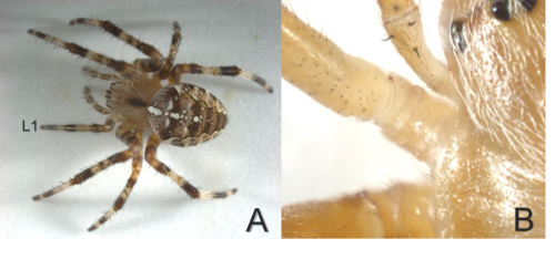
Fritz Vollrath’s work (Vollrath, 1990) describes the remarkable ability of juvenile A. diadematus (Figure 1, A) to quickly regenerate functional legs from the coxa-trochanter joint following autotomy (Figure 1, B). This functionality is crucial to their survival, as the spiders rely on the limbs’ sensory-motor abilities for weaving their geometrically complex webs, which are intricately designed to facilitate capture of prey (Reed et al 1965).
Regeneration is understood to be an ancestral trait in arachnid species (Goss, 1992) and although some lineages have lost this ability (Vollrath, 1990), the evolutionary relationships giving rise to these differences remain unclear. Research into the development of closely related spider species, with and without regenerative capabilities, offers potential insight into the changes that have led to the loss of this trait. Further, as outlined in Karl Ernst von Baer’s Laws of Embryology (Wanninger, 2015), studying the early development of a species offers essential contributions to the uncovering of evolutionary patterns and relationships of characteristics.
Figure 1. Fourth Instar Female Araneus diadematus (A) Overview of specimen with regenerated, left first walking leg (L1). (B) The coxa-trochanter joint.
The aim of my project was to improve understanding of the regeneration walking legs in the Araneus diadematus. Initially my Objectives comprised:
Generating a time series to document the species’ post-embryonic development
Comparing the regeneration of the first walking legs between different instars
Analysing the leg transcriptome of an A. diadematus leg, to compare with a species unable to regenerate lost limbs – Parasteatoda tepidarioum.
Due to impediments imposed by Covid19, in addition to further challenges thrown up by the unpredictable nature of scientific research, the outcomes and overall procedure of my project ultimately deviated significantly from the parameters of my original aims. Covid regulations limited my lab access early enough in the year, and as a result, I was unable to accurately document the beginning of the time series for the species, obstructing my first aim.
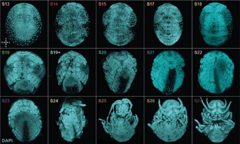
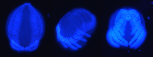
However, acquisition of A. diadematus embryos, contributed an additional branch to the project and supplemented my first objective. Embryos were frozen at different times in development, prior to being peeled, DAPI stained, and microscopically imaged (Figure 2). This contributed to the creation of a partial embryonic time-series, in place of the initially intended post-embryonic time-series.
The examination and comparison of the imaged A. diadematus embryos, with pre-existing images of the P. tepidarioum (Mittman and Wolff 2012) also presents the opportunity to identify significant disparities within the development of the two species, with the potential to propose relevant evolutionary relationships of regeneration, signposted through embryonic development.
Figure 2: The Prosomal, Lateral and Frontal View (left to right) of a DAPI Stained Araneus diadematus embryo
The findings from the second objective, analyzing and comparing the regeneration and emergent legs (Figure 3) of individuals within the third and fourth instar, prompted a secondary investigation, examining the effects and implications of the leg regeneration of individuals within the fifth instar. Throughout these analyses, I thoroughly enjoyed the weekly imaging and recording of my experimental subjects’ regenerative development, but nothing quite compared to the excitement of discovering the emergence of a new regenerated leg on my visits to the lab’s spider room first thing in the morning.
Figure 3: The Regenerated Left, First Walking Leg of a Third Instar Female, 21 Days (and 1 molt) After Leg Loss
The final objective (obtaining, analyzing, and comparing the A. diadematus leg transcriptome) is still ongoing and I am learning a lot about the patience required for bioinformatics!
My experiences in the lab have emphasized the fluid and unpredictable nature of scientific study, reinforcing the importance of patience, open mindedness, and flexibility. The Gurdon Summer Studentship has taught me so much that I would not otherwise have had access to in the ordinary course of my undergraduate program. I’ve been involved in lab meetings and journal clubs, worked alongside an amazing group of PhD students, and gained experience using cutting-edge equipment and techniques. I’ve developed lab skills, learned specialized spider husbandry techniques, and grown as a scientist, thanks to the lab team I have been privileged to be a small part of.
I did not underestimate the incredible opportunity being afforded to me and was excited before the project started, but I could not have imagined just how much I would enjoy the process. The unpredictable nature of the field of developmental biology has made for an exciting summer and I owe an enormous debt of gratitude to the BDSB. Further I am beyond grateful to Alistair McGregor and to Anna Schönauer for this incredible opportunity and for their support, encouragement, and training. Their passion for their subject is infectious and would inspire anyone to study further in the field of developmental biology!
References
Goss, R. J. (1992) “The Evolution of Regeneration: Adaptive or Inherent?,” Journal of theoretical biology, 159(2), pp. 241–60.
Lai, A. G. and Aboobaker, A. A. (2018) “Evoregen in Animals: Time to Uncover Deep Conservation or Convergence of Adult Stem Cell Evolution and Regenerative Processes,” Developmental Biology, 433(2), pp. 118–131. doi: 10.1016/j.ydbio.2017.10.010.
Mittmann, B. and Wolff, C. (2012) “Embryonic Development and Staging of the Cobweb Spider Parasteatoda Tepidariorum C. L. Koch, 1841 (syn.: Achaearanea Tepidariorum; Araneomorphae; Theridiidae),” Development genes and evolution, 222(4), pp. 189–216. doi: 10.1007/s00427-012-0401-0.
Reed, C. F. Witt, P. N. and Jones, R. L. (1965) “The Measuring Function of the First Legs of Araneus Diadematus Cl,” Behaviour, 25(1-2), pp. 98–119.
Vollrath, F. (1990) “Leg regeneration in web spiders and its implications for orb weaver phylogeny”. Basel (Suiza): Zoologisches Institute.
Wanninger, A. (2015) “Evolutionary developmental biology of invertebrates”. Wien: Springer (Online access: Springer (t). doi: 10.1007/978-3-7091-1868-9.
In our final SciArt profile of 2021 we meet Jessica Richardson, a final year PhD student in Kate Poole’s group at the University of New South Wales, Sydney. In addition to using her artistic skills in her lab work, Jessica works as a freelance illustrator and writer.You can find Jessica on Twitter and LinkedIn.
Where are you originally from and what do you work on now?
I grew up and still live in Sydney, Australia, where I completed my Bachelor’s degree in Neuroscience and Physiology. I am currently in the final year of my PhD in Physiology and Pharmacology at UNSW Sydney working in Associate Professor Kate Poole’s lab. For my PhD project, I am investigating how cells can sense mechanical forces using specialised proteins known as mechanically activated ion channels.
Outside of my research, I am a freelance illustrator and writer and am currently working on various projects, including contributing writing and drawings for high school science textbooks, as well as creating promotional material for commercial organisations in the STEMM space.
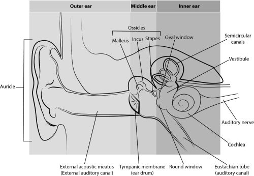
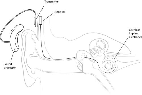
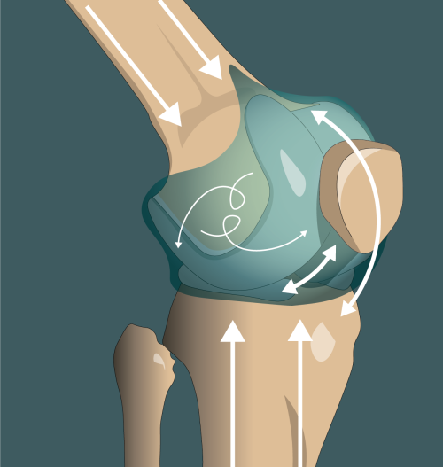
Left: Ear Anatomy – Anatomy of the human ear Middle: Cochlear Implant – Basic diagram of the components of a cochlear implant Right: Knee Joint – Illustration of the forces acting on cartilage within the knee joint, including compressive forces from weight bearing, shear forces of articulating surfaces gliding over one another, and movements of the synovial fluid within the joint capsule.
Were you always going to be a scientist?
I have always had a passion for science since childhood and was the annoying child that constantly asked ‘why?’ and ‘how does that work?’, so you could say so. However, my journey towards becoming a scientist hasn’t been a linear path! I’ve always struggled to choose between pursuing a career in STEMM or pursuing one in the creative industries. For a long time during high school, I really wanted to be creative writer and/or a comic artist. At some point, my love for science won out and I decided to pursue a degree in neuroscience. Neuroscience had always fascinated me as a subject, especially regarding how we sense the world around us and how that differs so greatly from person to person.
But that wasn’t the end of the story. Early during my degree, my university made a double-degree available: a Bachelor of Advanced Science alongside a Bachelor of Fine Arts. At around the same time, I also was introduced to the possibility of a career in science illustration which would combine two of my passions, so I immediately transferred into this degree.
Unfortunately, the Art and Design school was on a different campus from the main campus and trying to run across town between my lab tutorials and my art classes with my huge art book (and supplies) and safety glasses still on was too much. I made the sad decision to transfer to science only, knowing that while I could always find the time to make art on my own while completing my science degree, the reverse would not be true.
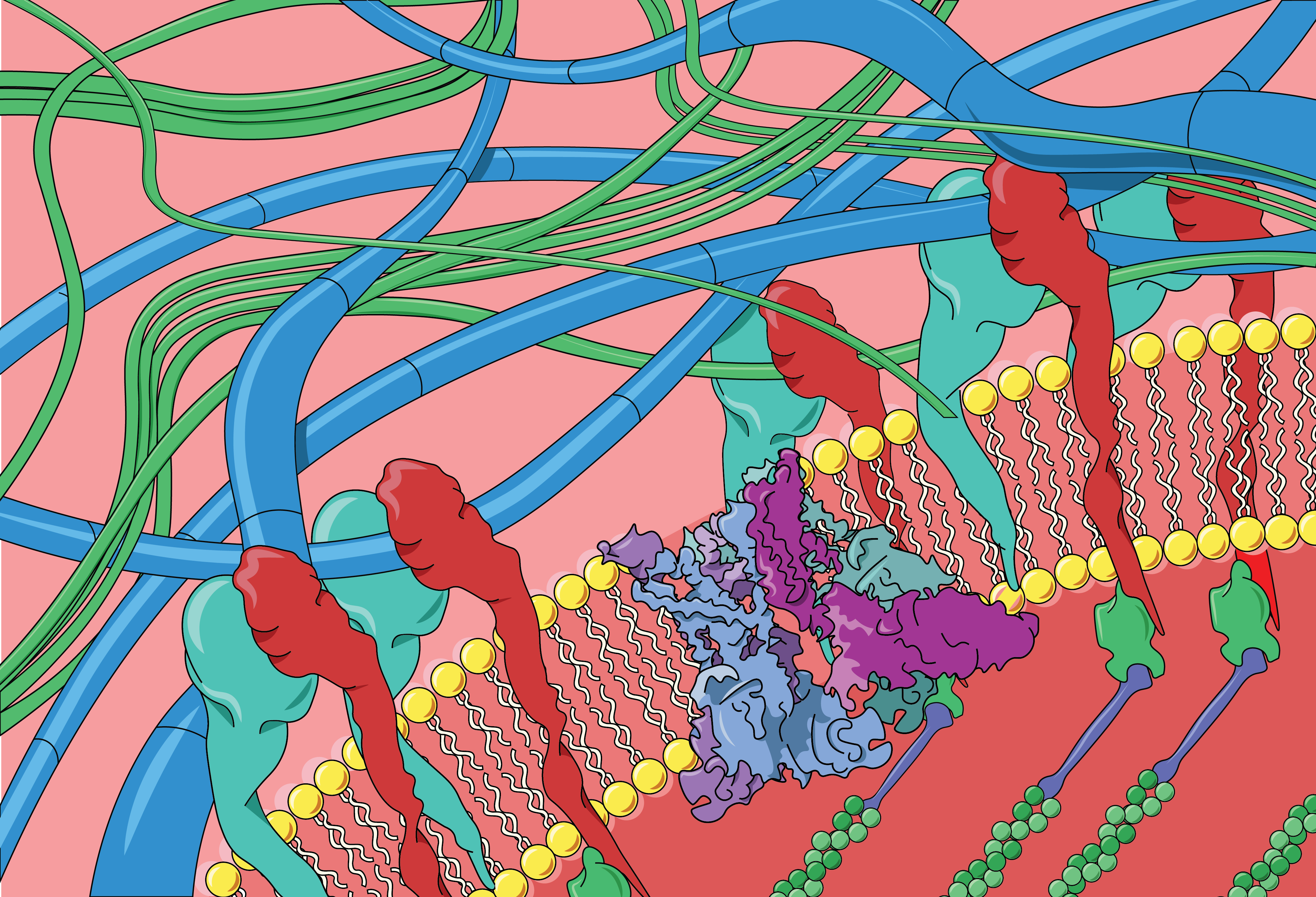
Ion Channels at the Cell-Substrate Interface – Illustration of the interface between a cell and its surrounding microenvironment including proteins which may be important for cellular sensing of mechanical forces. This diagram features the cytoskeleton inside the cell (bottom right), proteins such as integrins and mechanically activated ion channels within the cell membrane, and the extracellular matrix outside of the cell (top left).
And what about art – have you always enjoyed it?
I have always enjoyed making art since I was a very young child. I was always doodling in class in school and always had to have a sketch book on hand. Throughout primary school and high school, I experimented a lot with different mediums, and to this day, while I’ve found my preferred mediums, I still like to dabble occasionally in different techniques.
T-cell – Illustration of a T-cell (or T-lymphocyte)
What or who are your most important artistic influences?
Generally speaking, I have always been influenced by genres that use monochromatic black and white colours and heavy shadowing. Think the stark look of noir films and black and white comics, as well as high contrast black and white photography. Looking back to my childhood dreams of being a comic book artist, I was definitely inspired by comic book artists like Frank Miller and James O’Barr, and still am, in recent years, by newer comic artists like Nicola Scott and Fiona Staples. I definitely incorporate these influences into my science illustration work and figures, and often try to create a cartoon-like feel. However, especially for illustrating cellular environments at a molecular level, I am heavily inspired by David Goodsell, who creates absolutely stunning works of art to visualise such complex concepts about the microscopic world.
My absolute biggest artistic influence and inspiration, however, has to be my oldest sister, Sara, who first got me interested in drawing and gave me my very first lessons on proportions, perspective, shading, and drawing human faces. Looking through old sketchbooks from when I was a child, there are plenty of beautiful drawings by her and many more poor emulations from me. I really have her to thank for my artistic endeavours.
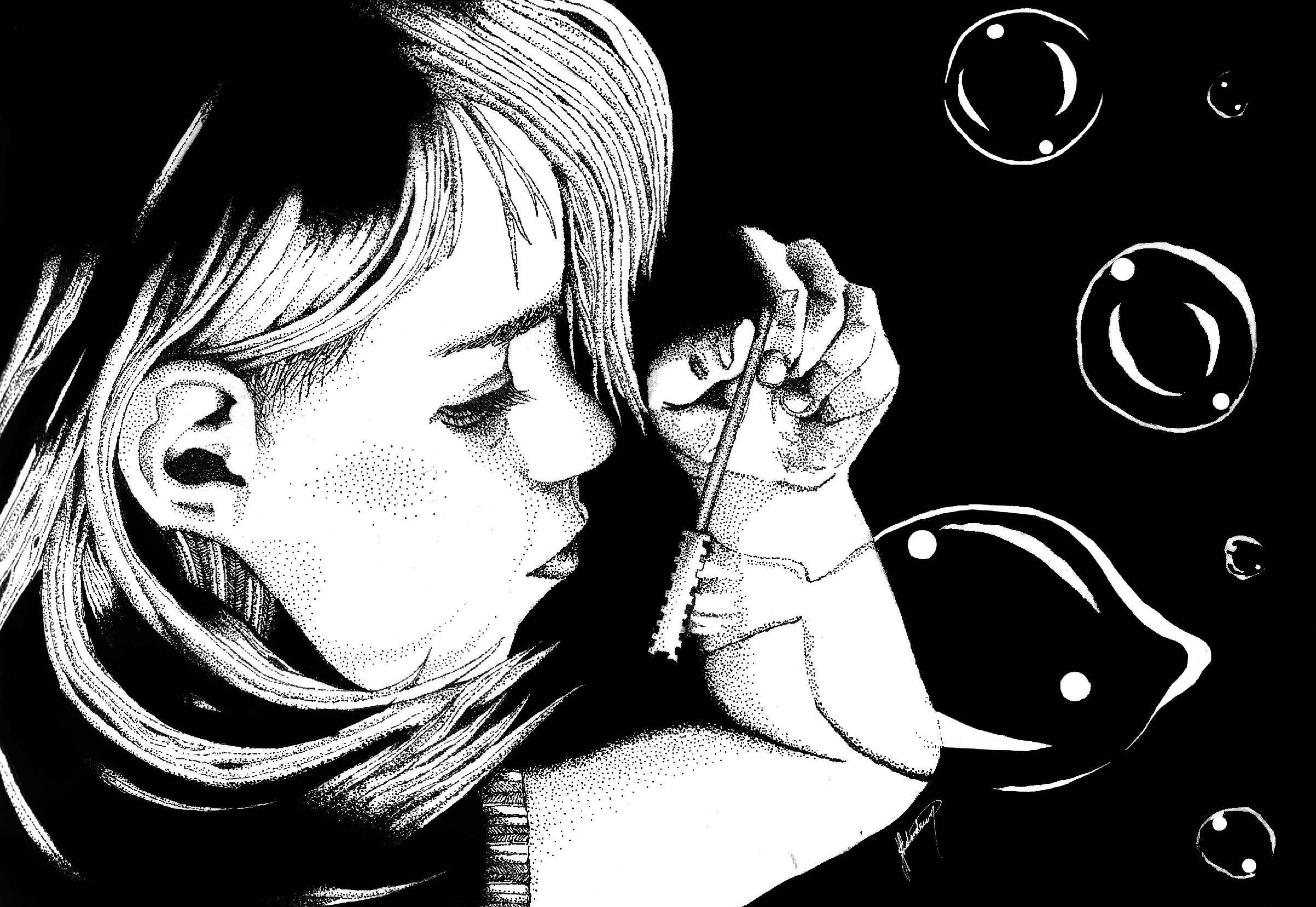
Bubbles – Illustration of my niece blowing bubbles
How do you make your art?
My favourite things to draw with are ink and fine-tipped pens. I like to use stippling and heavy shadows and contrast in my artworks and using a lot of black ink really gets this job done well! When making my more formal science illustrations and figures, however I like to use a graphics tablet and Adobe Illustrator and Photoshop.
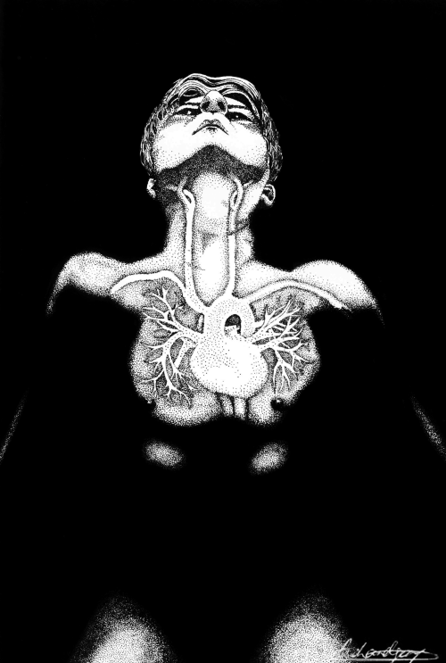
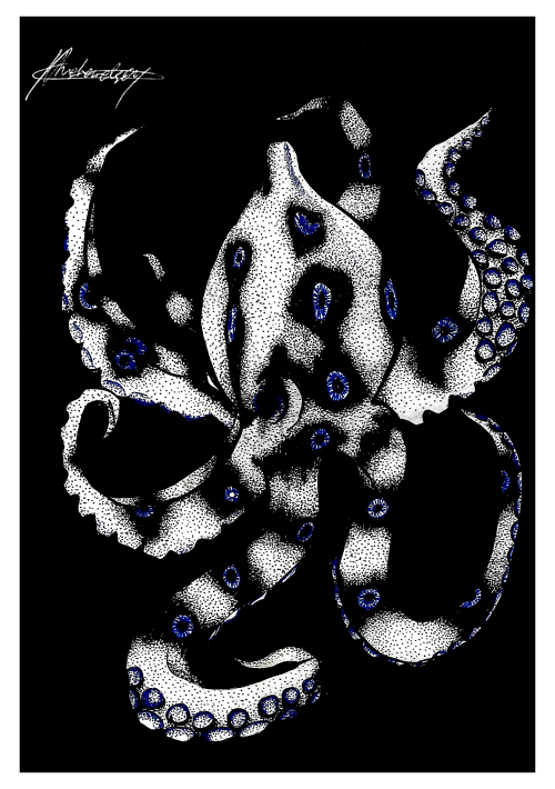
Left: Resonance – Drawing of the cardiovascular system, inspired by magnetic resonance imaging and other medical imaging techniques that allow us to see into the living human body. Right: Blue Ringed Octopus – Illustration of a blue ringed octopus
Does your art influence your science at all, or are they separate worlds?
I didn’t realise how much my art influenced my science until I began to present my scientific work. Trying to explain my results and ideas, and more importantly trying to communicate them quickly during my presentations and in publications made me focus on making figures and diagrams that could easily convey complex information. While it is a cliché that ‘a picture is worth a thousand words’, I strongly believe this to be true, and that really helps when you have a set word limit!
While it is a cliché that ‘a picture is worth a thousand words’, I strongly believe this to be true, and that really helps when you have a set word limit!
I think that the mental and physical act of trying to represent complicated ideas visually can teach you a lot about what is important in your own work and hence where to go next in your experiments. For instance, even having to ask simple questions like “what can I draw in a high level of detail? What aspects can I only draw very vaguely?” can give insight into where there are holes in your own knowledge, or even what might be unknown in the field and waiting to be discovered.
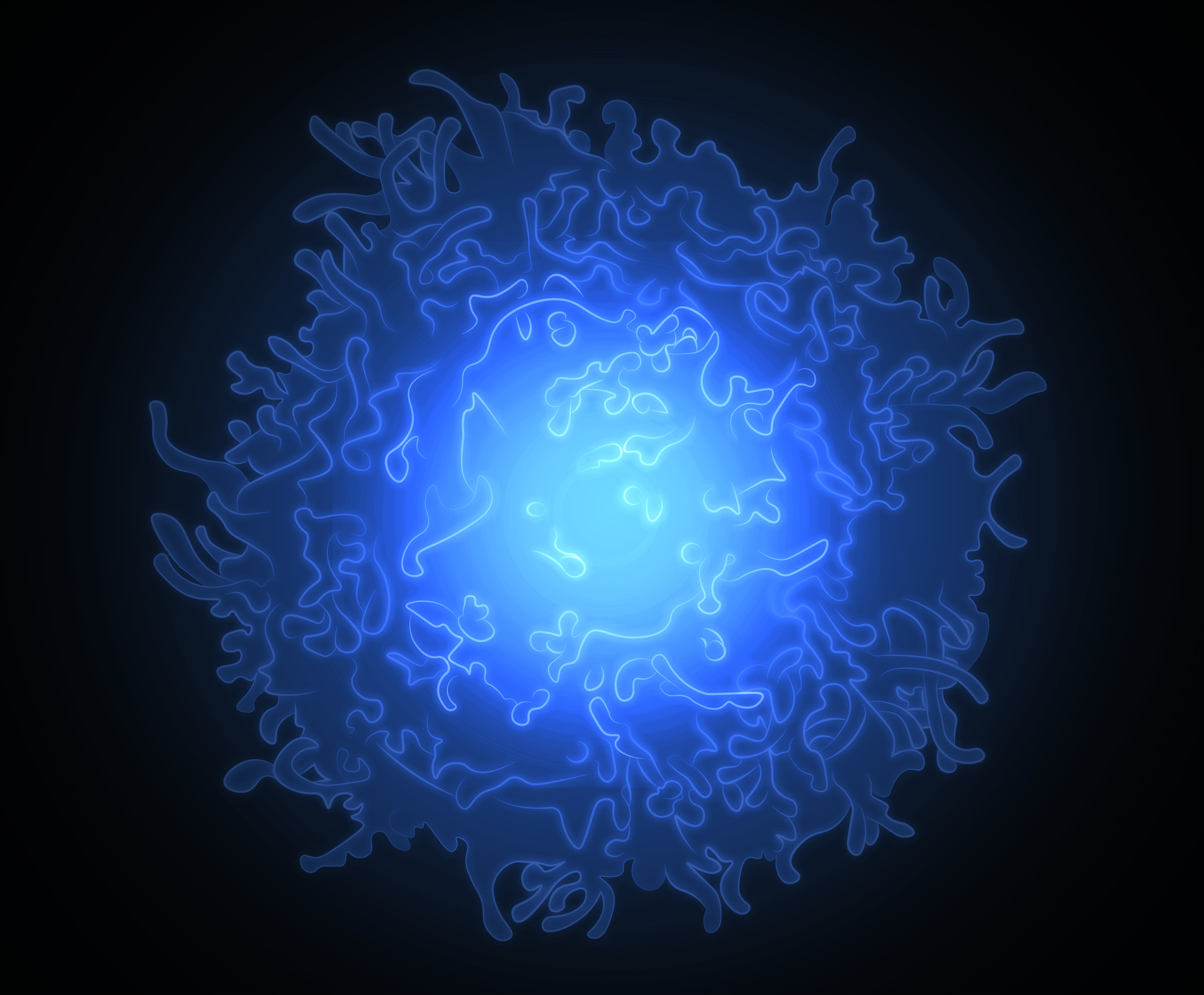
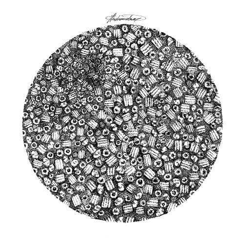
Freeze – Illustration of a cryo-electron microscopy sample investigating bacterial chaperonin proteins.
What are you thinking of working on next?
I am currently finishing the final stages of my PhD and am really excited to see what’s next for me in research. I hope to continue making illustrations to communicate my own research (and others!). In the future, I’d really love to start a science web comic as well as design online visual educational tools for learning STEMM subjects. I have long-term ambitions about creating or joining a consultancy for science communication, helping other scientists to create figures and drawings which are not only useful and accurate, but visually appealing too.
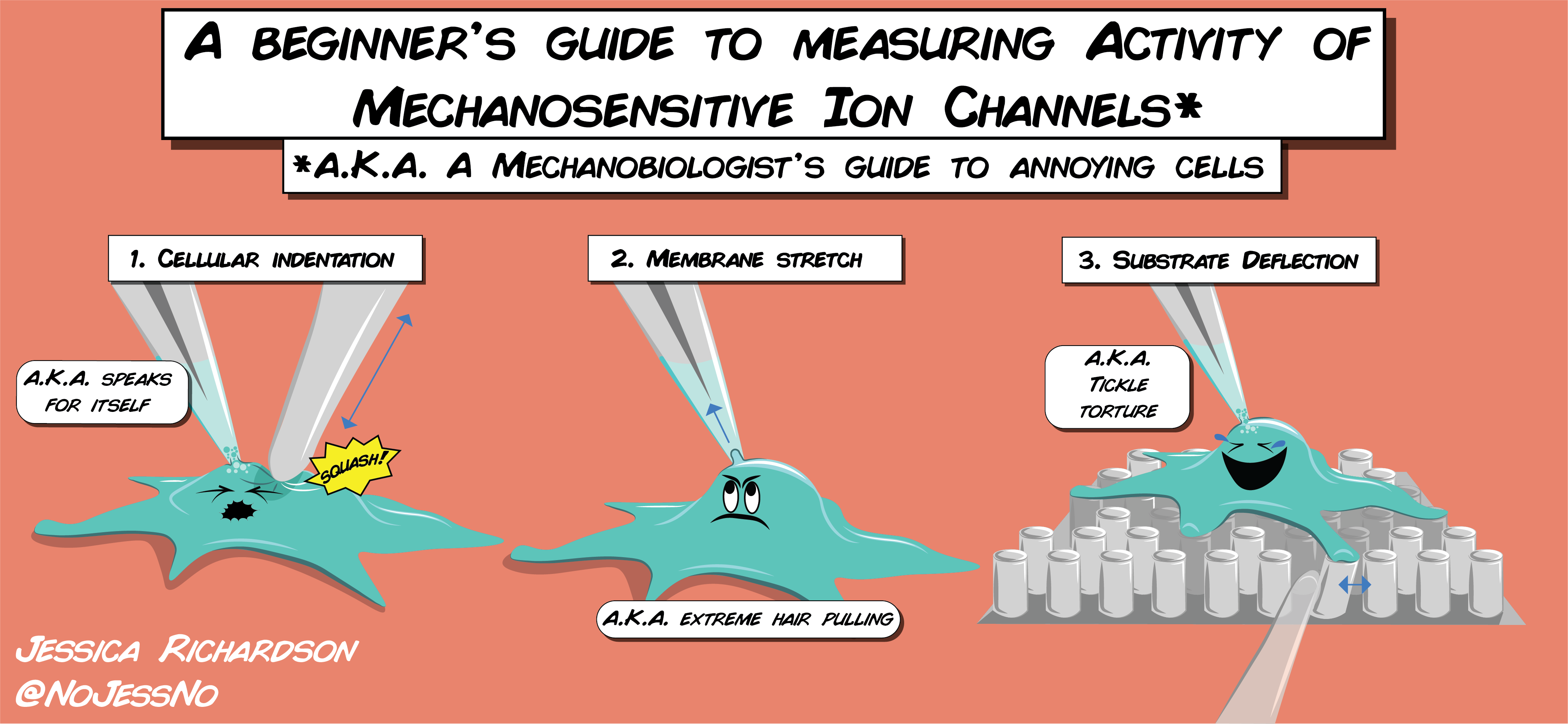
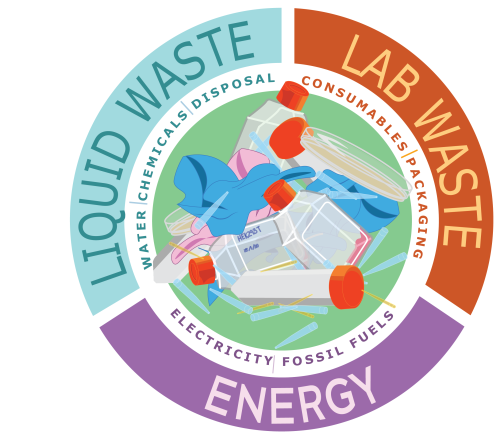
A Beginner’s Guide – A beginner’s guide to measuring mechanically activated ion channel activity in cells. Many mechanical stimuli can be applied experimentally to cells including: 1) cellular indentation, where the cell is physically compressed using a blunt glass probe 2) high speed pressure clamp which applies membrane stretch by applying pressure changes to the cell membrane via a micropipette 3) substrate deflections, where cells can be cultured onto a bed of elastic cylindrical pili which can be individually deflected to apply a mechanical stimulus at the base of cells All of these techniques can be combined with single-cell patch clamp electrophysiology to measure electrical activity in cells.Lab Waste – Poster illustration for a lab campaign to raise awareness about the environmental impact of life science research and areas of focus to decrease our footprint.
Thanks to Jessica and all the other SciArtists we have featured so far.We’re looking for new people to feature in this series – whatever kind of art you do, from sculpture to embroidery to music to drawing, if you want to share it with the community just email thenode@biologists.com (nominations are also welcome!)
On Wednesday 8 December, Development hosted three talks to celebrate the 25th anniversary of our zebrafish special issue.
Below you’ll find each of the talks, plus reflections on the impact of the special issue, 25 years on from its publication, from our guest chair Alex Schier.
Alex Schier (Harvard University and Biozentrum, University of Basel) Reflections on the 25th anniversary of the ‘zebrafish issue’
Caren Norden (Instituto Gulbenkian Ciência) ‘Lamination in the vertebrate retina: An interplay of diverse modes of cell migration’
You can read the preprint of the research presented by Caren here.
Frieda Leesch (Research Institute of Molecular Pathology) ‘A molecular network of conserved factors keeps ribosomes dormant in the egg’
The research presented by Frieda is available as a preprint on biorxiv
Brad Cairns (Huntsman Cancer Institute) ‘Maternally-inherited anti-sense piRNAs antagonize transposon expression in zebrafish and medaka embryos’
You can read the preprint of the research presented by Brad here.
 (No Ratings Yet)


 (1 votes)